. 2018 Jul 6:15:27.
doi: 10.1186/s12983-018-0271-z. eCollection 2018.

An atlas of larval organogenesis in the European shore crab Carcinus maenas L. (Decapoda, Brachyura, Portunidae)

Affiliations

An atlas of larval organogenesis in the European shore crab Carcinus maenas L. (Decapoda, Brachyura, Portunidae)

Franziska Spitzner et al. Front Zool. .

Abstract

Background: The life history stages of brachyuran crustaceans include pelagic larvae of the Zoea type which grow by a series of moults from one instar to the next. Zoeae actively feed and possess a wide range of organ systems necessary for autonomously developing in the plankton. They also display a rich behavioural repertoire that allows for responses to variations in environmental key factors such as light, hydrostatic pressure, tidal currents, and temperature. Brachyuran larvae have served as distinguished models in the field of Ecological Developmental Biology fostering our understanding of diverse ecophysiological aspects such as phenotypic plasticity, carry-over effects on life-history traits, and adaptive mechanisms that enhance tolerance to fluctuations in environmental abiotic factors. In order to link such studies to the level of tissues and organs, this report analyses the internal anatomy of laboratory-reared larvae of the European shore crab Carcinus maenas. This species has a native distribution extending across most European waters and has attracted attention because it has invaded five temperate geographic regions outside of its native range and therefore can serve as a model to analyse thermal tolerance of species affected by rising sea temperatures as an effect of climate change.

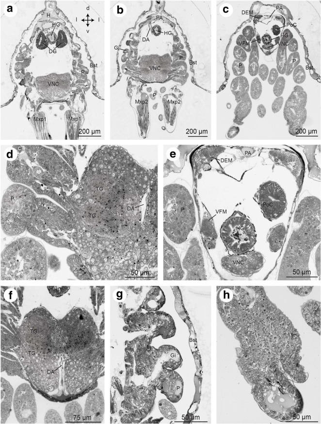
Results: Here, we used X-ray micro-computed tomography combined with 3D reconstruction to describe organogenesis in brachyuran larvae. We provide a detailed atlas of the larval internal organization to complement existing descriptions of its external morphology. In a multimethodological approach, we also used cuticular autofluorescence and classical histology to analyse the anatomy of selected organ systems.

Conclusions: Much of our fascination for the anatomy of brachyuran larvae stems from the opportunity to observe a complex organism on a single microscopic slide and the realization that the entire decapod crustacean bauplan unfolds from organ anlagen compressed into a miniature organism in the sub-millimetre range. The combination of imaging techniques used in the present study provides novel insights into the bewildering diversity of organ systems that brachyuran larvae possess. Our analysis may serve as a basis for future studies bridging the fields of evolutionary developmental biology and ecological developmental biology.

Keywords: 3D reconstruction; Central nervous system; Excretion; Locomotion; Metamorphosis; Micro-CT; Osmoregulation; Sensory systems.


Conflict of interest statement

The research presented in this paper complies with the guidelines from the directives 2010/63/EU of the European parliament and of the Council of 22nd September 2010 on the protection of animals used for scientific purposes.The authors declare that they have no competing interests.Springer Nature remains neutral with regard to jurisdictional claims in published maps and institutional affiliations.

Figures

Fig. 1
Fig. 1
a – Larval development of Carcinus maenas L. (Decapoda, Brachyura, Portunidae) modified from Rice and Ingle (1975). b – ontogenetic change of appendage function during the double metamorphosis (see text for further details). Abbreviations: Ey - compound eye, DS - dorsal spine, Md – mandibular segment, Mx1, Mx2 – segments of 1st and 2nd maxilla, P1–5 - pereiopod one to five, PM1–6 - pleomeres one to six, RS - rostral spine, T1-8 – thoracomeres one to eight
Fig. 2
Fig. 2
a - lateral view of a Zoea IV (autofluorescence of cuticle). b - higher magnification of specimen in A showing cephalic and thoracic appendages, the colour code corresponds to Fig. 1A. c: Zoea IV, 3D-reconstruction of selected organs (colour code see Fig. 3) to indicate the sectioning planes shown in Figs. 9, 10, 11, 12, 13, 14, 15, 16 and 17. Abbreviations: A1 - first antenna, A2 - second antenna, Da - dactylus of the first pereiopod (chela), Md - mandible, Mx1 - first maxilla, Mx2 - second maxilla, Mxp1 - first maxilliped, Mxp2 - second maxilliped, Mxp3 - third maxilliped, P - pereiopod anlagen, Pl - pleopod anlagen, Pr - Propodus of the first pereiopod (chela)
Fig. 3
Fig. 3
Larval morphology and organogenesis from the Zoea I to the Megalopa based on micro-CT analyses, dorsal views (left panels) and lateral views (right panels). Isosurface views of the cuticle merged with 3D reconstructions of selected organ systems. Crossed arrows indicate the orientation (a - anterior, d - dorsal, l - lateral, p - posterior, v - ventral)
Fig. 4
Fig. 4
Ontogeny of selected organs based on micro-CT analyses, lateral views of 3D reconstructions. Crossed arrows indicate the orientation (a - anterior, d - dorsal, p - posterior, v - ventral). Abbreviations: AC - anterior caeca, AL1-2 - anterior lobe 1-2, Br – median brain, CEC - circumoesophageal connective, CG - commissural ganglion, CS - cardiac stomach, DEM - dorsal extensor muscles, DL - dorsal lobe, Eso - oesophagus, EyNP - eyestalk neuropil, FG - foregut, HG - hindgut, MG - midgut, PlG - pleonal ganglia, PLM – pleopod muscles, PoC - posterior caecum, PosL1-2 - posterior lobe 1-2, PS - pyloric stomach, SEG - suboesophageal ganglia, TG - thoracic ganglia, VFM - ventral flexor muscles, VL - ventral lobe, VNC - ventral nerve cord
Fig. 5
Fig. 5
Development of selected organs from the Zoea I to the Megalopa based on micro-CT-analyses, dorsal views of 3D reconstructions. Crossed arrows indicate the orientation (a - anterior, l -lateral, p - posterior). Abbreviations: AC - anterior caeca, AL1, 2 - anterior lobes 1, 2, Br – median brain, CEC - circumoesophageal connective, CG - commissural ganglion, DA - descending artery, DEM - dorsal extensor muscles, DL - dorsal lobe, Eso - oesophagus, EyNP - eyestalk neuropil, FG - foregut, HG - hindgut, MG - midgut, PlG - pleonal ganglia, PoC - posterior caecum, PosL1-2 - posterior lobe 1-2, SEG - suboesophageal ganglia, TG - thoracic ganglia, VFM - ventral flexor muscles, VL - ventral lobe, VNC - ventral nerve cord
Fig. 6
Fig. 6
Zoea IV, consecutive horizontal sections from micro-CT analysis, from dorsal (a) to ventral (i). In a and c, the organs are coloured according to code used in Fig. 5. Crossed arrows in I indicate the orientation (a - anterior, l -lateral, p - posterior). Abbreviations: A1 - first antenna, A2 - second antenna, AC - anterior caecum, AG - antennal gland, Br – median brain, Bst - epithelium of the branchiostegite, CS - cardiac stomach, DA – descending artery, DCL - deutocerebral chemosensory lobe, DEM – dorsal extensor muscle, DG - digestive gland, Eso - oesophagus, EyNP - eyestalk neuropil, Gi - gill, H - heart, HG - hindgut, Lb - labrum, Lu - lumen, MA - mandibular adductor musculature, Md - mandible, Mxp1 - first maxilliped, Mxp2 - second maxilliped, PNT - projection neuron tract, P - pereiopod anlagen, Pl - pleopod anlagen, PoC - posterior caecum, PS - pyloric stomach, Ros - rostrum, SEG - suboesophageal ganglia, TG - thoracic ganglia, VFM - flexor muscles, VNC –ventral nerve cord. Scale bars represent 200 μm
Fig. 7
Fig. 7
Zoea IV, consecutive lateral sections (a - f) from micro-CT analysis. In e and f, the organs are coloured according to code used in Fig. 5. Crossed arrows in A indicate the orientation (a - anterior, d – dorsal, p – posterior, v - ventral). Abbreviations: A1 - first antenna, A2 - second antenna, AC - anterior caecum, AG - antennal gland, Br – median brain, Bst - epithelium of the branchiostegite, CS - cardiac stomach, CV – cardio-pyloric valve, DA - descending artery, DCL - deutocerebral chemosensory lobe, DEM – dorsal extensor muscle, DG - digestive gland, Eso - oesophagus, EyNP - eyestalk neuropil, Gi - gill, H - heart, HG - hindgut, Lb - labrum, Lu – lumen of digestive gland, MA - mandibular adductor musculature, Md - mandible, Mxp1 - first maxilliped, Mxp2 - second maxilliped, Mxp3 - third maxilliped, PNT - projection neuron tract, P - pereiopod anlagen, Pl - pleopod anlagen, PoC - posterior caecum, PS - pyloric stomach, Ros - rostrum, SEG - suboesophageal ganglia, TG - thoracic ganglia, VFM - flexor muscles, VNC - ventral nerve cord. Scale bars represent 200 μm
Fig. 8
Fig. 8
a - Zoea IV, isosurface of micro-CT-scans showing the external pores of the dorsal organ (black arrowheads) between the attachment sites of the mandibular adductor muscles (circles). Asterisks indicate the attachment of single muscle bundles. Crossed arrows indicate the orientation (a - anterior, d – dorsal, p – posterior, v - ventral). b – Zoea IV, consecutive frontal semi-thin sections through the pores (arrowheads) of the dorsal organ (anterior is towards the top). Abbreviations: AA - anterior aorta, Cu - cuticle, DS - dorsal spine, Ey - compound eye, MA - mandibular adductor musculature, RS - rostral spine
Fig. 9
Fig. 9
Frontal histological sections of Zoea IV (Holländer stain). a - overview of the anterior part of the head with stalked compound eyes and eyestalk neuropils (EyNP), see Fig. 2c for section plane; b - higher magnification of the right compound eye showing the visual neuropils lamina (La), medulla (Me), and lobula (Lo), proximally adjoined by the complex of the hemiellipsoid body neuropil and the medulla terminalis (HN/TM); b` - visual chiasm (arrow head) between medulla and lamina; c – the compound eye consists of many single ommatidia (one ommatidium surrounded by a line), note that the cornea is detached because of poor fixation (↔ double arrow); c`- cross sections of ommatidia show that the crystalline cones are composed of four cone cells. Each cell of one crystalline cone is indicated by an arrowhead; d - the ommatidia extend bundles of neurites (Neu) into the lamina; e – a branch of the ophthalmic artery (OA) extends towards the eyestalk neuropils; e`- the two main branches of the ophthalmic artery (OA) course towards the eyestalks; f – the protocerebral tract (PCT) connects the eyestalk neuropils and protocerebrum (PC) of median brain. Abbreviations: A1 - first antenna, Cap - capillary, CL - corneal lens, Co - cornea of compound eye, Cry - crystalline cone, Cu - cuticle, DP - distal pigment, Ey - compound eye, EyNP - eyestalk neuropils, HN/TM - complex of hemiellipsoid body neuropil and medulla terminalis, La - lamina, Lo - lobula, Me - medulla, Neu - neurites, OA - ophthalmic artery, Om - ommatidium of compound eye, PC - protocerebrum, PCT - protocerebral tract, Pi - shielding pigment, Rh - rhabdomeres of photoreceptor cell, Ros - rostrum, RS - rostral spine
Fig. 10
Fig. 10
Frontal histological sections of Zoea IV (Holländer stain). a - overview of the anterior part of the head showing the eyestalk neuropils (EyNP), and medially the most anterior part of the protocerebrum (PC), see Fig. 2C for section plane; b - section slightly posterior to A showing the ophthalmic artery (OA) dorsal to the protocerebrum (PC); c - clusters of olfactory sensory neurons (OSN, circled and labelled with black arrowheads) within the first antenna send bundles of dendrites (De, white arrowheads) into aesthetasc sensilla (AES); c`- autofluorescence image of first antenna (A1) with aesthetascs (AES); d - axons (Ax) of olfactory sensory neurons extend towards the median brain; e – putative anlage of the statocyst (St) at the base of the first antenna; f - tegumental gland (TeG) on the dorsal side of the rostrum (Ros); note that cuticle (Cu) and epidermis are detached (double arrow); g - anterior aorta (AA) in the rostrum (Ros); h – the anterior aorta (AA) opens into to the auxiliary heart, the cor frontale (CF), from which the ophthalmic artery (OA) emerges; i – the cephalic artery (CA) emerges from the cor frontale. The anterior dorsal cells (ADC) are located dorsal to the protocerebral neuropil. The stomatogastric ganglion (StG) within the cor frontale is flanked by the anterior gastric muscles (AGM); j – higher magnification slightly posteriorly than I showing the cor frontale muscles (CFM) ventral to the stomatogastric ganglion (StG); k – the cephalic artery (CA) is flanked by the anterior dorsal cells (ADC) of the protocerebrum as it penetrates into the protocerebrum. The stomatogastric nerve (StN) connects the stomatogastric ganglion to the protocerebrum (PC). The anterior part of the hematopoietic tissue (HPT) is located dorsally to the cor frontale that embraces the stomatogastric ganglion. Abbreviations: A1 - first antenna, AA - anterior aorta, ADC - anterior dorsal cells of the protocerebrum, AES - aesthetasc, AGM - anterior gastric muscle, Ax - axon, CA - cephalic artery, Cap - capillary, CF - cor frontale, CFM - cor frontale muscle, Cu - cuticle, De - dendrites, EyNP - eyestalk neuropils, HPT - hematopoietic tissue, OA - ophthalmic artery, OSN - olfactory sensory neurons, PC - protocerebrum, Ros - rostrum, St – putative statocyst anlage, StG - stomatogastric ganglion, StN - stomatogastric nerve, TeG - tegumental gland
Fig. 11
Fig. 11
Frontal histological sections of Zoea IV (Holländer stain). a - overview showing the median brain and anterior part of the cephalothorax, see Fig. 2C for section plane; b - overview slightly posterior to A showing the prominent mandibular adductor musculature (MA) dorsally, on the ventral side the labrum (Lb) and second antenna (A2). Hematopoietic tissue is associated dorsally and laterally with the wall of the cardiac stomach (CS); c – the unpaired central body (CB) is located in the centre of the protocerebrum that is dorsally surmounted by the anterior dorsal cells (ADC). The stomatogastric nerve (STN) is also visible. d – the deutocerebrum is characterized by spherical deutocerebral chemosensory lobes (DCL) on both sides. The projection neuron tract (PNT) emerges from the left DCL. e – rosette-shaped tegumental gland (TeG) in the labrum. Abbreviations: A1 - first antenna, A2 - second antenna, AA - anterior aorta, ADC - anterior dorsal cells of the protocerebrum, AGM - anterior gastric muscle, CA - cephalic artery, CB - central body, CF - cor frontale, CFM - cor frontale muscle, CS - cardiac stomach, Cu - cuticle, DC - deutocerebrum, DCL - deutocerebral chemosensory lobe, EyNP - eyestalk neuropil, HPT - hematopoietic tissue, Lb - labrum, MA - mandibular adductor musculature, PC - protocerebrum, PNT - projection neuron tract, Ros - rostrum, StG - stomatogastric ganglion, StN - stomatogastric nerve, TeG - tegumental gland
Fig. 12
Fig. 12
Frontal histological sections of Zoea IV (Holländer stain). a - section showing the median brain at the level of the deutocerebral chemosensory lobes and the cardiac stomach, see Fig. 2C for section plane; b - section slightly more posterior to A showing the tritocerebrum (TC) flanking the oesophagus (Eso), and the anterior lobes of the digestive gland with its lumen (Lu) and large lipid inclusions (black dots). The right antennal gland (AG) is also visible. c - mandibular adductor musculature (MA) displays the characteristic cross striation; d - the anterior part of the cardiac stomach (CS) inside is lined with cuticle (arrowheads); e - inner portion of the anlage of the mandibular palp located inside of the mandible; e`- autofluorescence of the mandible with the outer portion of the mandibular palp anlage (arrowhead). Abbreviations: AA - anterior aorta, AG - antennal gland, CS - cardiac stomach, DCL - deutocerebral chemosensory lobe, DG - digestive gland, Eso - oesophagus, HPT - hematopoietic tissue, Lb - labrum, Lu – lumen of digestive gland, MA - mandibular adductor musculature, Md - mandible, MdP – mandibular palp, TC - tritocerebrum, TeG - tegumental gland
Fig. 13
Fig. 13
Frontal histological sections of Zoea IV (Holländer stain). a - section through the median part of the cardiac stomach (CS), see Fig. 2c for section plane; b - higher magnification of the cardiac stomach with the gastric mill that comprises two lateral teeth (LT) and one median tooth (MT), note the thickened cuticle of the teeth as indicated by black arrowheads; c - higher magnification of the antennal gland; c` - the bladder (BL) of the antennal gland opens to the outside through a nephropore (No). Abbreviations: AA - anterior aorta, AC - anterior caecum, AG - antennal gland, Bl - bladder, CEC - circumoesophageal connective, CMsc - contractor muscles of the cardiac stomach, CoeSac - coelomosac, CS - cardiac stomach, CSac - cardiac sac, DG - digestive gland, DGM - dorsal gastric muscles, Eso - oesophagus, LT - lateral tooth, Ly - labyrinth, MA - mandibular adductor musculature, Md - mandible, MdP – mandibular palp, MT - median tooth, No - nephropore, TC – tritocerebrum
Fig. 14
Fig. 14
Frontal histological sections of Zoea IV (Holländer stain). a - section of the cephalothorax with the cardiac stomach (CS) in the centre. Dorsally, the paired anterior caeca (AC) are located. Ventrally the oesophagus (Eso) opens to the mouth field, laterally flanked by the mandibles (Md); compare Fig. 2c for section plane; b - posterior part of the cardiac stomach with sclerotized lateral teeth (LT). Ventrally, the ventral gastric muscles are attached (VGM). c - details of the pyloric stomach with gastric mill. d - higher magnification of the sclerotized mandible (Md), note the massive cuticle (Cu) of the molar process (MP); e - detail of the dilatator muscles (DMsc) attached to the oesophagus (Eso). Abbreviations: AA - anterior aorta, AC - anterior caecum, AG - antennal gland, AR - ampullary ridge, ASS - ampullary setal screen of the gland filter, CEC - circumoesophageal connective, CMsc - dilatator muscles of the cardiac stomach, CS - cardiac stomach, Cu - cuticle, DG - digestive gland, DGM - dorsal gastric muscles, DLC - dorsolateral pyloric channel, DMsc - dilatator muscles of the oesophagus, DPC - dorsal region of the pyloric chamber, DPF - dorsal pyloric fold, Eso - oesophagus, FP - filter press, IAR - interampullary ridge, IP – incisor process, LAC - lower ampullary chamber, MA - mandibular adductor musculature, Md - mandible, MdP – mandibular palp, MP – molar process, MT - median tooth, PPO - prepyloric ossicle, PSS - pyloric setal screen, UAC - upper ampullary chamber, VGM – ventral gastric muscle
Fig. 15
Fig. 15
Frontal histological sections of Zoea IV (Holländer stain). a - section showing the median portion of the pyloric stomach (PS) laterally flanked by the lobes of the midgut gland, see Fig. 2c for section plane; b - section of the anterior part of the midgut (MG) with the dorsal heart (H) and the ventral nerve cord (VNC) at the level of the suboesophageal ganglia. Dorsally, the VNC is crossed by muscles (Msc) associated with the maxillae; c - higher magnification of rosette-shaped tegumental glands of the mandible; d - the heart is surrounded by the pericardial cavity (PCav) into which the ostia (O) open. Asterisks identify putative trabeculae; E - cross section of musculature (asterisks and arrow) laterally to the heart (H) underneath the branchiostegites (Bst). Abbreviations: AA - anterior aorta, AC - anterior caecum, CEC - circumoesophageal connective, DG - digestive gland, H - heart, Lu – lumen of digestive gland, MA - mandibular adductor musculature, MG - midgut, Msc - musculature of the maxillae, PS - pyloric stomach, TeG - tegumental gland, VNC - ventral nerve cord
Fig. 16
Fig. 16
Frontal histological sections of Zoea IV (Holländer stain). a - cross-section showing the lobes of the digestive gland (DG) opening into the midgut (MG), see Fig. 2c for section plane; b - close-up of the interface of the digestive gland to the midgut (MG). Cell-types of the digestive gland identified include embryonic cells (E), blister like cells (B) and resorptive cells (R). c - section across the posterior lobes of the midgut gland and the anterior part of the hindgut (HG); d - the Y-organ (Y) is located underneath the lateral carapace close to the interface with the branchiostegites (Bst); e - distal part of a digestive gland lobe showing lumen (Lu) surrounded by epithelium composed of E- and F-cells. f - section through a lobe of the digestive gland showing epithelium with R- and B-cells surrounding the lumen (Lu). g - section of the digestive gland more posteriorly than A, the B-, F-, R-, and E-cells are identified. Abbreviations: B – blister like cell, Bst - epithelium of branchiostegite, DG - digestive gland, E - embryonic cell, F - fibrillary cell, H - heart, HG - hindgut, Lu – lumen of digestive gland, MG - midgut, R - resorptive cell, VNC - ventral nerve cord, Y - Y-organ
Fig. 17
Fig. 17
Frontal histological sections of Zoea IV (Holländer stain). a - section through the distal part of the digestive gland (DG) with the two posterior lobes flanking the hindgut (HG), see Fig. 2c for section plane. b - section showing the posterior artery (PA) as well as the descending artery (DA). c - section through the first pleon segment with the hindgut (HG) and its blindly ending posterior caecum (PoC) as well as pereiopod anlagen (P). d - higher magnification of the ventral nerve cord (VNC), penetrated by the descending artery (DA). e - higher magnification of the first pleon segment shown in B, the flexor muscles (VFM) and extensor muscles (DEM) of the pleon are identified. f - higher magnification of the ventral nerve cord (VNC) as it is penetrated by the descending artery (DA). g - undifferentiated tissue of pereiopod (P) and gill anlagen (Gi) surrounded by the lateral branchiostegites; h - close-up of undifferentiated tissue of a pereiopod anlage. Abbreviations: Bst - epithelium of branchiostegite, DA - descending artery, DEM - extensor muscle, DG - digestive gland, Gi - gill, H - heart, HG - hindgut, Mxp1 - first maxilliped, Mxp2 - second maxilliped, P - pereiopod anlagen 1 - 5, PA - posterior artery, PoC - posterior caecum, TG – thoracic ganglia, VFM - flexor muscle, VNC - ventral nerve cord

Similar articles

Cited by

References

    1. Williamson DI. Larval morphology and diversity. In: Abele LG, editor. The Biology of Crustacea: 2. Embryology, morphology and genetics. New York: Academic press; 1982. pp. 43–110.
    1. Ingle RW. Larval stages of north-eastern Atlantic crabs: an illustrated key. London: Chapman & Hall; 1992.
    1. Anger K. The Biology of Decapod Crustacean Larvae. 1. Lisse: A.A. Balkema Publishers; 2001.
    1. Anger K. Contributions of larval biology to crustacean research: a review. Invertebr Reprod Dev. 2006;49:175–205. doi: 10.1080/07924259.2006.9652207. - DOI
    1. Martin JW, Olesen J, Hoeg JT. Atlas of Crustacean Larvae. Maryland: JHU Press; 2014.

LinkOut - more resources