

Current understanding of trigeminal ganglion structure and function in headache
- PMID: 29989427
- PMCID: PMC7007999
- DOI: 10.1177/0333102418786261
Current understanding of trigeminal ganglion structure and function in headache
Abstract
Introduction: The trigeminal ganglion is unique among the somatosensory ganglia regarding its topography, structure, composition and possibly some functional properties of its cellular components. Being mainly responsible for the sensory innervation of the anterior regions of the head, it is a major target for headache research. One intriguing question is if the trigeminal ganglion is merely a transition site for sensory information from the periphery to the central nervous system, or if intracellular modulatory mechanisms and intercellular signaling are capable of controlling sensory information relevant for the pathophysiology of headaches.
Methods: An online search based on PubMed was made using the keyword "trigeminal ganglion" in combination with "anatomy", "headache", "migraine", "neuropeptides", "receptors" and "signaling". From the relevant literature, further references were selected in view of their relevance for headache mechanisms. The essential information was organized based on location and cell types of the trigeminal ganglion, neuropeptides, receptors for signaling molecules, signaling mechanisms, and their possible relevance for headache generation.
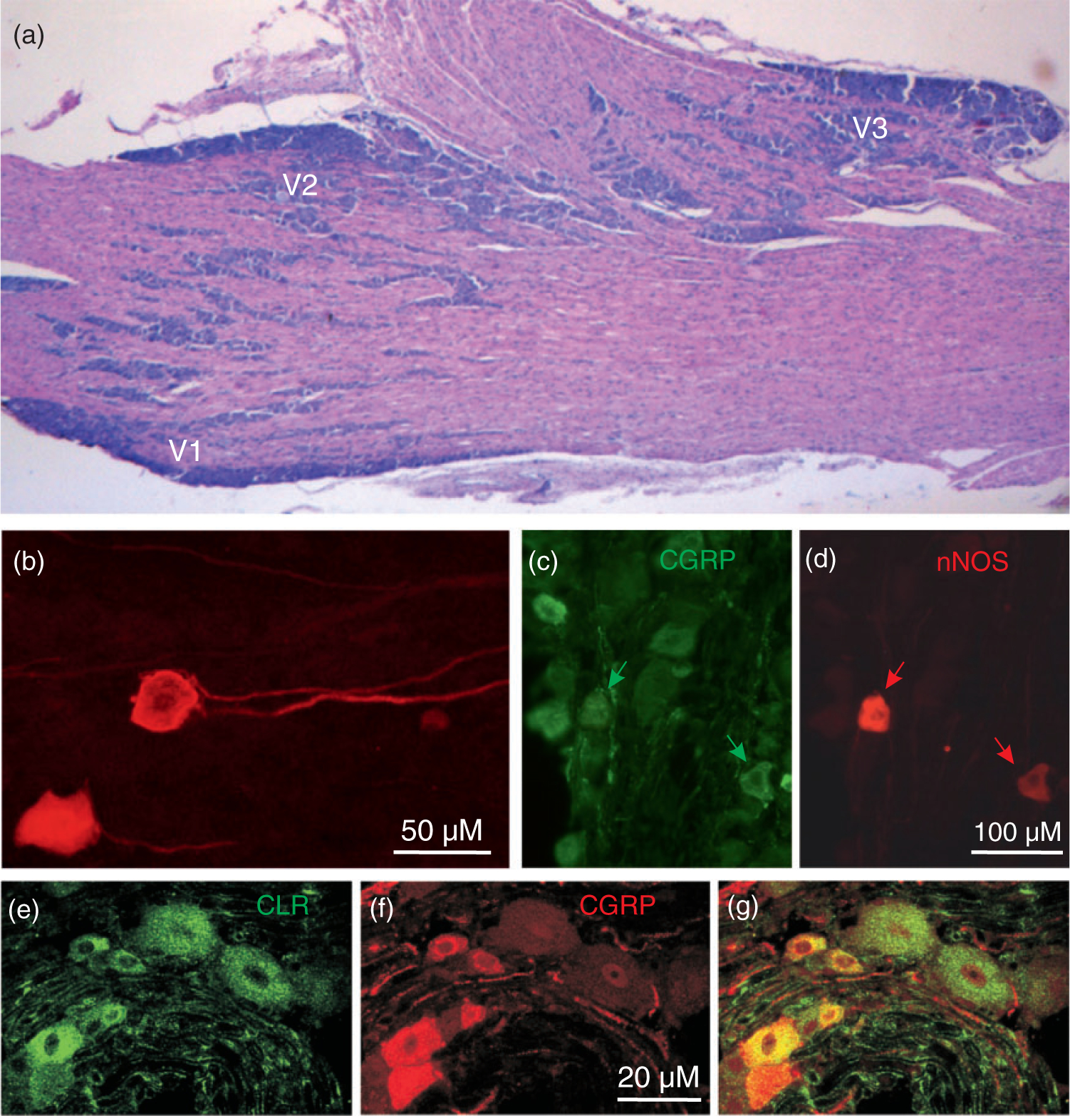
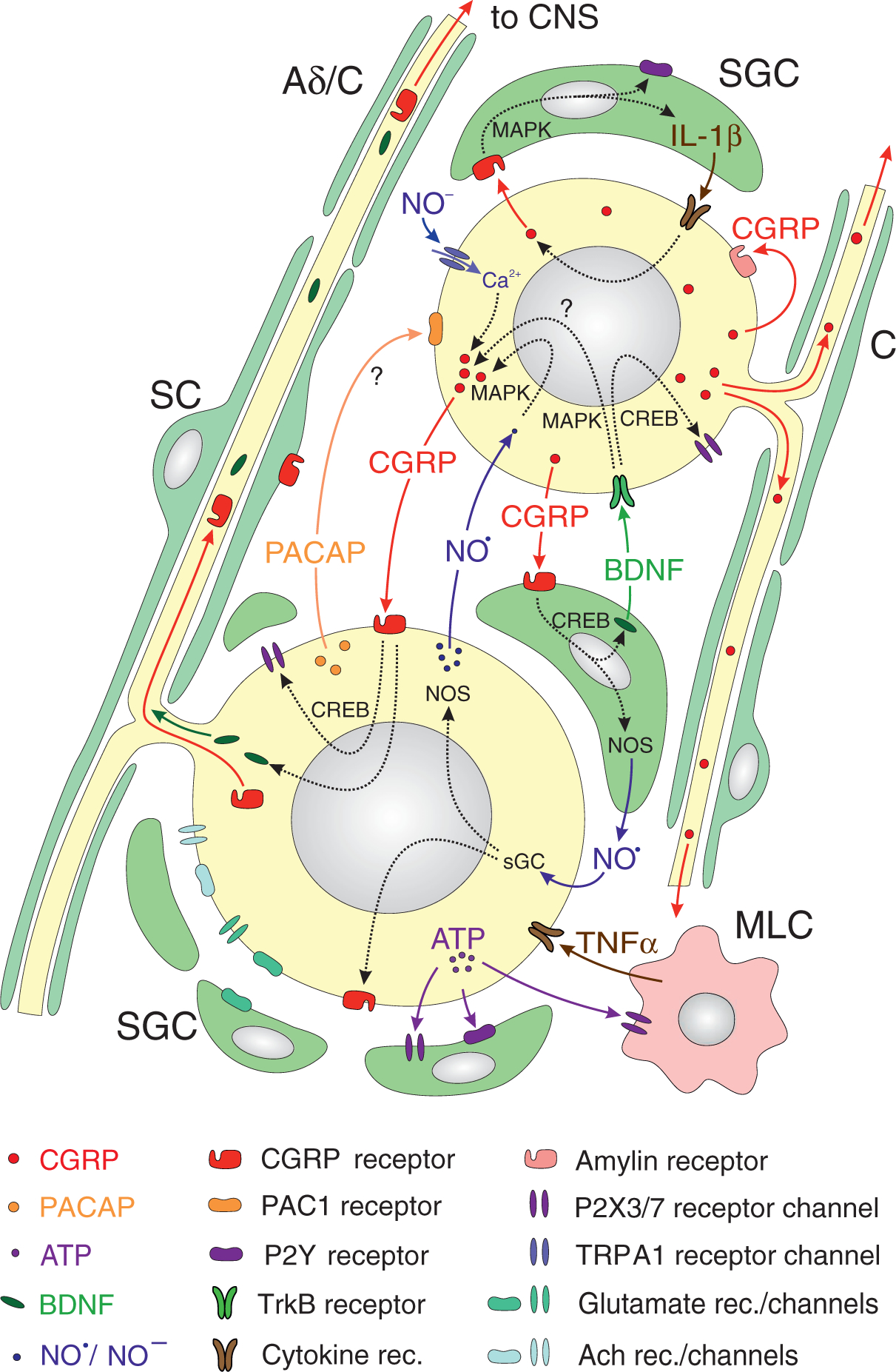
Results: The trigeminal ganglion consists of clusters of sensory neurons and their peripheral and central axon processes, which are arranged according to the three trigeminal partitions V1-V3. The neurons are surrounded by satellite glial cells, the axons by Schwann cells. In addition, macrophage-like cells can be found in the trigeminal ganglion. Neurons express various neuropeptides, among which calcitonin gene-related peptide is the most prominent in terms of its prevalence and its role in primary headaches. The classical calcitonin gene-related peptide receptors are expressed in non-calcitonin gene-related peptide neurons and satellite glial cells, although the possibility of a second calcitonin gene-related peptide receptor in calcitonin gene-related peptide neurons remains to be investigated. A variety of other signal molecules like adenosine triphosphate, nitric oxide, cytokines, and neurotrophic factors are released from trigeminal ganglion cells and may act at receptors on adjacent neurons or satellite glial cells.
Conclusions: The trigeminal ganglion may act as an integrative organ. The morphological and functional arrangement of trigeminal ganglion cells suggests that intercellular and possibly also autocrine signaling mechanisms interact with intracellular mechanisms, including gene expression, to modulate sensory information. Receptors and neurotrophic factors delivered to the periphery or the trigeminal brainstem can contribute to peripheral and central sensitization, as in the case of primary headaches. The trigeminal ganglion as a target of drug action outside the blood-brain barrier should therefore be taken into account.
Keywords: CGRP; Trigeminal neurons; neuromodulation; neuropeptides; satellite glial cells; signaling.
Conflict of interest statement
Declaration of conflicting interests
The authors declared no potential conflicts of interest with respect to the research, authorship, and/or publication of this article.
Figures
References
-
- MacIver MB and Tanelian DL. Free nerve ending terminal morphology is fiber type specific for A delta and C fibers innervating rabbit corneal epithelium. J Neurophysiol 1993; 69: 1779–1783. - PubMed
-
- Andres KH, von Düring M, Muszynski K, et al. Nerve fibres and their terminals of the dura mater encephali of the rat. Anat Embryol (Berl) 1987; 175: 289–301. - PubMed
-
- Messlinger K, Hanesch U, Baumgärtel M, et al. Innervation of the dura mater encephali of cat and rat: Ultrastructure and calcitonin gene-related peptide-like and substance P-like immunoreactivity. Anat Embryol (Berl) 1993; 188: 219–237. - PubMed
-
- Ray BS and Wolff HG. Experimental studies on headache: Pain sensitive structures of the head and their significance in headache. Arch Surg 1940; 1: 813–856.
-
- Young RF and Stevens R. Unmyelinated axons in the trigeminal motor root of human and cat. J Comp Neurol 1979; 183: 205–214. - PubMed
Publication types
MeSH terms
Substances
Grants and funding
LinkOut - more resources
Full Text Sources
Other Literature Sources
Medical
Research Materials
