. 2018;14(11):1898-1910.
doi: 10.1080/15548627.2018.1491489. Epub 2018 Aug 10.

The lysosomal membrane protein LAMP2A promotes autophagic flux and prevents SNCA-induced Parkinson disease-like symptoms in the Drosophila brain

Affiliations

The lysosomal membrane protein LAMP2A promotes autophagic flux and prevents SNCA-induced Parkinson disease-like symptoms in the Drosophila brain

Abdul-Raouf Issa et al. Autophagy. 2018.

Abstract

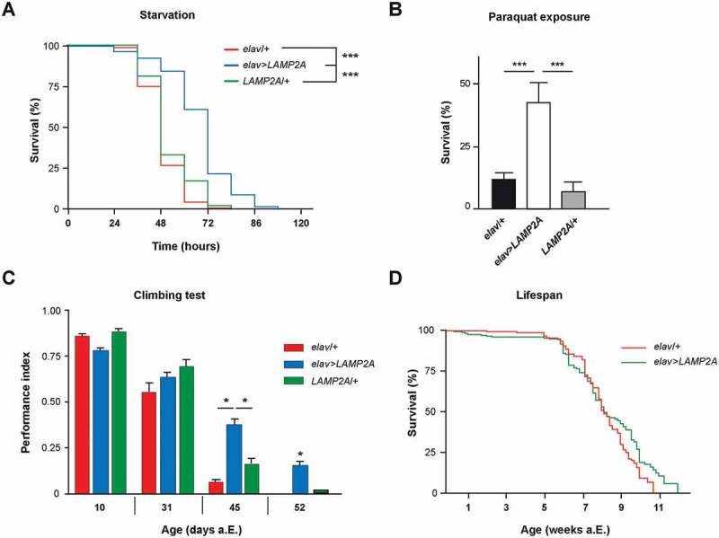
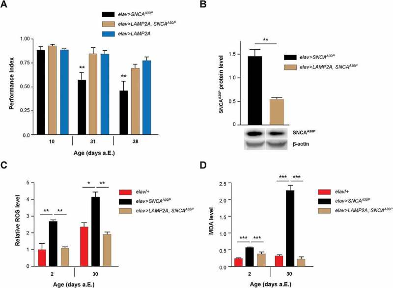
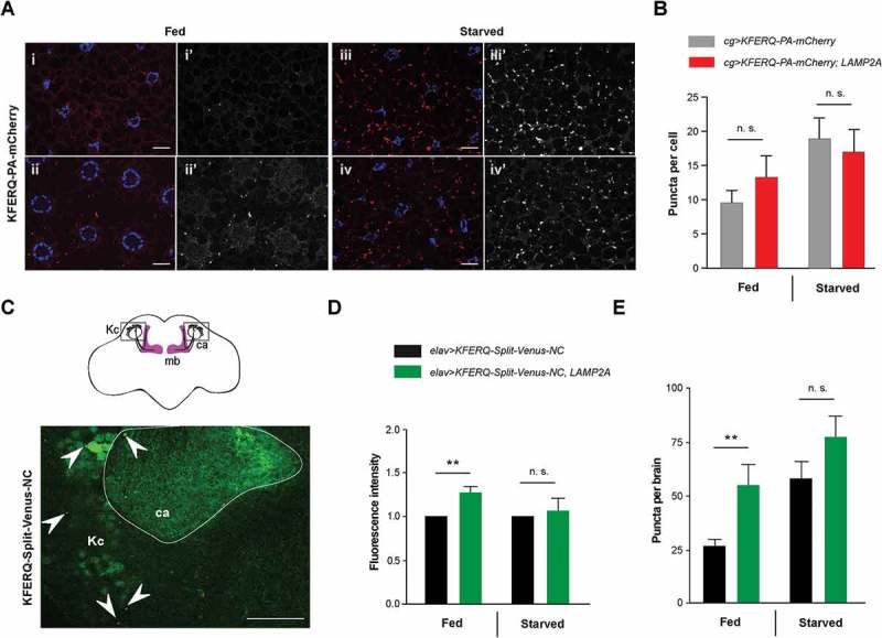
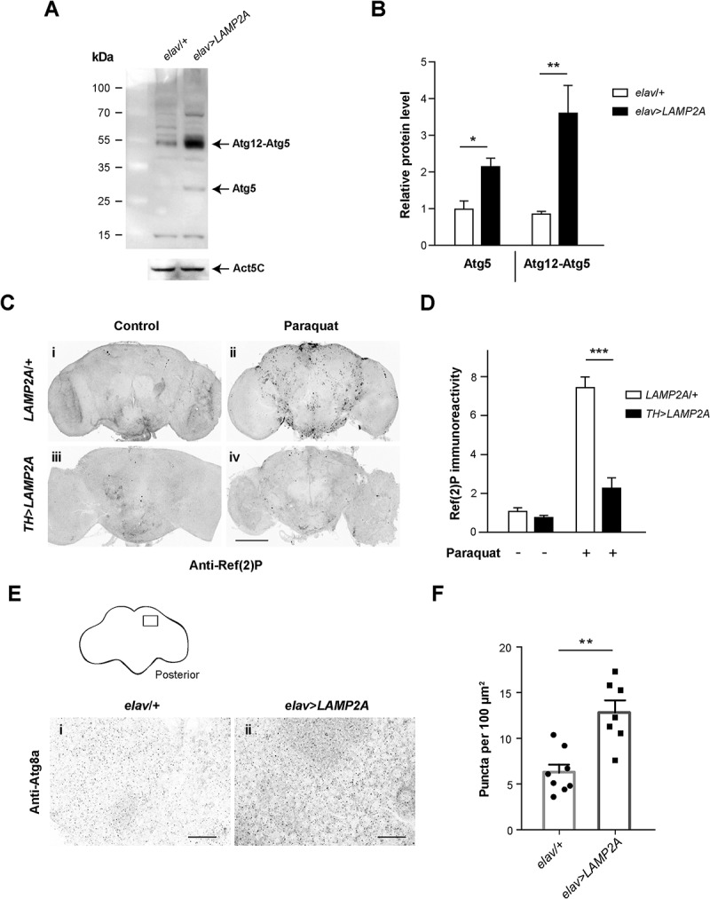
The autophagy-lysosome pathway plays a fundamental role in the clearance of aggregated proteins and protection against cellular stress and neurodegenerative conditions. Alterations in autophagy processes, including macroautophagy and chaperone-mediated autophagy (CMA), have been described in Parkinson disease (PD). CMA is a selective autophagic process that depends on LAMP2A (lysosomal-associated membrane protein 2A), a mammal and bird-specific membrane glycoprotein that translocates cytosolic proteins containing a KFERQ-like peptide motif across the lysosomal membrane. Drosophila reportedly lack CMA and use endosomal microautophagy (eMI) as an alternative selective autophagic process. Here we report that neuronal expression of human LAMP2A protected Drosophila against starvation and oxidative stress, and delayed locomotor decline in aging flies without extending their lifespan. LAMP2A also prevented the progressive locomotor and oxidative defects induced by neuronal expression of PD-associated human SNCA (synuclein alpha) with alanine-to-proline mutation at position 30 (SNCAA30P). Using KFERQ-tagged fluorescent biosensors, we observed that LAMP2A expression stimulated selective autophagy in the adult brain and not in the larval fat body, but did not increase this process under starvation conditions. Noteworthy, we found that neurally expressed LAMP2A markedly upregulated levels of Drosophila Atg5, a key macroautophagy initiation protein, and that it increased the density of Atg8a/LC3-positive puncta, which reflects the formation of autophagosomes. Furthermore, LAMP2A efficiently prevented accumulation of the autophagy defect marker Ref(2)P/p62 in the adult brain under acute oxidative stress. These results indicate that LAMP2A can potentiate autophagic flux in the Drosophila brain, leading to enhanced stress resistance and neuroprotection.

Abbreviations: Act5C: actin 5C; a.E.: after eclosion; Atg5: autophagy-related 5; Atg8a/LC3: autophagy-related 8a; CMA: chaperone-mediated autophagy; DHE: dihydroethidium; elav: embryonic lethal abnormal vision; eMI: endosomal microautophagy; ESCRT: endosomal sorting complexes required for transport; GABARAP: GABA typeA receptor-associated protein; Hsc70-4: heat shock protein cognate 4; HSPA8/Hsc70: heat shock protein family A (Hsp70) member 8; LAMP2: lysosomal associated membrane protein 2; MDA: malondialdehyde; PA-mCherry: photoactivable mCherry; PBS: phosphate-buffered saline; PCR: polymerase chain reaction; PD: Parkinson disease; Ref(2)P/p62: refractory to sigma P; ROS: reactive oxygen species; RpL32/rp49: ribosomal protein L32; RT-PCR: reverse transcription polymerase chain reaction; SING: startle-induced negative geotaxis; SNCA/α-synuclein: synuclein alpha; SQSTM1/p62: sequestosome 1; TBS: Tris-buffered saline; UAS: upstream activating sequence.

Keywords: Autophagy-lysosome pathway; Drosophila melanogaster; Parkinson disease; lysosomal-associated membrane protein 2A (LAMP2A); neuroprotection; synuclein alpha (SNCA).


Figures

Figure 1.
Figure 1.
The LAMP2A receptor promotes stress resistance and neuroprotection in Drosophila. (a) Starvation resistance. Expression of the human LAMP2A protein in all neurons significantly extended Drosophila survival upon prolonged starvation (elav>LAMP2A flies) compared to elav-Gal4/+ (elav/+) driver and UAS-LAMP2A/+ (LAMP2A/+) effector controls. (b) Paraquat exposure. Survival of elav>LAMP2A flies fed with 20 mM paraquat for 72 h was markedly increased compared to the elav/+ and LAMP2A/+ controls. (c) Effect on age-related locomotor decline. Pan-neuronal LAMP2A expression (elav>LAMP2A flies) significantly delayed age-related decrements in climbing performance (SING assay) compared to driver and effector controls that behaved like the wild type. (d) Lifespan assay. elav/+ and elav>LAMP2A flies showed similar longevity curves (median lifespan 57 and 55 days, respectively) indicating that neuronal LAMP2A expression does not affect Drosophila lifespan.
Figure 2.
Figure 2.
LAMP2A prevents SNCA-induced behavioral and oxidative defects in Drosophila. (a) LAMP2A coexpression fully prevented the progressive locomotor defects induced by pan-neuronal SNCAA30P. Climbing ability (SING assay) of elav>LAMP2A, SNCAA30P flies was compared to that of elav>SNCAA30P and elav>LAMP2A flies at 10, 31 and 38 days after a.E. (b) Human LAMP2A reduced neuronal SNCA accumulation. Western blots of head protein extracts from 30-day-old elav>SNCAA30P flies compared to elav>LAMP2A, SNCAA30P probed with anti-SNCA antibody. Act5C was used as a loading control. Quantification of SNCAA30P protein level from 3 independent experiments. Coexpression of LAMP2A reduced SNCAA30P accumulation without decreasing its mRNA level (see Fig. S2A). (c) ROS levels in the brain of elav>LAMP2A, SNCAA30P flies were lower than those of elav>SNCAA30Pand comparable to the elav/+ control at both 2 and 30 days a.E. Representative pictures of DHE-labeled brains are shown in Figure S2B. (d) MDA concentration assayed in the brain of 2- and 30-day-old adult Drosophila as an index of lipid peroxidation. Brain MDA level was markedly increased in elav>SNCAA30P flies but not in elav>LAMP2A, SNCAA30P flies that show similar levels as the elav/+ control.
Figure 3.
Figure 3.
Effect of LAMP2A on selective autophagy in the larval fat body and adult brain. (a) In 3rd-instar larval fat body cells, LAMP2A expression (LAMP2A, bottom panels) did not increase KFERQ-PA-mCherry fluorescent sensor puncta formation 25 h after photoactivation, either under fed (i, ii) or starvation (iii, iv) conditions, compared to controls (top panels). In composite images, mCherry fluorescence is in red and DAPI-stained nuclei are in blue. i’-iv’ monochromatic images show the KFERQ-PA-mCherry single channel. Scale bars: 20 µm. (b) Quantification of puncta number per cell in larvae expressing photoactivated KFERQ-PA-mCherry in fat body with or without (control) LAMP2A under fed or starvation conditions. Starvation-induced sensor puncta formation was not further increased by LAMP2A expression. Similar results were obtained using a different eMI biosensor (KFERQ-Split-Venus) (shown in Figure S3). (c) Localization of reconstituted KFERQ-Split-Venus sensor fluorescent puncta (arrowheads) in a posterior region of the adult brain of elav>KFERQ-Split-Venus-NC flies expressing the eMI sensor in all neurons. The square in the scheme (top inset) shows localization of the magnified brain region that surrounds the calyx of the mushroom body, where fluorescence was prominent and in which puncta were scored. mb, mushroom body; Kc, cell bodies of the mushroom body Kenyon cells; ca, calyx. Representative scans of whole brain and calyx region for the different genotypes and feeding conditions are shown in Figure S4a and b, respectively. (d, e) Reconstitution of KFERQ-Split-Venus eMI sensor was increased in adult brain of fed, but not starved, flies expressing LAMP2A in all neurons (elav>KFERQ-Split-Venus-NC, LAMP2A) (LAMP2A, right panel), as indicated by higher overall fluorescence level (d) and increased density of eMI-positive puncta in the calyx region (e), compared to elav>KFERQ-Split-Venus-NC controls. Quantification from 3 independent experiments.
Figure 4.
Figure 4.
Human LAMP2A enhances macroautophagy in the Drosophila brain. (a, b) Effect of LAMP2A on Atg5 expression. (a) Western blot of head protein extracts from 10-day old control flies (elav/+) and flies expressing human LAMP2A in neurons (elav>LAMP2A) probed with anti-Atg5 antibody. LAMP2A expression markedly increased levels of Atg5 and of the Atg12-Atg5 complex that is required for autophagosome formation. Act5C served as a loading control. (b) Quantification of Atg5 protein and the Atg12-Atg5 complex from 3 independent western blot experiments. (c, d) Effect of LAMP2A on paraquat-induced Ref(2)P accumulation. (c) Anti-Ref(2)P immunostaining in whole-mount adult brains of LAMP2A/+ (panels i and ii) and TH>LAMP2A (panels iii and iv) flies exposed to paraquat (panels ii and iv) or not (panels i and iii). LAMP2A expression prevented paraquat-induced Ref(2)P accumulation (black puncta) suggesting that the human protein is able to maintain efficient autophagic flux under oxidative stress. Scale bar: 100 μm. (d) Quantification of Ref(2)P immunostaining in the central brain region normalized to LAMP2A/+ control not exposed to paraquat (n = 4 or 5 independent brains per condition). (e, f) Effect of LAMP2A on the number of Atg8a-positive puncta. (e) Anti-Atg8a immunostaining in whole-mount adult brains of elav/+ (panel i) and elav>LAMP2A (panel ii) flies. The inset scheme on top shows the posterior neuropil region that was magnified in panels i and ii and in which Atg8a puncta were counted. Scale bars: 10 µm. (f) Quantification of Atg8a-positive dots. Each black circle (elav/+) or square (elav>LAMP2A) represents the score for a different brain. The number of Atg8a puncta that reflect autophagosome formation was markedly increased in LAMP2A-expressing flies. Similar results were obtained in 3 independent experiments.

Similar articles

Cited by

References

    1. Boya P, Reggiori F, Codogno P.. Emerging regulation and functions of autophagy. Nat Cell Biol. 2013;15(7):713–720. - PMC - PubMed
    1. Parzych KR, Klionsky DJ. An overview of autophagy: morphology, mechanism, and regulation. Antioxid Redox Signal. 2014;20(3):460–473. - PMC - PubMed
    1. Menzies FM, Fleming A, Rubinsztein DC. Compromised autophagy and neurodegenerative diseases. Nat Rev Neurosci. 2015;16(6):345–357. - PubMed
    1. Bento CF, Renna M, Ghislat G, et al. Mammalian autophagy: how does it work? Annu Rev Biochem. 2016;85:685–713. - PubMed
    1. Feng Y, He D, Yao Z, et al. The machinery of macroautophagy. Cell Res. 2014;24(1):24–41. - PMC - PubMed

Publication types

MeSH terms

Substances