Practice Guideline
. 2018 Jul;20(7):602-634.
doi: 10.1177/1098612X18781391.

AAFP Feline Anesthesia Guidelines

Affiliations
Practice Guideline

AAFP Feline Anesthesia Guidelines

Sheilah A Robertson et al. J Feline Med Surg. 2018 Jul.

Abstract

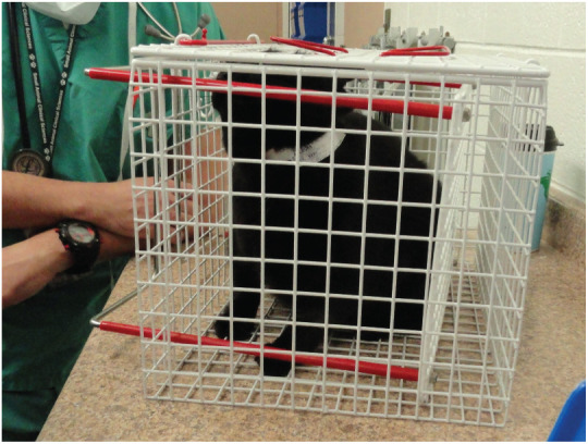
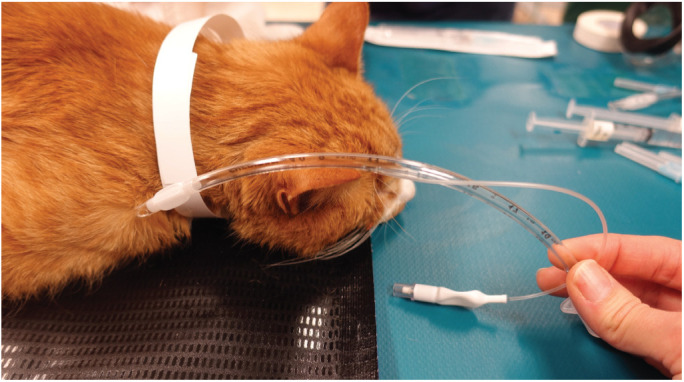
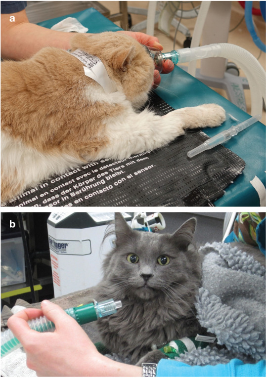
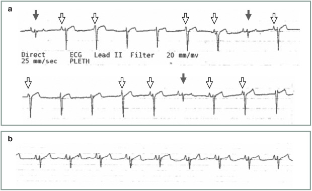
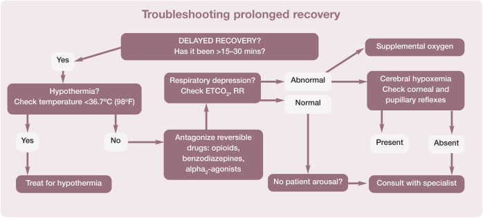
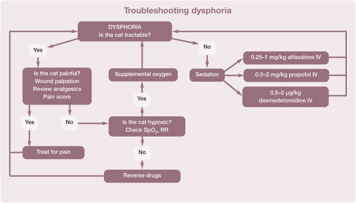
The overarching purpose of the AAFP Anesthesia Guidelines (hereafter referred to as the 'Guidelines') is to make anesthesia and sedation safer for the feline patient. Scope and accessibility: It is noteworthy that these are the first exclusively feline anesthesia guidelines authored by an expert panel, making them particularly useful as an extensively referenced, practical resource for veterinary practice teams. Because much of the key content is presented in tabular or visual format, the Guidelines have a high level of accessibility and convenience that invites regular usage. While the recommendations in the Guidelines focus primarily on client-owned cats, the content is also applicable to community-sourced animals with an unknown medical history.

Keywords: Anesthesia; airway management; anesthetic equipment; comorbidities; monitoring; sedation.


Conflict of interest statement

The Panel members have no conflicts of interest to declare.

Figures

Figure 1
Figure 1
Bain non-rebreathing circuit (NRC) adapter with in-circuit manometer (white arrow) and safety pop-off valve (black arrow). Courtesy of Heidi Shafford
Figure 2
Figure 2
Example of a battery-powered high pressure alarm with an adapter (white arrow) that is inserted into the expiratory limb of a Bain NRC. Courtesy of Heidi Shafford
Figure 3
Figure 3
Dead space (in red; black arrows) associated with the patient end of various types of anesthetic breathing circuit: (a) Bain circuit; (b) T piece circuit; (c) Norman elbow circuit; (d) rebreathing circuit; and (e) rebreathing circuit with divider. Courtesy of Peter Pascoe
Figure 4
Figure 4
Dead space related to various capnograph adapters. (a) Side-stream adapter for use with a removable sampling line; arrow shows diameter width. (b) An ‘all-in-one’ side-stream adapter; arrow shows diameter width. (c) A pediatric adapter; note the small diameter of the lumen, as shown by the arrow. The diameter of the capnograph adapter should always exceed the internal diameter of the ETT. (d) Low dead space ETT adapters are available for attachment to side-stream sampling lines. Courtesy of Gregg Griffenhagen
Figure 5
Figure 5
Multi-step protocol for the management of fearful cats for anesthesia
Figure 6
Figure 6
Key recommendations for minimizing anxiety and facilitating handling of feline patients presented for general anesthesia. Photograph courtesy of Sheilah Robertson
Figure 7
Figure 7
Specific considerations for each feline life stage.,,– ASA = American Society of Anesthesiologists. Photographs courtesy of Sheilah Robertson (neonatal/pediatric/kitten) and Susan Gogolski (junior, adult/prime, mature and senior/geriatric)
Figure 8
Figure 8
A cat can be transferred from a carrier to a humane trap for IM injection. (a) The door of the carrier has been removed and the carrier placed next to the humane trap. When the cat moves from one to the other, the acrylic door is slid into place. For injection, an injection guard (pictured, with holes and a handle, in [b]) is used to squeeze the cat against the back of the cage, and the IM injection can be made through one of the holes. Courtesy of Sheilah Robertson
Figure 9
Figure 9
When ready to inject, one person pulls the handles (red) towards themselves so the cat cannot turn around and is gently held in place; another person can then inject the cat IM. Squeeze cages are available with both top and end doors. Courtesy of Sheilah Robertson
Figure 10
Figure 10
The ETT is measured before placement; the tip should lie at the point of the shoulder when inserted. Mark (eg, with tape) or note the number on the tube that lines up with the incisors, and when intubating stop at this point. The tube can be cut to shorten it. Courtesy of Sheilah Robertson
Figure 11
Figure 11
The tip of the ETT should lie near the point of the shoulder. Proper placement can be assured by measuring the tube, as described in Figure 10. Tracheal tears can occur in cats when the cuff is overinflated and carry a poorer prognosis if they are intrathoracic (for example at the location of the ETT cuff shown in this radiograph). Courtesy of Sheilah Robertson
Figure 12
Figure 12
(a) Preoxygenation for 3 mins using a face mask is recommended if tolerated by the cat. (b) If the cat does not tolerate a face mask, a flow-by technique can be used. Courtesy of Sheilah Robertson (a) and Heidi Shafford (b)
Figure 13
Figure 13
(a) View of cat’s larynx with the mouth wide open and the cat in sternal recumbency. Under normal circumstances the ventral surface of the epiglottis would be dorsal to the soft palate, but opening the mouth and pulling the tongue produces this view. The tip of the laryngoscope should be placed just rostral to the epiglottis and pressed down to bring the opening of the larynx into view. (b) The same view as Figure 13a in a clinical patient. Courtesy of Chrisoula A Toupadakis Skouritakis (a) and Sheilah Robertson (b)
Figure 14
Figure 14
A 1 ml syringe being advanced over the top of the laryngeal opening and 0.2 ml of 2% lidocaine being dropped on to the top of the arytenoids. This will take 60–90 s to work so, once the lidocaine has been applied, the cat should be put back on oxygen until this time has elapsed. Courtesy of Chrisoula A Toupadakis Skouritakis
Figure 15
Figure 15
ETT being advanced into the larynx. Note that the bevel is vertical so that the end of the tube can act like a wedge to open up the vocal folds. The tube is advanced to the laryngeal opening and kept as ventral as possible. Advancing the tube on inspiration, when the laryngeal opening is at its widest, will provide the best conditions for ETT placement. Courtesy of Chrisoula A Toupadakis Skouritakis
Figure 16
Figure 16
(a) A soft ‘all-in-one’ mask that conforms to the cat’s face (a) and a mask with a removable rubber seal (b). Both are available in several sizes. Courtesy of Sheilah Robertson
Figure 17
Figure 17
A supraglottic airway device (v-gel) specifically designed for the cat’s pharyngeal and laryngeal anatomy. Courtesy of Docsinnovent, UK
Figure 18
Figure 18
The tip of the supraglottic airway device is lodged in the esophagus and the opening lies over the laryngeal opening; (a) overall view, (b) close-up view. Courtesy of Docsinnovent, UK
Figure 19
Figure 19
Recommended procedure for troubleshooting bradycardia in cats during anesthesia. An HR of <100 bpm may be associated with a decrease in cardiac output even in the face of normal blood pressure. Note that bradycardia may be accompanied by hypotension. The two algorithms (Figures 19 and 22) may need to be used together to achieve the best outcome, but bradycardia should usually be treated first. This algorithm may be downloaded; for details and key to abbreviations, see box on page 628
Figure 20
Figure 20
Recommended procedure for troubleshooting tachycardia (HR >180 bpm) in cats during anesthesia. This algorithm may be downloaded; for details and key to abbreviations, see box on page 628
Figure 21
Figure 21
Using the correct size of blood pressure cuff is important for accuracy. The cuff width should be 40% of the limb circumference, as shown in (a). The cuff is then placed around the most cylindrical portion of the limb (b). Courtesy of Sheilah Robertson
Figure 22
Figure 22
Recommended method for the treatment of hypotension during anesthesia. This algorithm may be downloaded; for details and key to abbreviations, see box on page 628
Figure 23
Figure 23
Recommended responses in the face of hypoxemia (SpO2 <90%). This algorithm may be downloaded; for details and key to abbreviations, see box on page 628
Figure 24
Figure 24
Recommendations for responding to abnormal capnograph readings during anesthesia. This algorithm may be downloaded; for details and key to abbreviations, see box on page 628
Figure 25
Figure 25
(a) ECG tracing from an anesthetized cat demonstrating synchronous atrioventricular dissociation. Note the largely negative QRS complexes that are junctional in origin, as well as the smaller, normally conducted QRS complexes (solid arrows). The P waves (open arrows) are seen to be masked by junctional escape beats, indicating a slightly faster rate of junctional depolarization as compared with sinus node depolarization. (b) ECG tracing from the same patient after administration of an anticholinergic agent (glycopyrrolate 0.01 mg/kg IV). Note the normal sinus rhythm, with slightly increased rate of sinus node depolarization and lack of junctional escape beats. ECG tracings in (a) and (b) were recorded at a paper speed of 25 mm/s. Courtesy of Gregg Griffenhagen
Figure 26
Figure 26
Steps that should be followed if the cat’s recovery is prolonged. This algorithm may be downloaded; for details and key to abbreviations, see box below
Figure 27
Figure 27
Recommended treatment steps for dealing with a dysphoric cat. This algorithm may be downloaded; for details and key to abbreviations, see box above
None
None
None
None
None
None
None
None
None
None
None
None
None
None

Comment in

References

    1. Dyson DH, Maxie MG, Schnurr D. Morbidity and mortality associated with anesthetic management in small animal veterinary practice in Ontario. J Am Anim Hosp Assoc 1998; 34: 325–335. - PubMed
    1. Brodbelt DC, Pfeiffer DU, Young LE, et al. . Risk factors for anaesthetic-related death in cats: results from the confidential enquiry into perioperative small animal fatalities (CEP-SAF). Br J Anaesth 2007; 99: 617–623. - PubMed
    1. Epstein ME, Rodan I, Griffenhagen G, et al. . AAHA/AAFP pain management guidelines for dogs and cats. J Feline Med Surg 2015; 17: 251–272. - PMC - PubMed
    1. Paepe D, Verjans G, Duchateau L, et al. . Routine health screening: findings in apparently healthy middle-aged and old cats. J Feline Med Surg 2013; 15: 8–19. - PMC - PubMed
    1. Vogt AH, Rodan I, Brown M, et al. . AAFP–AAHA feline life stage guidelines. J Am Anim Hosp Assoc 2010; 46: 70–85. - PubMed

Publication types

LinkOut - more resources