Practice Guideline
. 2018;96(1):1-11.
doi: 10.1159/000490193. Epub 2018 Jul 10.

Endobronchial Coils for Endoscopic Lung Volume Reduction: Best Practice Recommendations from an Expert Panel

Affiliations
Practice Guideline

Endobronchial Coils for Endoscopic Lung Volume Reduction: Best Practice Recommendations from an Expert Panel

Dirk-Jan Slebos et al. Respiration. 2018.

Abstract

Endobronchial coils are an additional treatment option for lung volume reduction in patients with severe emphysema. Patient selection should be focused on patients with severe emphysema on optimal medical therapy and with evidence of severe hyperinflation. The technique is suitable in a broad range of patients with emphysema; however, patients with paraseptal emphysema, large focal (giant) bullae, significant co-morbidity and airway-predominant disease should be avoided. Treatment involves placing between 10 and 14 coils by bronchoscopy in the selected treatment lobe, with 2 lobes being treated sequentially. Lobe selection for treatment should be based on quantitative computed tomography, and the lobes with the greatest destruction should be targeted (excluding the right middle lobe). The treatment results in an improvement in pulmonary function, exercise performance and quality of life, particularly in patients with severe hyperinflation (residual volume > 200% predicted) and upper-lobe heterogeneous emphysema, but will also be of benefit in lower-lobe predominant and homogeneous emphysema. Finally, it has an acceptable safety profile, although special attention has to be paid to coil-associated opacity which is an inflammatory response that occurs in some patients treated with endobronchial coils.

Keywords: Bronchoscopy; COPD; Endobronchial coils; Endoscopic lung volume reduction; Severe emphysema.


Figures

Fig. 1
Fig. 1
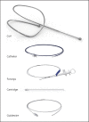
The PneumRx endobronchial coil system. The system consists of a single-patient use delivery system with a cartridge, catheter, guidewire, forceps and coils (with permission from PneumRx, CA, USA). The coil is available in 3 lengths (100, 125 and 150 mm) to accommodate the different airways. The distal and proximal ends of the coil are designed to reside in sub-segmental airways.
Fig. 2
Fig. 2
Coils implanted in homogeneous emphysema. Chest X-ray in a patient with homogeneous emphysema and severe hyperinflation. Twelve coils have been implanted in the right upper lobe and 10 in the left upper lobe. The distribution of the coils is around the upper part of the hilum, keeping enough distance to the pleurae. A small coil-associated opacity is seen around the most inferiorly located coil on the left side.
Fig. 3
Fig. 3
Optimal position of the bronchoscope and guidewire. a The bronchoscope is placed at the ostium of the target segment in a fixated manner. b The guidewire is advanced until the first marker is just outside the bronchoscope. In this position, mostly a coil of 125 mm can be placed in the target area of the lung (see Fig. 4).
Fig. 4
Fig. 4
Target area for coils. The ideal target area of coils is in the mid third of the lung; not too peripherally (the best distance to the pleura is 25–40 mm) and not too centrally (not proximally of the segmental ostium).
Fig. 5
Fig. 5
Coil deployment. Fluoroscopic series of a coil placement in the left upper lobe. Note that the bronchoscope is positioned all the time at the same position at the ostium of the segment. a Introduction of the guidewire up to one marker plus 1/3 length of the guidewire and 25–40 mm away from the pleura. Now, also the appropriate coil length can be chosen (in this case 125 mm). b The catheter is advanced over the guidewire (which is held in a stable position) just up to the distal end of the guidewire. c The guidewire is removed, leaving the catheter in place with just a radio-opaque marker being visible at the distal end. d Introduction of the coil, which is being pushed forward using the biopsy forceps until it reaches the distal end (marker) of the catheter. e The coil is still being pushed forward until the first half loop of the coil has been deployed. f From this point, the catheter is withdrawn, keeping the coil in the exact position, which requires gentle feeding/pushing of the biopsy forceps in the distal direction. g The catheter is now fully withdrawn (allowing full coil deployment), also over the biopsy forceps head to allow release of the coil. h Gentle backward force is applied to both the catheter and biopsy forceps, thereby retracting the coil system about 2 cm back, also to facilitate coil release. i In this position, the biopsy forceps is opened, and the coil will jump out, back to the previous position. Repeat these steps for the next coils to be placed.
Fig. 6
Fig. 6
Coil removal. The coil placement process can be reversed to reposition or retrieve the coil. First, grasp the proximal ball with the forceps. After aligning the forceps and coil by slight traction, the catheter can be gently advanced until the end of the coil but not distally from the coil. Afterwards, the coil and catheter can be withdrawn. Theoretically, the coil can be left at a more proximal position within the same retrieval procedure.
Fig. 7
Fig. 7
Don'ts during the coil placement. The most important don't is to advance the catheter distally beyond the guidewire (left picture). The distal end of the catheter is too rigid and too sharp and may lead to haemorrhage and pneumothorax. Another important don't is to use the catheter to push the guidewire in front (right picture). Allow the guidewire to do the job of finding the right distal bronchi.
Fig. 8
Fig. 8
Coil-associated opacity. Coil-associated opacities are dense consolidations on the X-ray, mimicking an organizing pneumonia. On the left chest X-ray, the consolidations are in the left upper lobe, both proximally near the hill and distally extending to the pleura. On the right picture, the coil-associated opacities are larger and more dense. The coil-associated opacity in the left upper lobe shows also pleural involvement. Sometimes, an extra side view or computed tomography scanning is necessary to demonstrate the coil-associated position.

References

    1. van Geffen WH, Kerstjens HAM, Slebos DJ. Emerging bronchoscopic treatments for chronic obstructive pulmonary disease. Pharmacol Ther. 2017;179:96–101. - PubMed
    1. Shah PL, Herth FJ, van Geffen WH, Deslee G, Slebos DJ. Lung volume reduction for emphysema. Lancet Respir Med. 2017;5:147–156. - PubMed
    1. Slebos DJ, Hartman JE, Klooster K, Blaas S, Deslee G, Gesierich W, et al. Bronchoscopic coil treatment for patients with severe emphysema: a meta-analysis. Respiration. 2015;90:136–145. - PubMed
    1. Herth FJF, Slebos DJ, Criner GJ, Shah PL. Endoscopic lung volume reduction: an expert panel recommendation - update 2017. Respiration. 2017;94:380–388. - PubMed
    1. Slebos DJ, Shah PL, Herth FJ, Valipour A. Endobronchial valves for endoscopic lung volume reduction: best practice recommendations from Expert Panel on Endoscopic Lung Volume Reduction. Respiration. 2017;93:138–150. - PMC - PubMed

Publication types