Case Reports
. 2018 Jun 4:30:37.
doi: 10.1186/s40557-018-0248-6. eCollection 2018.

Pneumoconiosis in a polytetrafluoroethylene (PTFE) spray worker: a case report with an occupational hygiene study

Affiliations
Case Reports

Pneumoconiosis in a polytetrafluoroethylene (PTFE) spray worker: a case report with an occupational hygiene study

Namhoon Lee et al. Ann Occup Environ Med. .

Abstract

Background: Using analysis of air samples from the workplace, we report on one case of pneumoconiosis in an individual who has been working in a polytetrafluoroethylene (PTFE) spraying process for 28 years.

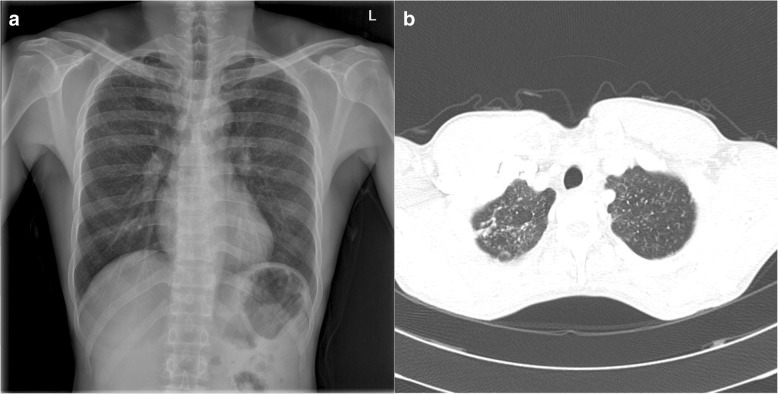
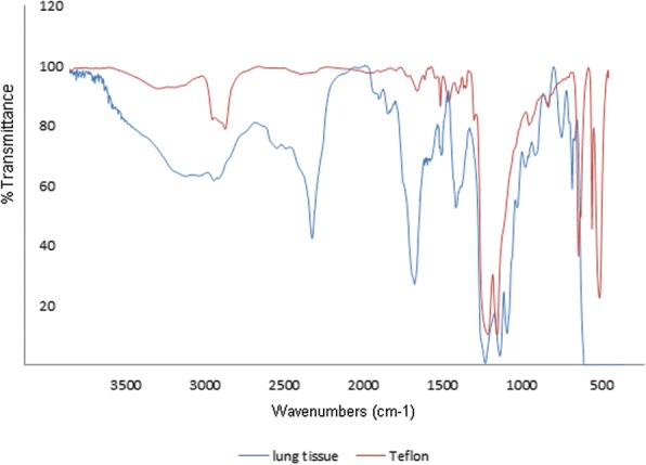
Case presentation: The patient was diagnosed with granulomatous lung disease caused by PTFE using computed tomography (CT), lung biopsy and electron microscopy. To assess the qualitative and quantitative exposure to PTFE in workplace, Fourier transform infrared spectroscopy (FT-IR), energy-dispersive X-ray spectroscopy (EDX) and thermogravimetric analysis (TGA) were performed on air samples from the workplace. The presence of PTFE particles was confirmed, and the airborne concentration of PTFE was estimated to be 0.75 mg/m3.

Conclusions: This case demonstrates that long-term exposure to PTFE spraying can cause granulomatous lung lesions such as pneumoconiosis; such lesions appear to be caused not by the degradation products of PTFE from high temperatures but by spraying the particles of PTFE. Along with air-sampling analysis, we suggest monitoring the concentration of airborne PTFE particles related to chronic lung disease.

Keywords: Occupational diseases; Pneumoconiosis; Polytetrafluoroethylene.


Conflict of interest statement

This study was approved by Keimyung university Dongsan medical center institutional review board. The number of approval is 2016–02–024-008.The authors declare that they have no competing interests.Springer Nature remains neutral with regard to jurisdictional claims in published maps and institutional affiliations.

Figures

Fig. 1
Fig. 1
a Chest radiograph showing multiple bilateral nodules in both lung fields. b CT scan of the chest showing numerous tiny nodules with perilymphatic distribution without lymph-node calcification
Fig. 2
Fig. 2
Histologic features of a case of polytetrafluoroethylene (PTFE) exposure-induced pulmonary granulomatous lesions (a and b). a The specimen shows mainly small airway- centered granulomatous micronodular lesions (arrow). b Higher magnification of the specimen shows multinucleated giant cells containing amorphous transparent particles (black arrows). c Polarizing microscopic finding of the same multinucleated giant cell found in Fig. b reveals the birefringent particle (white arrows). a to c Hematoxylin-Eosin stain (Magnification: A: × 40, B and C: × 400). d Transmission electron microscopic feature show intracytoplasmic electron lucent amorphous materials (asterisks) in the histiocyte. e and f Scanning electron microscopic feature (e) and comparing element mapping of fluorine (yellow) reveals the presence of fluorine in the pulmonary lesion. g The scanning electron microscopic feature of the lesion revealing fluorine in Fig. f shows multiple round to oval granular material measuring 2–6 μm. (Original magnification, d: × 3500, e and f: × 500, g × 5000). h: Energy-dispersive X-ray spectroscopy (EDS) spectrum of a particle in a round particle (target 1 shown in Fig. g) found in multinucleated giant cell showing a prominent peak for fluorine (F) but with other associated elements such as carbon (C), and oxygen (O)
Fig. 3
Fig. 3
FT-IR analysis of the lung tissue of the patient shows the presence of PTFE compared with the standard PTFE peak result
Fig. 4
Fig. 4
a Thermogravimetric analysis of the patient’s personal air sample shows prominent weight loss at 550–660 °C. A total of 0.40994 mg of the sample weight was reduced at 550–600 °C. b Thermogravimetric analysis of the regional sample shows weight loss near 450 °C, and no prominent weight loss was found at 550–600 °C. c Thermogravimetric analysis of the PTFE spray solution shows prominent weight loss at 550–600 °C

Similar articles

Cited by

References

    1. Hays HL, Spiller H. Fluoropolymer-associated illness. Clin Toxicol (Phila) 2014;52(8):848–855. doi: 10.3109/15563650.2014.946610. - DOI - PubMed
    1. Teng H. Overview of the development of the fluoropolymer industry. Appl Sci. 2012;2(2):496–512. doi: 10.3390/app2020496. - DOI
    1. Harris DK. Polymer-fume fever. Lancet. 1951;2(6692):1008–1011. doi: 10.1016/S0140-6736(51)93401-0. - DOI - PubMed
    1. Johnston CJ, Finkelstein JN, Mercer P, Corson N, Gelein R, Oberdorster G. Pulmonary effects induced by ultrafine PTFE particles. Toxicol Appl Pharmacol. 2000;168(3):208–215. doi: 10.1006/taap.2000.9037. - DOI - PubMed
    1. Heinzer R, Ribordy V, Kuzoe B, Lazor R, Fitting J. Recurrence of acute respiratory failure following use of waterproofing sprays. Thorax. 2004;59(6):541–542. - PMC - PubMed

Publication types

LinkOut - more resources