

The utility of ultra-widefield fluorescein angiography in pediatric retinal diseases
- PMID: 29992045
- PMCID: PMC5987662
- DOI: 10.1186/s40942-018-0122-2
The utility of ultra-widefield fluorescein angiography in pediatric retinal diseases
Abstract
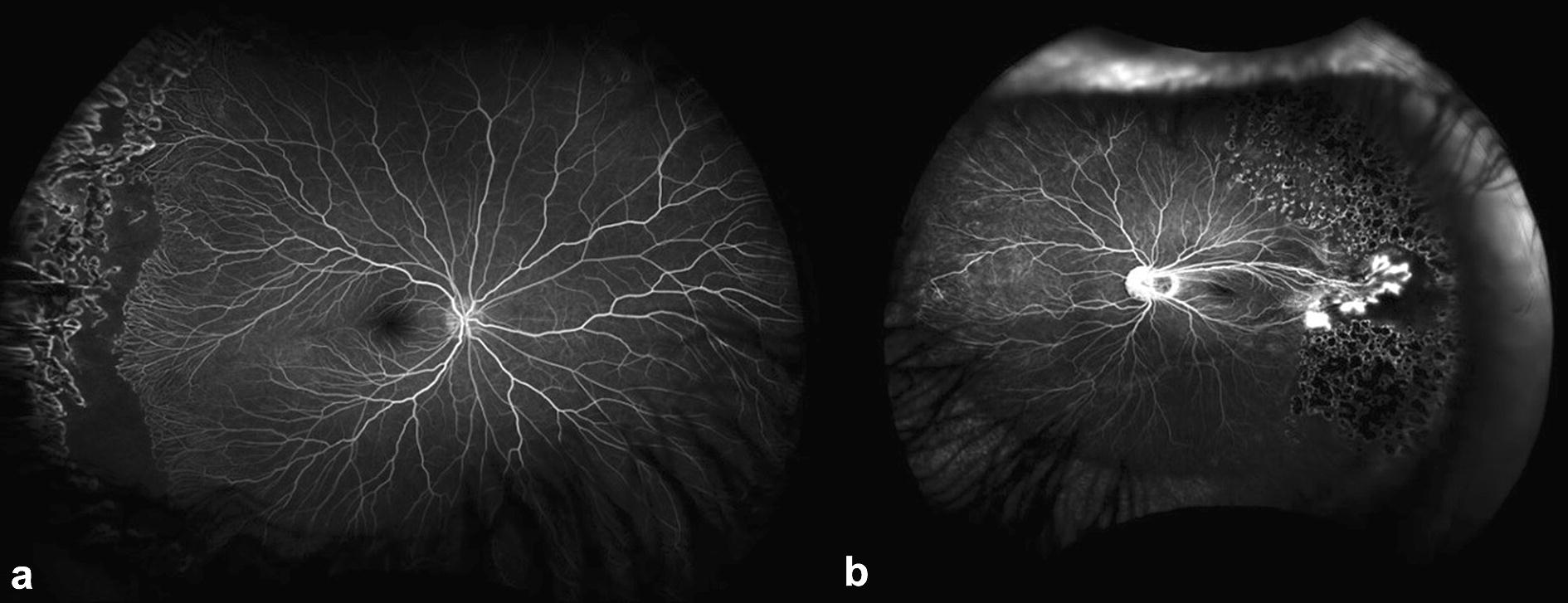
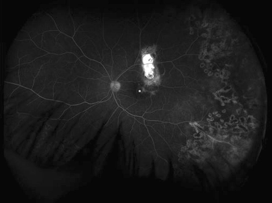
Background: Ultra-widefield angiography is the latest technology in the evolution of fundus fluorescein angiography. With the ability to capture up to 200° of the fundus in a single image, far peripheral retinal pathology can be imaged. Generally, obtaining high-quality fundus fluorescein angiography in a child without sedation in the outpatient setting is exceedingly challenging. Therefore, there are advantages to imaging platforms that can capture the peripheral retina in young children without anesthesia. Often pediatric retinal diseases have pathology localized to the far periphery, which further validates the utility of ultra-widefield angiography. Ultra-widefield angiography has been successfully used without sedation for evaluation of children with various pediatric retinal diseases such as Coats disease, familial exudative vitreoretinopathy, and retinopathy of prematurity.
Conclusion: This non-contact, non-mydriatic modality has been utilized in the evaluation of pediatric retinal diseases and demonstrated to have benefits over conventional fluorescein angiography techniques.
Keywords: Coats disease; Familial exudative vitreoretinopathy; Incontentia pigmenti; Pediatric retina; Retinopathy of prematurity; Ultra-wide field imaging; Ultra-widefield fluorescein angiography.
Figures
References
-
- Shulman J, Hartnett ME. Clinical trials and management of severe retinopathy of prematurity. In: Hartnett ME, editor. Pediatric retina. 2. Philadelphia: Wolters Kluwer; 2015.
-
- Drenser KA, Trese MT, Capone A. Familial exudative vitreoretinopathy (FEVR) In: Harnett ME, editor. Pediatric retina. 2. Philadelphia: Wolters Kluwer; 2015.
-
- Recchia FM. Coats disease. In: Hartnett ME, editor. Pediatric retina. 2. Philadelphia: Wolters Kluwer; 2015.
-
- Grading diabetic retinopathy from stereoscopic color fundus photographs—an extension of the modified Airlie House classification. ETDRS report number 10. Early Treatment Diabetic Retinopathy Study Research Group. Ophthalmology 1991;98(5 Suppl):786–806. - PubMed
Publication types
LinkOut - more resources
Full Text Sources
Other Literature Sources
