

A 36-Year-Old Renal Transplant Recipient Female with Leg Ulcer: A Case Report and Brief Review
- PMID: 29992064
- PMCID: PMC5821986
- DOI: 10.1155/2018/5086501
A 36-Year-Old Renal Transplant Recipient Female with Leg Ulcer: A Case Report and Brief Review
Abstract
Background: Opportunistic infections are common in organ transplant recipients. After 6 months of transplantation, patients have the highest risk of opportunistic infections such as cryptococcosis.
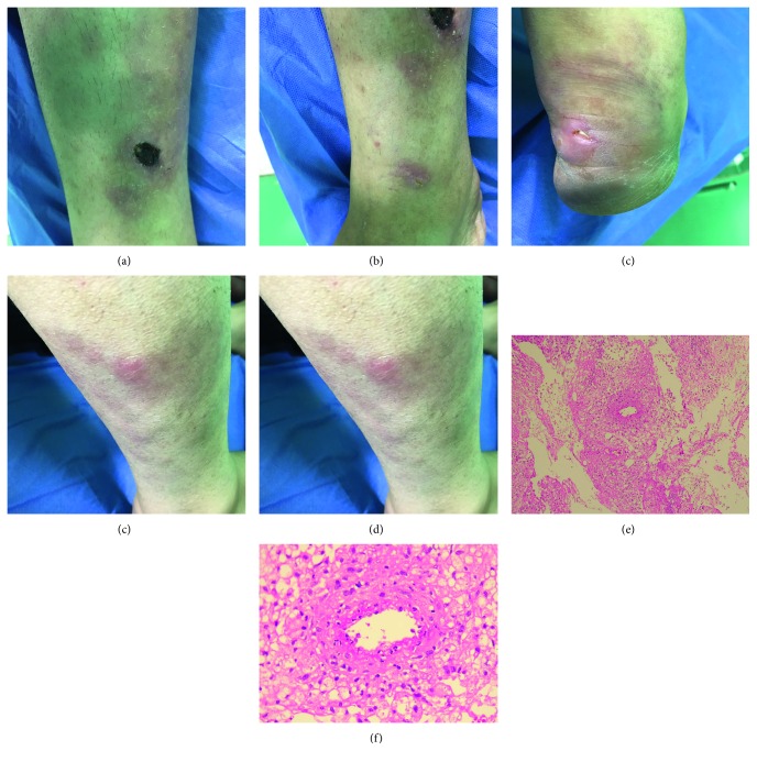
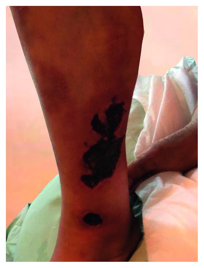
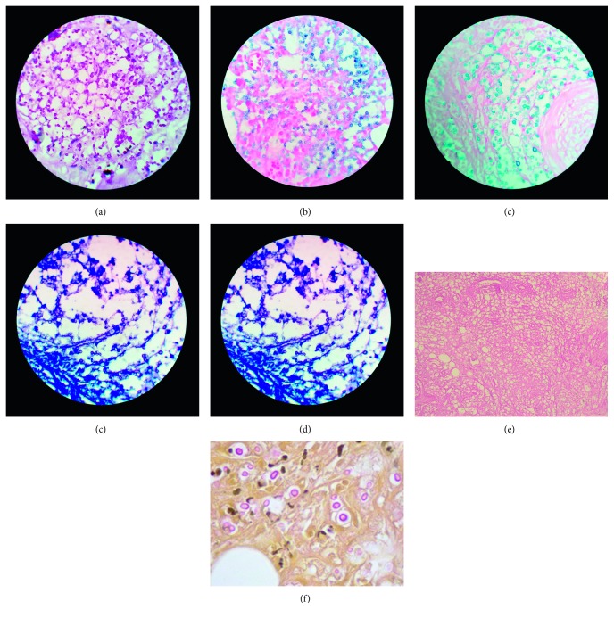
Case presentation: The report presents the case of a 36-year-old female renal transplant recipient, with complaints of few subcutaneous painful and warm nodules and large, warm, erythematous, nontender plaques on the mildly edematous right leg and ankle. Incisional biopsy of the subcutaneous nodule over the leg showed panniculitis with small- to medium-sized vasculitis associated with round yeast forms, and culture of the fragments revealed C. neoformans var. grubii.
Conclusions: This article also reviews in brief the treatment of this rare complication. Reviewing the literature showed that since the cryptococcal cutaneous lesions are often nonspecific, the clinical picture solely is not enough to construct a definite diagnosis and there must be a high clinical suspicion.
Figures
Similar articles
-
Cryptococcal panniculitis in a renal transplant recipient: case report and review of literature.J Dermatol Case Rep. 2015 Sep 30;9(3):76-80. doi: 10.3315/jdcr.2015.1205. eCollection 2015 Sep 30. J Dermatol Case Rep. 2015. PMID: 26512304 Free PMC article.
-
Cryptococcal panniculitis in an immunocompromised patient: a case report and review of the literature.Cutis. 2010 Jun;85(6):303-6. Cutis. 2010. PMID: 20666191
-
Unilateral leg pain caused by cryptococcal myositis: An unusual presentation of disseminated cryptococcosis in a kidney transplant recipient.Transpl Infect Dis. 2021 Apr;23(2):e13491. doi: 10.1111/tid.13491. Epub 2020 Oct 27. Transpl Infect Dis. 2021. PMID: 33040432
-
Pleural effusion as the initial clinical presentation in disseminated cryptococcosis and fungaemia: an unusual manifestation and a literature review.BMC Infect Dis. 2015 Sep 22;15:385. doi: 10.1186/s12879-015-1132-4. BMC Infect Dis. 2015. PMID: 26395579 Free PMC article. Review.
-
Disseminated Cryptococcosis Presenting as Cellulitis Diagnosed by Laser Capture Microdissection: A Case Report and Literature Review.Mycopathologia. 2021 Jun;186(3):423-433. doi: 10.1007/s11046-021-00543-3. Epub 2021 Apr 3. Mycopathologia. 2021. PMID: 33813690 Review.
References
-
- Kasiske B. L., Zeier M. G. KDIGO clinical practice guideline for the care of kidney transplant recipients. 2009. http://kdigo.org/wp-content/uploads/2017/02/KDIGO-2009-Transplant-Recipi.... - PubMed
-
- Skorecki K., Chertow G., Marsden P., Taal M., Yu A. Brener and Rectors the Kidney. 10th. Elsevier; 2016.
Publication types
LinkOut - more resources
Full Text Sources
Other Literature Sources
