

Case Reports
doi: 10.1155/2018/6125676.
eCollection 2018.
The Benefits of an In-Office Arthroscopy in the Diagnosis of Unresolved Knee Pain
Affiliations
- PMID: 29992071
- PMCID: PMC5827882
- DOI: 10.1155/2018/6125676
Item in Clipboard
Case Reports
The Benefits of an In-Office Arthroscopy in the Diagnosis of Unresolved Knee Pain
Case Rep Orthop.
.
Abstract
We report a patient who developed persistent knee pain with mechanical symptoms after an uncomplicated patellofemoral arthroplasty. The etiology of his knee pain remained inconclusive following magnetic resonance imaging due to metallic artifact image distortion. With the use of an in-office needle arthroscopy, an immediate and definitive diagnosis was obtained, preventing an unnecessary surgery for a diagnostic arthroscopy. We discovered a lateral meniscus tear, an anterior cruciate ligament tear, and a medial femoral condyle chondral defect for which the patient underwent arthroscopic partial meniscectomy, ligament reconstruction, and osteochondral allograft transplantation, with resolution of his knee pain.
Figures
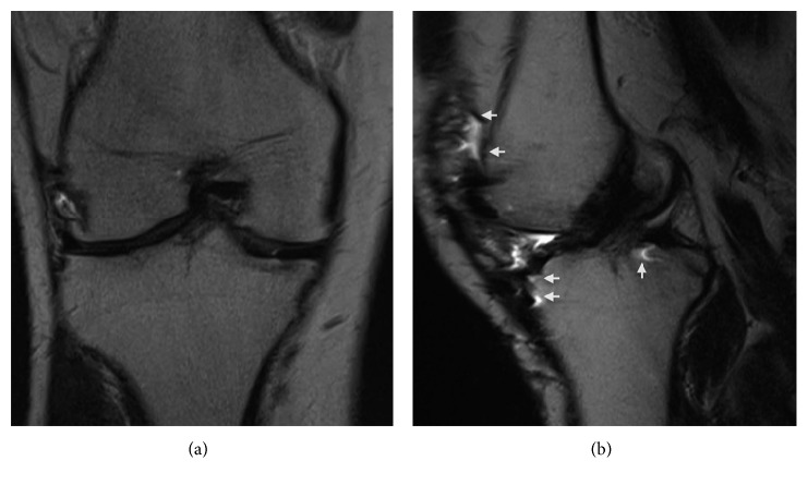
Coronal (a) and sagittal (b) T2-weighted metal reduction magnetic resonance images of the right knee, demonstrating an apparently intact ACL. Note the presence of metallic artifacts (white arrows) throughout the anterior and posterior aspect of the knee on the sagittal image.
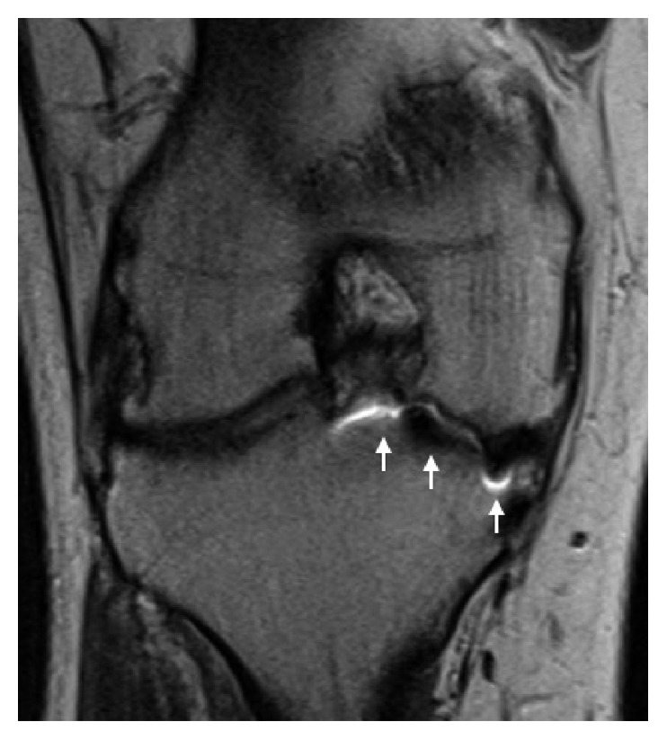
Coronal proton density with metal reduction MRI of the right knee demonstrates significant image distortion (white arrows) from metallic artifacts, obscuring accurate evaluation of the medial femoral condyle articular surface.
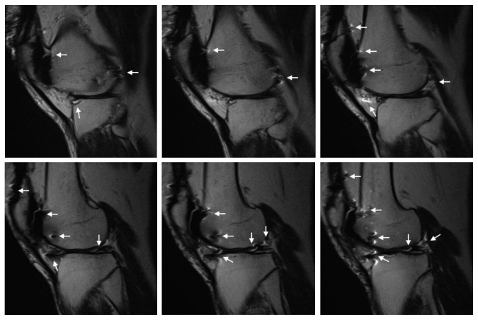
Sequential sagittal T2-weighted metal reduction MR images of the right knee lateral compartment. The presence of metal artifacts (white arrows) obscures accurate evaluation of the lateral meniscus.
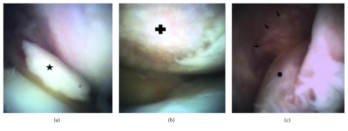
Arthroscopic images of the right knee obtained with mi-eye 2. (a) An intra-articular loose body ( ) is visualized in the anterior knee. (b) A large chondral defect (
 ) on the weight-bearing surface of the medial femoral condyle with complete loss of the articular cartilage and exposed subchondral bone. (c) A view of the intercondylar notch showing a tear of the ACL (
 ) with the remnant fibers of the femoral origin (arrows) along the lateral wall of the notch.
) is visualized in the anterior knee. (b) A large chondral defect () on the weight-bearing surface of the medial femoral condyle with complete loss of the articular cartilage and exposed subchondral bone. (c) A view of the intercondylar notch showing a tear of the ACL () with the remnant fibers of the femoral origin (arrows) along the lateral wall of the notch.References
-
- Kim S., Bosque J., Meehan J. P., Jamali A., Marder R. Increase in outpatient knee arthroscopy in the United States: a comparison of National Surveys of Ambulatory Surgery, 1996 and 2006. Journal of Bone and Joint Surgery-American Volume. 2011;93(11):994–1000. doi: 10.2106/JBJS.I.01618. - DOI - PubMed
-
- Cullen K. A., Hall M. J., Golosinskiy A. Ambulatory surgery in the United States. National Health Statistics Reports. 2009;28(11):1–25. - PubMed
Publication types
LinkOut - more resources
Full Text Sources
Other Literature Sources
Medical
