

The PIEPEAR Workflow: A Critical Care Ultrasound Based 7-Step Approach as a Standard Procedure to Manage Patients with Acute Cardiorespiratory Compromise, with Two Example Cases Presented
- PMID: 29992144
- PMCID: PMC6016228
- DOI: 10.1155/2018/4687346
The PIEPEAR Workflow: A Critical Care Ultrasound Based 7-Step Approach as a Standard Procedure to Manage Patients with Acute Cardiorespiratory Compromise, with Two Example Cases Presented
Abstract
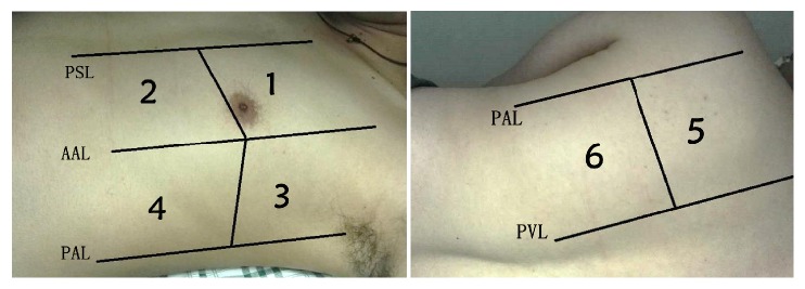
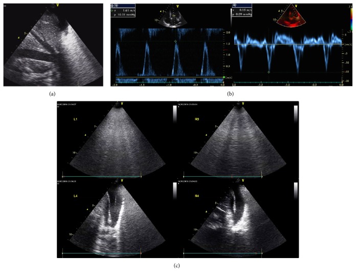
Critical care ultrasound (CCUS) has been widely used as a useful tool to assist clinical judgement. The utilization should be integrated into clinical scenario and interact with other tests. No publication has reported this. We present a CCUS based "7-step approach" workflow-the PIEPEAR Workflow-which we had summarized and integrated our experience in CCUS and clinical practice into, and then we present two cases which we have applied the workflow into as examples. Step one is "problems emerged?" classifying the signs of the deterioration into two aspects: acute circulatory compromise and acute respiratory compromise. Step two is "information clear?" quickly summarizing the patient's medical history by three aspects. Step three is "focused exam launched": (1) focused exam of the heart by five views: the assessment includes (1) fast and global assessment of the heart (heart glance) to identify cases that need immediate life-saving intervention and (2) assessing the inferior vena cava, right heart, diastolic and systolic function of left heart, and systematic vascular resistance to clarify the hemodynamics. (2) Lung ultrasound exam is performed to clarify the predominant pattern of the lung. Step four is "pathophysiologic changes reported." The results of the focused ultrasound exam were integrated to conclude the pathophysiologic changes. Step five is "etiology explored" diagnosing the etiology by integrating Step two and Step four and searching for the source of infection, according to the clues extracted from the focused ultrasound exam; additional ultrasound exams or other tests should be applied if needed. Step six is "action" supporting the circulation and respiration sticking to Step four. Treat the etiologies according step five. Step seven is "recheck to adjust." Repeat focused ultrasound and other tests to assess the response to treatment, adjust the treatment if needed, and confirm or correct the final diagnosis. With two cases as examples presented, we insist that applying CCUS with 7-step approach workflow is easy to follow and has theoretical advantages. The coming research on its value is expected.
Figures
References
-
- Beaulieu Y. Bedside echocardiography in the assessment of the critically ill. Critical Care Medicine. 2007;35(5, supplement):S235–S249. doi: 10.1097/01.ccm.0000260673.66681.af. - DOI - PubMed
-
- Frankel H. L., Kirkpatrick A. W., Elbarbary M., et al. Guidelines for the appropriate use of bedside general and cardiac ultrasonography in the evaluation of critically ill patients-part I: General ultrasonography. Critical Care Medicine. 2015;43(11):2479–2502. doi: 10.1097/CCM.0000000000001216. - DOI - PubMed
-
- Via G., Hussain A., Wells M., et al. nternational Liaison Committee on Focused Cardiac UltraSound (ILC-FoCUS); International Conference on Focused Cardiac UltraSound (IC-FoCUS).. International Evidence-Based Recommendations for Focused Cardiac Ultrasound. Journal of the American Society of Echocardiography. 2014;27(7):683 e681–683 e633. doi: 10.1016/j.echo.2014.05.001. - DOI - PubMed
MeSH terms
LinkOut - more resources
Full Text Sources
Other Literature Sources
Medical
