

Study of locust bean gum reinforced cyst-chitosan and oxidized dextran based semi-IPN cryogel dressing for hemostatic application
- PMID: 29992195
- PMCID: PMC6035369
- DOI: 10.1016/j.bioactmat.2017.11.005
Study of locust bean gum reinforced cyst-chitosan and oxidized dextran based semi-IPN cryogel dressing for hemostatic application
Abstract
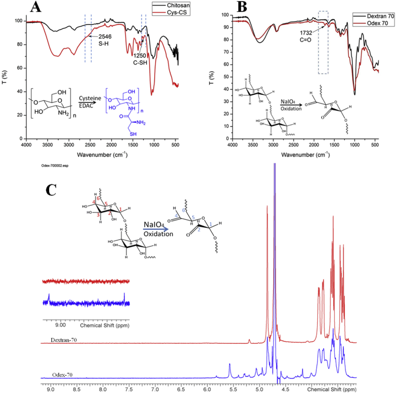
Severe blood loss due to traumatic injuries remains one of the leading causes of death in emergency settings. Chitosan continues to be the candidate material for hemostatic applications due to its inherent hemostatic properties. However, available chitosan-based dressings have been reported to have an acidic odor at the wound site due to the incorporation of acid based solvents for their fabrication and deformation under compression owing to low mechanical strength limiting its usability. In the present study semi-IPN cryogel was fabricated via Schiff's base cross-linking between the polyaldehyde groups of oxidized dextran and thiolated chitosan in presence of locust bean gum (LBG) known for its hydrophilicity. Polymerization at -12 °C yielded macroporous semi-IPN cryogels with an average pore size of 124.57 ± 20.31 μm and 85.46% porosity. The hydrophobicity index of LBG reinforced semi-IPN cryogel was reduced 2.42 times whereas the swelling ratio was increased by 156.08% compare to control cryogel. The increased hydrophilicity and swelling ratio inflated the compressive modulus from 28.1 kPa to 33.85 for LBG reinforced semi-IPN cryogel. The structural stability and constant degradation medium pH were also recorded over a period of 12 weeks. The cryogels demonstrated lower adsorption affinity towards BSA. The cytotoxicity assays (direct, indirect) with 3T3-L1 fibroblast cells confirmed the cytocompatibility of the cryogels. The hemolysis assay showed <5% hemolysis confirming blood compatibility of the fabricated cryogel, while whole blood clotting and platelet adhesion assays confirmed the hemostatic potential of semi-IPN cryogel.
Keywords: Chitosan; Cryogel; Hemostasis; Locust bean gum; Oxidized dextran.
Figures


References
-
- Chen J.-P., Chang G.-Y., Chen J.-K. Electrospun collagen/chitosan nanofibrous membrane as wound dressing. Colloids Surf. Physicochem. Eng. Asp. 2008;313:183–188.
-
- Voormolen J.H., Ringers J., Bots G.T., van der Heide A., Hermans J. Hemostatic agents: brain tissue reaction and effectiveness. A comparative animal study using collagen fleece and oxidized cellulose. Neurosurgery. 1987;20:702–709. - PubMed
-
- Yang J., Tian F., Wang Z., Wang Q., Zeng Y.J., Chen S.Q. Effect of chitosan molecular weight and deacetylation degree on hemostasis. J. Biomed. Mater. Res. B Appl. Biomater. 2008;84:131–137. - PubMed
-
- VandeVord P.J., Matthew H.W., DeSilva S.P., Mayton L., Wu B., Wooley P.H. Evaluation of the biocompatibility of a chitosan scaffold in mice. J. Biomed. Mater. Res. 2002;59:585–590. - PubMed
LinkOut - more resources
Full Text Sources
Other Literature Sources
