Review
. 2018 Sep 1;24(5):556-576.
doi: 10.1093/humupd/dmy021.

Epigenetic regulation in development: is the mouse a good model for the human?

Affiliations
Review

Epigenetic regulation in development: is the mouse a good model for the human?

Courtney W Hanna et al. Hum Reprod Update. .

Abstract

Background: Over the past few years, advances in molecular technologies have allowed unprecedented mapping of epigenetic modifications in gametes and during early embryonic development. This work is allowing a detailed genomic analysis, which for the first time can answer long-standing questions about epigenetic regulation and reprogramming, and highlights differences between mouse and human, the implications of which are only beginning to be explored.

Objective and rationale: In this review, we summarise new low-cell molecular methods enabling the interrogation of epigenetic information in gametes and early embryos, the mechanistic insights these have provided, and contrast the findings in mouse and human.

Search methods: Relevant studies were identified by PubMed search.

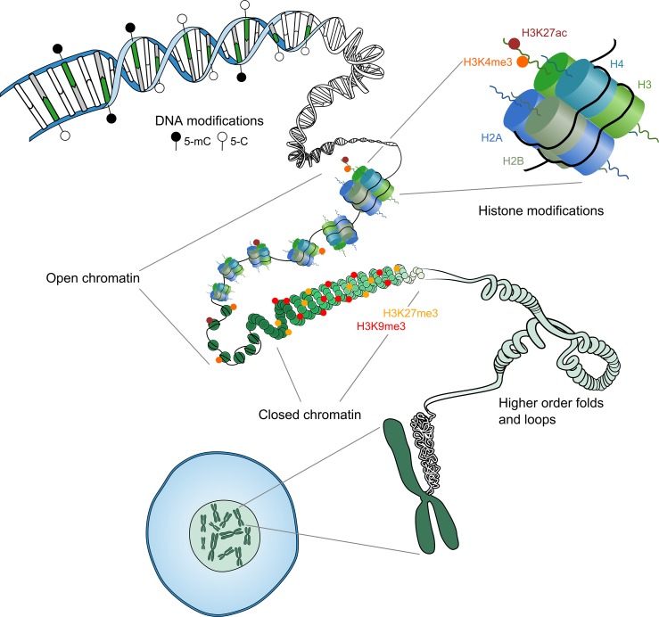
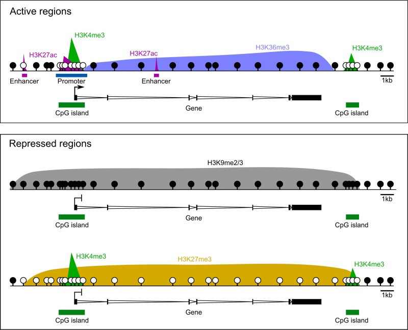
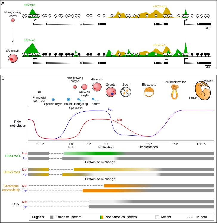
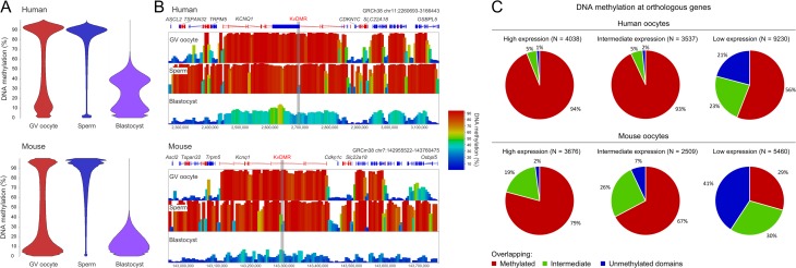
Outcomes: We discuss the levels of epigenetic regulation, from DNA modifications to chromatin organisation, during mouse gametogenesis, fertilisation and pre- and post-implantation development. The recently characterised features of the oocyte epigenome highlight its exceptionally unique regulatory landscape. The chromatin organisation and epigenetic landscape of both gametic genomes are rapidly reprogrammed after fertilisation. This extensive epigenetic remodelling is necessary for zygotic genome activation, but the mechanistic link remains unclear. While the vast majority of epigenetic information from the gametes is erased in pre-implantation development, new insights suggest that repressive histone modifications from the oocyte may mediate a novel mechanism of imprinting. To date, the characterisation of epigenetics in human development has been almost exclusively limited to DNA methylation profiling; these data reinforce that the global dynamics are conserved between mouse and human. However, as we look closer, it is becoming apparent that the mechanisms regulating these dynamics are distinct. These early findings emphasise the importance of investigations of fundamental epigenetic mechanisms in both mouse and humans.

Wider implications: Failures in epigenetic regulation have been implicated in human disease and infertility. With increasing maternal age and use of reproductive technologies in countries all over the world, it is becoming ever more important to understand the necessary processes required to establish a developmentally competent embryo. Furthermore, it is essential to evaluate the extent to which these epigenetic patterns are sensitive to such technologies and other adverse environmental exposures.


Figures

Figure 1
Figure 1
Levels of epigenetic regulation. The DNA sequence can be methylated at cytosine residues in a CpG context, termed DNA methylation. DNA is wrapped around the histone octamer to form the nucleosome. Variants and post-translational modifications of these histone proteins form another layer of epigenetic regulation. The state of these epigenetic modifications together determines whether the chromatin will be organised in an accessible ‘open’ or an inaccessible ‘closed’ state. Higher order folds and loops organise the chromatin into active and inactive compartments.
Figure 2
Figure 2
Canonical epigenetic patterns. H3K4me3 is associated with actively transcribed promoters, as well as CpG islands, irrespective of transcription. H3K27ac demarks active promoters and enhancers, while associated transcribed genes bodies are enriched for H3K36me3. Repressed regions of the genome are typically associated with either H3K9me2/3 or H3K27me3. DNA is generally highly methylated throughout the genome, with the exception of regulatory regions marked by H3K4me3 and/or H3K27ac, and H3K27me3- domains. Methylated CpGs are depicted as closed circles and unmethylated CpGs are open circles.
Figure 3
Figure 3
Epigenetic reprogramming in mouse development. (A) Epigenetic patterns are shown for non-growing oocytes and fully grown germinal vesicle (GV) oocytes. In non-growing oocytes, DNA is almost completely unmethylated, H3K4me3 is exclusively enriched at active promoters and H3K27me3 is spanning broad non-canonical domains. By the fully grown GV stage, DNA across transcribed gene bodies is fully methylated and H3K4me3 has accumulated in broad domains throughout untranscribed regions. (B) Schematic of epigenetic reprogramming events during gametogenesis and embryogenesis. DNA methylation is erased in primordial germ cells and re-established earlier in the sperm of males and after birth in oocytes in females. Oocytes acquire lower overall methylation than sperm, with non-canonical genome-wide distribution. After fertilisation, the paternal DNA is rapidly demethylated, while maternal DNA methylation is passively lost over several cell divisions. DNA methylation is re-acquired in canonical patterns in the post-implantation embryo, concomitant with lineage specification. H3K4me3 is non-canonically distributed in the oocyte, is rapidly erased after fertilisation, and becomes canonically enriched at CpG islands and active promoters. Very few domains retain H3K4me3-marked histones in the protamine exchange in sperm and subsequently through the re-acquisition of histones in the zygote. H3K27me3 acquires a non-canonically broad distribution in PGCs in the absence of other repressive epigenetic marks. This pattern is relatively maintained throughout oogenesis, while very few H3K27me3-marked histones are retained in the sperm protamine exchange. In the pre-implantation embryo, H3K27me3-transmitted from the gametes is progressively lost, with pronounced loss at CpG-rich regions. H3K27me3 is then re-established in a canonical pattern in the post-implantation embryo. Chromatin accessibility is contrastingly and exceptionally open in the oocyte and compact in the sperm. The open chromatin state of maternal DNA is gradually resolved in the pre-implantation embryo, while the compact packaging of paternal DNA is rapidly resolved with incorporation of histones in the zygote. Topological associated domains (TADs) are nearly absent in the mature oocyte and become gradually re-instated in the pre-implantation embryo.
Figure 4
Figure 4
Comparison of DNA methylation in human and mouse development. (A) Beanplots showing the distribution of DNA methylation percentages of 100-CpG running windows (minimum coverage of 10 CpGs) in human (top) and mouse (bottom) GV oocytes, sperm and blastocysts, with human oocytes and blastocysts being notably more methylated than mouse oocytes and blastocysts, respectively. (B) Screenshot of DNA methylation at the KvDMR imprinted locus in human (top) and mouse (bottom) GV oocytes, sperm and blastocysts. The locus illustrates the increased number of regions that are fully methylated in human compared to mouse oocytes. Additionally, the human blastocyst retains the maternal pattern of methylation more substantially than the mouse blastocyst. (C) Proportion of orthologous genes that are methylated in human and mouse oocytes. Orthologous genes were defined by ENSEMBL BioMart and categorised as highly expressed (FPKM>10), intermediately expressed (1<FPKM<10) or lowly expressed (FPKM<1). These genes were then evaluated for overlap with fully methylated (>75%) and intermediately methylated (25–75%) 100-CpG windows; genes that did not overlap a methylated window were defined as unmethylated. This analysis demonstrates that the increase in methylated domains in human oocytes reflects an increased number of genes becoming fully methylated compared to mouse. Publically available data was used for these analyses, including RNA-seq data for mouse and human oocytes (GSE44183) (Xue et al., 2013) and DNA methylation data from mouse (Kobayashi et al., 2012) and human (Okae et al., 2014) oocytes, sperm and blastocyst embryos.

References

    1. Ahmed K, Dehghani H, Rugg-Gunn P, Fussner E, Rossant J, Bazett-Jones DP. Global chromatin architecture reflects pluripotency and lineage commitment in the early mouse embryo. PLoS One 2010;5:e10531. - PMC - PubMed
    1. Allis CD, Jenuwein T. The molecular hallmarks of epigenetic control. Nat Rev Genet 2016;8:487–500. - PubMed
    1. Amouroux R, Nashun B, Shirane K, Nakagawa S, Hill PW, D’Souza Z, Nakayama M, Matsuda M, Turp A, Ndjetehe E et al. . De novo DNA methylation drives 5hmC accumulation in mouse zygotes. Nat Cell Biol 2016;2:225–233. - PMC - PubMed
    1. Ancelin K, Syx L, Borensztein M, Ranisavljevic N, Vassilev I, Briseno-Roa L, Liu T, Metzger E, Servant N, Barillot E et al. . Maternal LSD1/KDM1A is an essential regulator of chromatin and transcription landscapes during zygotic genome activation. Elife 2016;5:e08851. - PMC - PubMed
    1. Andergassen D, Dotter CP, Wenzel D, Sigl V, Bammer PC, Muckenhuber M, Mayer D, Kulinski TM, Theussl HC, Penninger JM et al. . Mapping the mouse Allelome reveals tissue-specific regulation of allelic expression. Elife 2017;6:e25125. - PMC - PubMed

Publication types