. 2018 Sep;22(9):4366-4376.
doi: 10.1111/jcmm.13728. Epub 2018 Jul 10.

The expression of MMP-1 and MMP-9 is up-regulated by smooth muscle cells after their cross-talk with macrophages in high glucose conditions

Affiliations

The expression of MMP-1 and MMP-9 is up-regulated by smooth muscle cells after their cross-talk with macrophages in high glucose conditions

Razvan Daniel Macarie et al. J Cell Mol Med. 2018 Sep.

Abstract

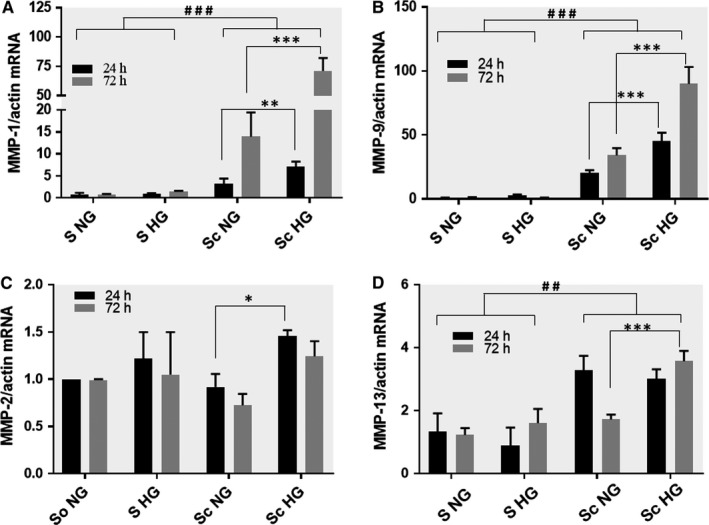
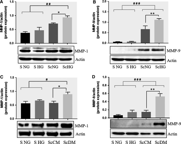
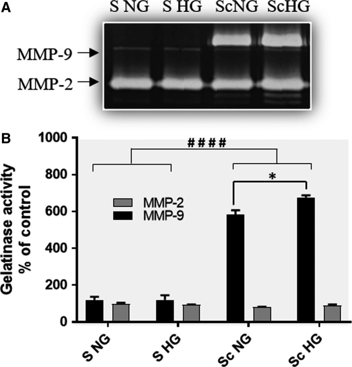
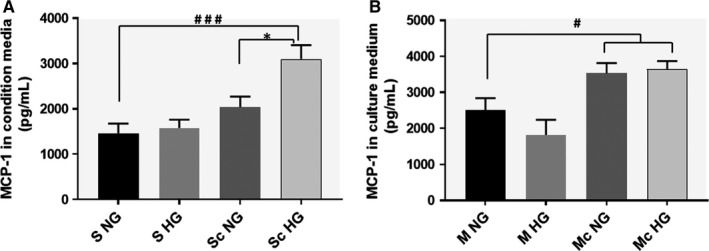
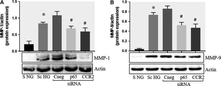
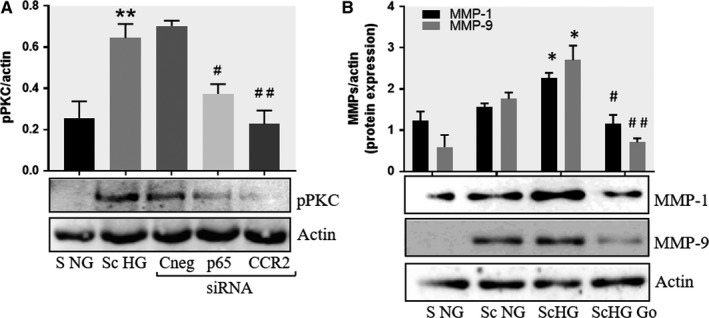
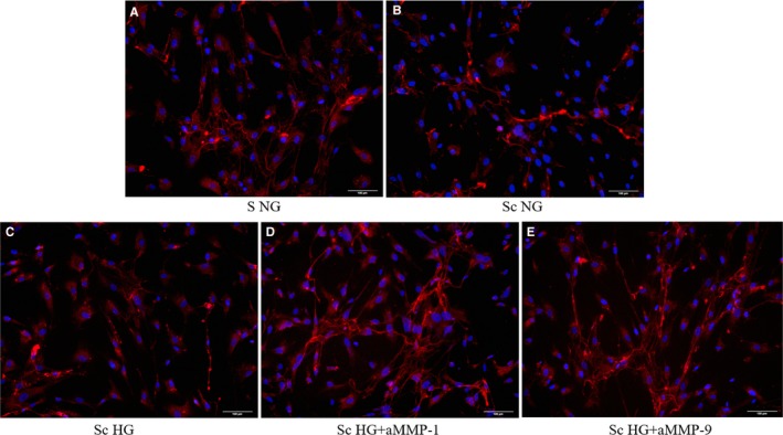
Patients with diabetes mellitus have an increased risk of myocardial infarction and coronary artery disease-related death, exhibiting highly vulnerable plaques. Many studies have highlighted the major role of macrophages (MAC) and smooth muscle cells (SMC) and the essential part of metalloproteases (MMPs) in atherosclerotic plaque vulnerability. We hypothesize that in diabetes, the interplay between MAC and SMC in high glucose conditions may modify the expression of MMPs involved in plaque vulnerability. The SMC-MAC cross-talk was achieved using trans-well chambers, where human SMC were grown at the bottom and human MAC in the upper chamber in normal (NG) or high (HG) glucose concentration. After cross-talk, the conditioned media and cells were isolated and investigated for the expression of MMPs, MCP-1 and signalling molecules. We found that upon cross-talk with MAC in HG, SMC exhibit: (i) augmented expression of MMP-1 and MMP-9; (ii) significant increase in the enzymatic activity of MMP-9; (iii) higher levels of soluble MCP-1 chemokine which is functionally active and involved in MMPs up-regulation; (iv) activated PKCα signalling pathway which, together with NF-kB are responsible for MMP-1 and MMP-9 up-regulation, and (v) impaired function of collagen assembly. Taken together, our data indicate that MCP-1 released by cell cross-talk in diabetic conditions binds to CCR2 and triggers MMP-1 and MMP-9 over-expression and activity, features that could explain the high vulnerability of atherosclerotic plaque found at diabetic patients.

Keywords: atherosclerosis; cell cross-talk; high glucose; matrix metalloproteinases; protein kinase C.


Figures

Figure 1
Figure 1
Gene expression of MMP‐1 (A), MMP‐9 (B), MMP‐2 (C) and MMP‐13 (D) in smooth muscle cells (SMC) subsequent to their co‐culture with macrophages (MAC) in normal and high‐glucose conditions. After 24 or 72 h, mRNA was obtained from cell homogenates and subjected to RTPCR. The mRNA of MMPs were normalized to actin mRNA. S NG, control SMC grown in normal glucose (5 mmol/L) concentration; S HGSMC exposed to high (25 mmol/L) glucose concentration in the culture medium; Sc NGSMC after their co‐culture with MAC in normal glucose, Sc HGSMC after their co‐culture with MAC in high glucose conditions. Note the significant increase of MMP‐1 and MMP‐9 gene expression in SMC after their co‐culture with MAC in HG conditions compared to SMC co‐cultured with MAC in normal glucose or controls (SMC not co‐cultured, exposed to NG or HG conditions) at both, 24 or 72 h. n = 4, *P < .05, **P < .01, ***P < .001, ## P < .01, ### P < .001
Figure 2
Figure 2
Co‐culture of SMC and MAC in high glucose conditions (24 h) induces a significant increase in the protein expression of MMP‐1 and MMP‐9. A, B: Protein expression of MMP‐1 (A) and MMP‐9 (B) was assessed in SMC after co‐culture with THP‐derived macrophages and evaluated by Western blot assay. C, D: Protein expression of MMP‐1 (C) and MMP‐9 (D) in SMC after co‐culture with human macrophages derived from freshly isolated monocytes from control subjects (CM) or diabetic patients with ACS (DM). S NG –control SMC, S HGSMC exposed to HG, Sc NG or Sc CMSMC which were co‐cultured with MAC in normal glucose, Sc HG or Sc DMSMC which were co‐cultured with MAC in high glucose. n = 3, *P < .05, **P < .01, ***P < .001, # P < .05, ## P < .01, ### P < .001
Figure 3
Figure 3
The enzymatic activity of MMP‐2 and MMP‐9 gelatinases assessed by SDSPAGE zymography of the conditioned media collected from SMC that were previously co‐cultured with MAC in normal or high glucose (Sc NG, Sc HG) conditions and from control cells (S NG or S HG). Note that the enzymatic activity of MMP‐9 released by SMC after cross‐talk with MAC is significantly increased in HG compared with NG. n = 4, *P < .05; #### P < .001
Figure 4
Figure 4
SMCMAC cross talk in high glucose conditions induces a significant increased level of MCP‐1 released in the conditioned medium, both in SMC (A) and MAC (B). The MCP‐1 released by macrophages was quantified in the conditioned media collected from the upper insert (of trans‐well system), while the MCP‐1 released from SMC was quantified in the conditioned media collected from the bottom chamber. As controls, MCP‐1 was quantified in the conditioned media collected from macrophages or SMC grown independently (no co‐culture) in normal or high glucose conditions. Note that consequent to the cell co‐culture, MCP‐1 is significantly released by SMC after cross‐talk with MAC in HG vs NG conditions (A). n = 4, *P < .05, # P < .05, ### P < .001
Figure 5
Figure 5
Effect of CCR2 and p65 silencing in the regulation of MMP‐1 and MMP‐9 expression in SMC. Protein expression of MMP‐1 (A) and MMP‐9 (B) determined by Western blot in control SMC (S NG), SMC which were co‐cultured with MAC in HG conditions (Sc HG), and SMC subjected to silencing of CCR‐2 and p65 and co‐cultured with MAC in HG conditions (Cneg, p65, CCR2 siRNA). Note that silencing of CCR2 or p65 decreases significantly the protein expression of MMP‐1 and MMP‐9 induced by SMCMAC co‐culture in high glucose conditions. n = 3, *P < .05, # P < .05
Figure 6
Figure 6
Inhibition of PKCα decreases the protein expression of MMP‐1 and MMP‐9 induced by cell cross‐talk in SMC. A, Evaluation of PKCα activation in control SMC (S NG) vs SMC previously co‐cultured with MAC in HG (Sc HG). Note that, the phosphorylated form of PKCα is significantly increased in SMC that were before exposed to MAC in HG conditions. Silencing of CCR2 or p65 decreases the phosphorylation of PKCα to control levels. B, Effect of PKCα inhibitor – Go 6976 on the MMP‐1 and MMP‐9 protein expression induced by SMCMAC cross‐talk. S NG – control SMC, Sc NGSMC which were co‐cultured with MAC in normal glucose, Sc HGSMC which were co‐cultured with MAC in high glucose, Sc HGGo ‐ SMC which were co‐cultured with MAC in high glucose in presence of 1 μmol/L Go 6976. n = 3, *P < .05, **P < .01, # P < .05, ## P < .01
Figure 7
Figure 7
Formation of collagen fibres from soluble collagen labelled with Texas Red by SMC. A‐ control SMC cultured in normal glucose conditions (S NG), B‐ SMC after their co‐culture with MAC in normal glucose (Sc NG) or under high glucose conditions (C ‐ Sc HG), (D and E) ‐ SMC after their co‐culture with MAC in HG and pre‐incubated with blocking anti‐MMP‐1 or 9, respectively, before collagen addition; red ‐ stained collagen fibers; blue ‐ nuclei of SMC stained with Hoechst

References

    1. Martín‐Timón I, Sevillano‐Collantes C, Segura‐Galindo A, Del Cañizo‐Gómez FJ. Type 2 diabetes and cardiovascular disease: have all risk factors the same strength? World J Diabetes. 2014;5:444‐470. - PMC - PubMed
    1. Moreno V, Fuster PR. New aspects in the pathogenesis of diabetic atherothrombosis. J Am Coll Cardiol. 2004;44:2293‐2300. - PubMed
    1. Booth GL, Kapral MK, Fung K, Tu JV. Relation between age and cardiovascular disease in men and women with diabetes compared with non‐diabetic people: a population‐based retrospective cohort study. Lancet. 2006;368:29‐36. - PubMed
    1. Amin M, Pushpakumar S, Muradashvili N, Kundu S, Tyagi SC, Sen U. Regulation and involvement of matrix metalloproteinases in vascular diseases. Front Biosci (Landmark Ed.). 2016;21:89‐118. - PMC - PubMed
    1. Varani J, Perone P, Fligiel SEG, Fisher GJ, Voorhees JV. Inhibition of type I procollagen production in photodamage: correlation between presence of highmolecular weight collagen fragments and reduced procollagen synthesis. J Investig Dermatol. 2002;119:122‐129. - PubMed

Publication types

MeSH terms

Associated data