. 2018 Jul 11;8(1):10432.
doi: 10.1038/s41598-018-28706-1.

The influenza virus hemagglutinin head evolves faster than the stalk domain

Affiliations

The influenza virus hemagglutinin head evolves faster than the stalk domain

Ericka Kirkpatrick et al. Sci Rep. .

Abstract

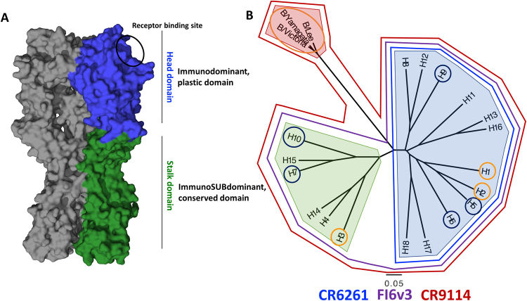
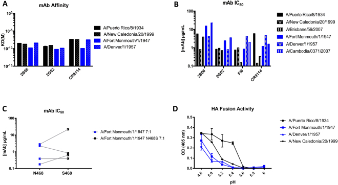
The limited ability of current influenza virus vaccines to protect from antigenically drifted or shifted viruses creates a public health problem that has led to the need to develop effective, broadly protective vaccines. While current influenza virus vaccines mostly induce an immune response against the immunodominant and variable head domain of the hemagglutinin, the major surface glycoprotein of the virus, the hemagglutinin stalk domain has been identified to harbor neutralizing B-cell epitopes that are conserved among and even between influenza A virus subtypes. A complete understanding of the differences in evolution between the main target of current vaccines and this more conserved stalk region are missing. Here, we performed an evolutionary analysis of the stalk domains of the hemagglutinin of pre-pandemic seasonal H1N1, pandemic H1N1, seasonal H3N2, and influenza B viruses and show quantitatively for the first time that the stalk domain is evolving at a rate that is significantly slower than that of the head domain. Additionally, we found that the cross-reactive epitopes in the stalk domain targeted by broadly neutralizing monoclonal antibodies are evolving at an even slower rate compared to the full head and stalk regions of the protein. Finally, a fixed-effects likelihood selection analysis was performed for these virus groups in both the head and stalk domains. While several positive selection sites were found in the head domain, only a single site in the stalk domain of pre-pandemic seasonal H1 hemagglutinin was identified at amino acid position 468 (H1 numbering from methionine). This site is not located in or close to the epitopes of cross-reactive anti-stalk monoclonal antibodies. Furthermore, we found that changes in this site do not significantly impact virus binding or neutralization by human anti-stalk antibodies, suggesting that some positive selection in the stalk domain is independent of immune pressures. We conclude that, while the stalk domain does evolve over time, this evolution is slow and, historically, is not directed to aid in evading neutralizing antibody responses.


Conflict of interest statement

The Icahn School of Medicine at Mount Sinai has filed patents regarding influenza virus vaccines.

Figures

Figure 1
Figure 1
Structure and classification of influenza virus hemagglutinins (HA). (A) The homotrimeric structure of the A/Puerto Rico/8/1934 hemagglutinin (PDB ID 1RU7,). A monomer of the stalk domain is colored in green, while a monomer of the head domain is colored in blue. The receptor binding site is circled in black. (B) Phylogenetic tree of all known hemagglutinin subtypes of influenza A viruses and influenza B virus HA lineages. The light blue shading shows influenza A virus group 1 HAs, light green influenza A virus group 2 HAs, and light red shows influenza B virus HAs. HAs circled in orange are currently circulating in humans (or have in the past like H2) while those in dark blue have infected humans, but mostly reside in avian hosts. The binding breadth of broadly neutralizing anti-stalk mAbs CR6261, CR9114 and FI6v3 is outlined.
Figure 2
Figure 2
Determining the evolutionary rates of head antigenic sites and stalk monoclonal antibody footprints for influenza A viruses. (A) Linear schematic of the H1 HA molecule and partitions used for analysis. The stalk domain amino acid demarcations are shown on the figure (D18 to L58 and N293 to Q529) in green. The head domain (amino acids 59–292) is shown in blue and the signal peptide, cytoplasmic, and transmembrane domains (STC) are shown in grey (amino acids 1–17 and 530–566). Approximate estimation of the stalk mAb footprints are shown in light green while approximate estimates of head antigenic sites are shown in cyan. On the right is the 3D representation of an H1 HA (1RU7,) with the head in blue, head antigenic sites in cyan, stalk in green, and stalk mAb epitopes in light green. (B) Linear schematic of the H3 HA molecule and partitions used for analysis. The stalk domain amino acid demarcations are shown on the figure (Q17 to I67 and I294 to W530) in green. The head domain (amino acids 68–293) is shown in blue and the signal peptide, cytoplasmic, and transmembrane domains (STC) are shown in grey (amino acids 1–17 and 531–565). Approximate estimation of the stalk mAb footprints are shown in light green while approximate estimates of head antigenic sites are shown in cyan. On the right is the 3D representation of a H3 HA (2YPG,) with the head in blue, head antigenic sites in cyan, stalk in green, and stalk mAb epitopes in light green. (C) Evolutionary rates of sH1N1, pH1N1, and H3N2 virus hemagglutinin head (blue), stalk (dark green), head antigenic sites (cyan) and stalk mAb footprints (green). The mean and 95% credible intervals of BEAST runs (using a single dataset) are shown in amino acid substitutions/site/year (a/s/t). (D) Linear schematic of the influenza B virus HA molecule and partitions used for analysis. The stalk domain amino acid demarcations are shown on the figure (D16 to F56 and D307 to T548) in green. The head domain (amino acids 57–306) is shown in blue and the signal, cytoplasmic, and transmembrane domains (STC) are shown in grey (amino acids 1–15 and 549–583). On the right is the 3D representation of an influenza B virus HA (4M40,) with the head in blue and the stalk in green. (E) Evolutionary rates of B/Victoria/2/87-like and B/Yamagata/16/88-like virus hemagglutinin head (blue) and stalk (dark green). The mean and 95% credible intervals of BEAST runs (using a single dataset) are shown in amino acid substitutions/site/year (a/s/t).
Figure 3
Figure 3
Data Monkey fixed-effects likelihood selection analysis results. (A) sH1N1 HA protein (PDB ID 1RU7,) showing 4 positive selection sites in the head domain and 1 positive selection site in the stalk domain. (B) H3N2 HA (PDB ID 2YPG,) showing 11 positive selection sites in the head domain and no selection sites in the stalk domain. (C) B/Victoria HA (PDB ID 4M40,) showing the 2 positive selection sites for B/Victoria in the head domain and 1 positive selection site for B/Yamagata (in yellow) and no selection sites in the stalk domain. Some sites are on the interior of the HA and are listed as “not visible”.
Figure 4
Figure 4
Location and phenotype of stalk positive selection site aa468 in sH1N1 influenza virus HAs. (A) Amino acid phenotype of aa468 during sH1N1 circulation. The bottom of the figure shows the timeline of circulation (1918–1957, 1976–2009). The total number of sequences used for the amino acid percentages is shown in the center while the percent of these sequences containing a specific amino acid at site 468 is shown at the top. The most prevalent amino acids in this position are serine (S468, in black) and asparagine (N468, in blue). These lines show that aa468 shifted between the two amino acids many times during circulation, including years of fixation of a particular amino acid or years of co-circulation. (B) Phylogenetic tree of stalk sequences of sH1N1 showing the amino acid 468 phenotype. S468 is in black and N468 is in blue. This tree illustrates the predominance of S468 in aa468 and the periodic fixation of N468 (1946, 1991) or the co-circulation of S468 and N468 in 2007. The scale bar shows the percent change at the nucleotide level. This tree is rooted to A/South Carolina/1/1918 and was generated using BEAST. (C) Three-dimensional representation of the location of aa468 on the sH1N1 HA (PDB ID 1RU7). The head domain and antigenic sites are in blue and cyan, stalk domain and mAb footprints are in light and dark green, and the fusion peptide is in orange. Amino acid 468 is shown in magenta and indicated by an arrow. It is not located within the region of stalk cross-reactive mAb footprints but near the fusion machinery, hinting that its role may be in aiding in fusion of the HA.
Figure 5
Figure 5
Characterizing viruses containing polymorphisms at aa468 with cross-reactive anti-stalk mAbs. (A) Affinity of mAbs to various sH1N1 viruses containing either an S468 (in black) or N451 (in blue). (B) IC50s of mAbs to various sH1N1 viruses containing S468 (in black) or N468 (in blue). (C) IC50s of reverse genetics virus containing seven A/Puerto Rico/8/1934 genomic segments and the wild type HA of A/Fort Monmouth/1/1947 (N) or a mutated version (S). The blue symbols represent the wild type N468 virus and the black represent N468S. Each square represents a single antibody (mAbs 2B06, 2G02, FI6, CR9114). The lines connect the data for the same antibody against the two viruses. (D) The HA fusion activity of purified viruses measured by detecting hemolysis via optical density (OD). Viruses containing a S468 are in black while viruses containing N468 are in blue.

References

    1. Cox, N. & Subbarao, K. Global epidemiology of influenza: past and present. Annu Rev Med51, 10.1146/annurev.med.51.1.407 (2000). - PubMed
    1. Office of the Associate Director for Communication, D. M. B., Division of Public Affairs. Seasonal Influenza Vaccine Effectiveness, 2005–2016 (2016).
    1. Krammer F, Palese P. Advances in the development of influenza virus vaccines. Nat Rev Drug Discov. 2015;14:167–182. doi: 10.1038/nrd4529. - DOI - PubMed
    1. Shaw, M. & Palese, P. (eds Howley, P. M. & Knipe, D. M.) Ch. Orthomyxoviridae, 1151–1185 (Lippincott-Raven, 2013).
    1. Hause BM, et al. Isolation of a novel swine influenza virus from Oklahoma in 2011 which is distantly related to human influenza C viruses. PLoS Pathog. 2013;9:e1003176. doi: 10.1371/journal.ppat.1003176. - DOI - PMC - PubMed

Publication types

MeSH terms