. 2018 Sep;22(9):4474-4485.
doi: 10.1111/jcmm.13749. Epub 2018 Jul 11.

Dynamin-related protein 1-mediated mitochondrial fission contributes to IR-783-induced apoptosis in human breast cancer cells

Affiliations

Dynamin-related protein 1-mediated mitochondrial fission contributes to IR-783-induced apoptosis in human breast cancer cells

Qin Tang et al. J Cell Mol Med. 2018 Sep.

Erratum in

  • Corrigendum.
    [No authors listed] [No authors listed] J Cell Mol Med. 2022 Jun;26(11):3309-3310. doi: 10.1111/jcmm.17363. J Cell Mol Med. 2022. PMID: 35668048 Free PMC article. No abstract available.

Abstract

IR-783 is a kind of heptamethine cyanine dye that exhibits imaging, cancer targeting and anticancer properties. A previous study reported that its imaging and targeting properties were related to mitochondria. However, the molecular mechanism behind the anticancer activity of IR-783 has not been well demonstrated. In this study, we showed that IR-783 inhibits cell viability and induces mitochondrial apoptosis in human breast cancer cells. Exposure of MDA-MB-231 cells to IR-783 resulted in the loss of mitochondrial membrane potential (MMP), adenosine triphosphate (ATP) depletion, mitochondrial permeability transition pore (mPTP) opening and cytochrome c (Cyto C) release. Furthermore, we found that IR-783 induced dynamin-related protein 1 (Drp1) translocation from the cytosol to the mitochondria, increased the expression of mitochondrial fission proteins mitochondrial fission factor (MFF) and fission-1 (Fis1), and decreased the expression of mitochondrial fusion proteins mitofusin1 (Mfn1) and optic atrophy 1 (OPA1). Moreover, knockdown of Drp1 markedly blocked IR-783-mediated mitochondrial fission, loss of MMP, ATP depletion, mPTP opening and apoptosis. Our in vivo study confirmed that IR-783 markedly inhibited tumour growth and induced apoptosis in an MDA-MB-231 xenograft model in association with the mitochondrial translocation of Drp1. Taken together, these findings suggest that IR-783 induces apoptosis in human breast cancer cells by increasing Drp1-mediated mitochondrial fission. Our study uncovered the molecular mechanism of the anti-breast cancer effects of IR-783 and provided novel perspectives for the application of IR-783 in the treatment of breast cancer.

Keywords: Drp1; IR-783; apoptosis; breast cancer; mitochondrial fission.


Figures

Figure 1
Figure 1
IR‐783 inhibits cell viability and induces apoptosis in human breast cancer cells. A, The structural formula of IR‐783. B and C, Human breast cancer MDAMB‐231 cells were treated with various IR‐783 concentrations for 24 h or with 40 μmol/L IR‐783 for different time intervals and then cell viability was detected using MTT assays. The results were counted in 3 independent experiments (n = 3). Data are expressed as a percentage of the control, which was set at 100%. D and E, MDAMB‐231 cells were treated with various concentrations of IR‐783 for 24 h. The percentage of apoptotic cells was measured by flow cytometry using annexin V‐FITC/PE staining. The results were counted in 3 independent experiments. F, MDAMB‐231 cells were exposed to various concentrations of IR‐783 for 24 h and the expression of apoptosis‐related proteins cleaved‐ poly ADP‐ribose polymerase (C‐PARP) and cleaved‐caspase 3 (C‐Caspase‐3) were detected by western blot analysis. Actin was used as the loading control (*< .05, **< .01, ***< .001 compared to control cells)
Figure 2
Figure 2
IR‐783 induces mitochondrial injury in MDAMB‐231 cells. A, MDAMB‐231 cells were treated with various concentrations of IR‐783 for 24 h. The mitochondrial membrane potential (MMP) was measured by JC‐1 staining and detected by a fluorescence microscope. Scale bars: 80 μm. B, MMP was measured by rhodamine‐123 staining and analysed by a fluorescence microplate reader. C, Measurement of intracellular content of ATP by a Luminometer Microplate reader. D, The opening of mitochondrial permeability transition pores (mPTP) was analysed with calcein‐AM+CoCl2 staining and detected by a fluorescence microplate reader. The calcium retention capacity (CRC) in contrast to the control group is an index of the opening of mPTP. E, Mitochondrial (Mito) and cytosolic (Cyto) fractions were prepared and subjected to western blot analysis using antibodies against cytochrome c (Cyto C). Actin (cytosolic fraction) and Cox IV (mitochondrial fraction) were used as the loading controls. Values represent the mean±SD for 3 separate experiments. Data are expressed as a percentage of the control, which was set at 100% (*< .05, **< .01 compared to the control group)
Figure 3
Figure 3
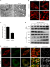
IR‐783 causes mitochondrial fission in MDAMB‐231 cells. A, MDAMB‐231 cells were treated with 40 μM IR‐783 for 24 h and mitochondria were imaged by transmission electron microscope. Scale bars: 2 μm. B, MDAMB‐231 cells were exposed to 40 μmol/L IR‐783 for 24 h. Mitochondria were then stained with MitoTracker Red CMXRos (red) and observed under a confocal microscope. Scale bars: 20 μm. C, Average mitochondrial length was counted in 30 cells. Error bars represent the mean ± SD, ***< .001. D, MDAMB‐231 cells were treated with IR‐783 with various concentrations as indicated, and mitochondrial fractions and whole cell lysates were subjected to western blot analysis using anti‐Drp1, OPA1, Mfn1, MFF and Fis1 antibodies. Cox IV (mitochondrial fraction) and actin (whole cell lysates) were used as the loading controls. E, MDAMB‐231 cells were treated with 40 μmol/L IR‐783 for 24 h and then the mitochondria were stained with MitoTracker Red CMXRos (red) after immunostaining with Drp1 (Alexa Fluor 488, green) and cells were visualized using confocal microscopy. The colocalization of Drp1 and mitochondria were analysed by ImageJ software. Scale bars: 20 μm
Figure 4
Figure 4
Knockdown of Drp1 blocked IR‐783‐induced mitochondrial fission, loss of MMP, ATP depletion, mPTP opening and apoptosis. A, MDAMB‐231 cells stably expressing non‐target shRNA (shCon) or Drp1 shRNA (shDrp1) were lysed and analysed by western blot. Actin was used as the loading control. B, shCon and shDrp1 cells were treated with or without 40 μmol/L IR‐783 for 24 h. Mitochondria were then stained using MitoTracker Red CMXRos (red) and observed under a confocal microscope. Scale bars: 20 μm. C, Average mitochondrial length was counted in 30 cells. D, shCon and shDrp1 cells were treated with or without 40 μmol/L IR‐783 for 24 h then the cells were stained with rhodamine‐123. The MMP was measured by fluorescence microplate. E, Measurement of intracellular content of ATP by Luminometer Microplate reader. F, Cells were stained with calcein‐AM+CoCl2 and tested by fluorescence microplate. The calcium retention capacity (CRC) contrast to control group is an index of the opening of mPTP. G, Cells were stained with annexin V‐FITC/PE and the percentage of apoptotic cells was measured by flow cytometry. The results were counted in 3 independent experiments (n = 3). Error bars represent the mean ± SD (*< .05, **< .01, ***< .001)
Figure 5
Figure 5
IR‐783 inhibited tumour growth in vivo by induction of the mitochondrial translocation of Drp1. A, Tumour volumes in xenograft mice were measured every week in the control and IR‐783 group *< .05. B, The bodyweight of mice after 4 weeks of IR‐783 treatment. C, Representative liver and kidney tissues were sectioned and subjected to haematoxylin‐eosin (H&E) staining. Scale bars: 100 μm. D, Representative tumour tissues were sectioned and subjected to H&E, TUNEL analysis and immunohistochemical staining for C‐Caspase‐3. Scale bars: 100 μm. E, The mitochondrial fractions of representative tumour tissues were prepared and subjected to western blot analysis using an anti‐Drp1 antibody. Cox IV was used as the loading control for mitochondrial fractions. F, The representative tumour tissues were sectioned and subjected to immunofluorescence using the anti‐Drp1 (green) and anti‐TOM20 (red, a mitochondrial marker) antibody. Scale bars: 20 μm

References

    1. Zhang B, Wang H, Shen S, et al. Fibrin‐targeting peptide CREKA‐conjugated multi‐walled carbon nanotubes for self‐amplified photothermal therapy of tumor. Biomaterials. 2016;79:46‐55. - PubMed
    1. Deng K, Li C, Huang S, et al. Recent progress in near infrared light triggered photodynamic therapy. Small. 2017;13:1702299. - PubMed
    1. Ishizawa T, Saiura A, Kokudo N. Clinical application of indocyanine green‐fluorescence imaging during hepatectomy. Hepatobiliary Surg Nutr. 2016;5:322‐328. - PMC - PubMed
    1. Tobis S, Knopf JK, Silvers CR, et al. Near infrared fluorescence imaging after intravenous indocyanine green: initial clinical experience with open partial nephrectomy for renal cortical tumors. Urology. 2012;79:958‐964. - PubMed
    1. Holt D, Okusanya O, Judy R, et al. Intraoperative near‐infrared imaging can distinguish cancer from normal tissue but not inflammation. PLoS ONE. 2014;9:e103342. - PMC - PubMed

Publication types

MeSH terms