. 2018 Dec;37(12):2718-2728.
doi: 10.1109/TMI.2018.2851150. Epub 2018 Jun 27.

Deep Learning Global Glomerulosclerosis in Transplant Kidney Frozen Sections

Deep Learning Global Glomerulosclerosis in Transplant Kidney Frozen Sections

Jon N Marsh et al. IEEE Trans Med Imaging. 2018 Dec.

Abstract

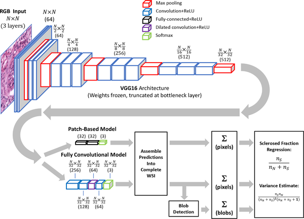
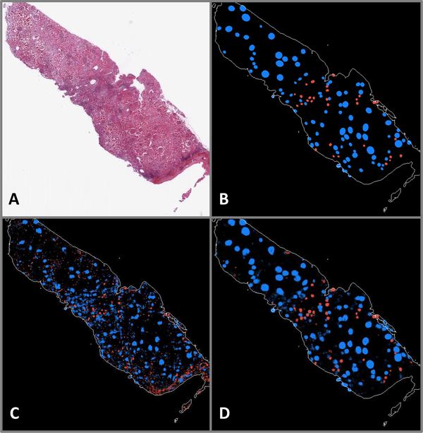
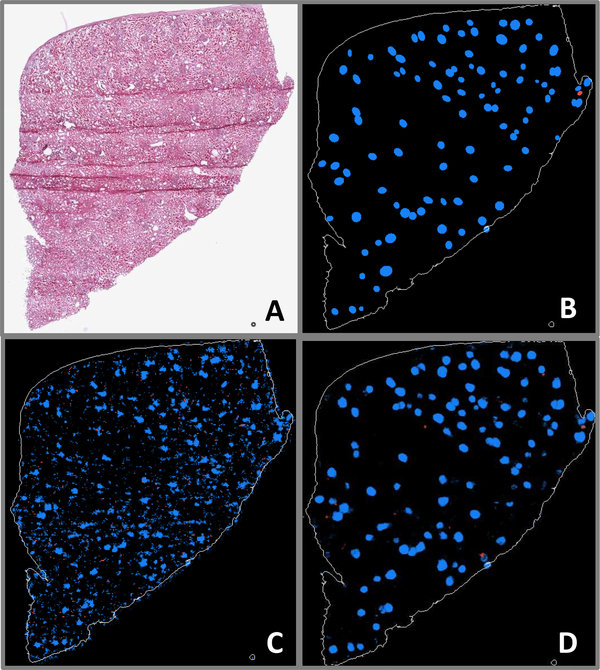
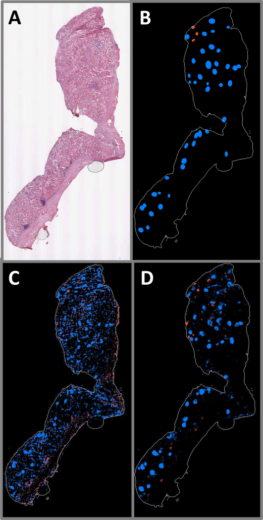
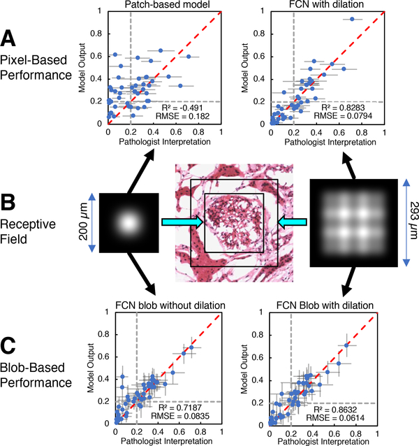
Transplantable kidneys are in very limited supply. Accurate viability assessment prior to transplantation could minimize organ discard. Rapid and accurate evaluation of intra-operative donor kidney biopsies is essential for determining which kidneys are eligible for transplantation. The criterion for accepting or rejecting donor kidneys relies heavily on pathologist determination of the percent of glomeruli (determined from a frozen section) that are normal and sclerotic. This percentage is a critical measurement that correlates with transplant outcome. Inter- and intra-observer variability in donor biopsy evaluation is, however, significant. An automated method for determination of percent global glomerulosclerosis could prove useful in decreasing evaluation variability, increasing throughput, and easing the burden on pathologists. Here, we describe the development of a deep learning model that identifies and classifies non-sclerosed and sclerosed glomeruli in whole-slide images of donor kidney frozen section biopsies. This model extends a convolutional neural network (CNN) pre-trained on a large database of digital images. The extended model, when trained on just 48 whole slide images, exhibits slide-level evaluation performance on par with expert renal pathologists. Encouragingly, the model's performance is robust to slide preparation artifacts associated with frozen section preparation. The model substantially outperforms a model trained on image patches of isolated glomeruli, in terms of both accuracy and speed. The methodology overcomes the technical challenge of applying a pretrained CNN bottleneck model to whole-slide image classification. The traditional patch-based approach, while exhibiting deceptively good performance classifying isolated patches, does not translate successfully to whole-slide image segmentation in this setting. As the first model reported that identifies and classifies normal and sclerotic glomeruli in frozen kidney sections, and thus the first model reported in the literature relevant to kidney transplantation, it may become an essential part of donor kidney biopsy evaluation in the clinical setting.


Figures

Fig. 1.
Fig. 1.
Example whole-slide image (WSI) of H&E-stained human renal frozen wedge biopsy scanned at 20X, with inset showing normal (yellow) and sclerosed (cyan) glomeruli as labeled by trained observers. Note the variability of appearance of glomeruli between and within categories.
Fig. 2.
Fig. 2.
Example image patches (at 20X magnification) of globally sclerosed glomeruli from frozen (left) and formalin-fixed (right) H&E slide preparations. Variability in glomerular appearance and stain intensity is greater in frozen preparations. Also note variability in stain intensity in frozen samples, typical of the dataset used in this study.
Fig. 3.
Fig. 3.
Data path for computation of sclerosed glomeruli fraction. Both patch-based and fully convolutional models utilize pretrained VGG16 architecture with frozen weights, truncated before bottleneck. Input patch sizes were 448×448 pixels for patch-based model, and 1024×1024 pixels for fully convolutional model.
Fig. 4.
Fig. 4.
Examples of patch-based model predictions on image patches containing isolated glomeruli. Top: Highest scored correctly identified patches. The model correctly identified sclerosed and non-sclerosed glomeruli, even in the presence of variable stain intensity and glomerular appearance. Bottom: Lowest scored incorrectly labeled patches; predicted label is shown in quotes beneath each image.
Fig. 5.
Fig. 5.
A: WSI exhibiting a large number of sclerosed glomeruli. B: Ground truth annotations indicating positions and shapes of non-sclerosed (blue) and sclerosed (red) glomeruli. C: Patch-based model prediction probability map. D: Fully convolutional model prediction probability map.
Fig. 6.
Fig. 6.
A: WSI exhibiting very few sclerosed glomeruli, as well as folding artifacts. B: Ground truth annotations indicating positions and shapes of nonsclerosed (blue) and sclerosed (red) glomeruli. C: Patch-based model prediction probability map. D: Fully convolutional model prediction probability map.
Fig. 7.
Fig. 7.
A: WSI exhibiting large region of renal capsule. B: Ground truth annotations indicating positions and shapes of non-sclerosed (blue) and sclerosed (red) glomeruli. C: Patch-based model prediction probability map. D: Fully convolutional model prediction probability map.
Fig. 8.
Fig. 8.
A: Pixel-wise model predictions of sclerosed glomerulus fraction vs. pathologists’ assessment for patch-based model (left) and fully convolutional model (right). Horizontal error bars indicate 95% confidence level for pathologist assessments. Dotted grey lines indicate a hypothetical clinical cutoff for rejection at 20% global glomerulosclerosis. B: Receptive field intensity map for fully convolutional model without dilated convolution layer (left) and with dilated convolution layer (right). Receptive field extent for both models are drawn to scale on image of normal glomerulus extracted from a WSI (center). C: Predictions of sclerosed glomerulus fraction vs pathologists assessment for fully convolutional model without dilated convolution layer (left) and with dilated convolution layer (right) after blob-detection postprocessing. Error bars indicate 95% confidence level, assuming the sclerosed and non-sclerosed glomeruli population is characterized by a beta distribution. Dotted grey lines indicate a hypothetical clinical cutoff for rejection at 20% global glomerulosclerosis.
Fig. 9.
Fig. 9.
Panels A, C, E: WSI annotations indicating positions and shapes of non-sclerosed (blue) and sclerosed (red) glomeruli. Panels B, D, F: Corresponding blob detection results using fully convolutional model prediction label maps as input. Solid circles indicate confirmed matches with annotations, X’s mark incorrect glomerulus detections, and open rectangles indicate annotated glomeruli that were overlooked by the model.
Fig. 10.
Fig. 10.
Free-response ROC curves for blob-detection algorithm applied to predicted probability maps generated by the fully convolutional model. Sensitivity vs false positive detections per image are shown for all glomeruli (left), nonsclerosed glomeruli (middle), and sclerosed glomeruli (right). Arrow indicate the selected operating point threshold (0.25).
Fig. 11.
Fig. 11.
An example blob detection error highlighted within dotted cyan circle. A: Image patch extracted from WSI annotation map, indicating positions and shapes of non-sclerosed (blue) and sclerosed (red) glomeruli. B: Probability map for the same image patch. C: Blob detection results using fully convolutional model prediction label map as input. Solid circles indicate confirmed matches with annotations, X’s mark incorrect glomerulus detections, and open rectangles indicate annotated glomeruli that were overlooked by the model. The blob detection algorithm fails to differentiate the model’s correctly-identified adjoining sclerosed glomeruli in this instance.

References

    1. Jochmans I and Pirenne J, “Graft quality assessment in kidney transplantation: not an exact science yet!” Current Opinion in Organ Transplantation, vol. 16, no. 2, pp. 174–179, April 2011. - PubMed
    1. Remuzzi G, Cravedi P, Perna A, Dimitrov BD, Turturro M, Locatelli G, Rigotti P, Baldan N, Beatini M, Valente U, Scalamogna M, and Ruggenenti P, “Long-term outcome of renal transplantation from older donors.” The New England Journal of Medicine, vol. 354, no. 4, pp. 343–52, January 2006. - PubMed
    1. Munivenkatappa RB, Schweitzer EJ, Papadimitriou JC, Drachenberg CB, Thom KA, Perencevich EN, Haririan A, Rasetto F, Cooper M, Campos L, Barth RN, Bartlett ST, and Philosophe B, “The Maryland Aggregate Pathology Index: A deceased donor kidney biopsy scoring system for predicting graft failure,” American Journal of Transplantation, vol. 8, no. 11, pp. 2316–2324, November 2008. - PubMed
    1. Rege A, Irish B, Castleberry A, Vikraman D, Sanoff S, Ravindra K, Collins B, and Sudan D, “Trends in Usage and Outcomes for Expanded Criteria Donor Kidney Transplantation in the United States Characterized by Kidney Donor Profile Index.” Cureus, vol. 8, no. 11, p. e887, November 2016. - PMC - PubMed
    1. Furness PN and Taub N, “International variation in the interpretation of renal transplant biopsies: Report of the CERTPAP project,” Kidney International, vol. 60, no. 5, pp. 1998–2012, November 2001. - PubMed

Publication types