. 2018 Sep 1;29(18):2213-2227.
doi: 10.1091/mbc.E17-12-0703. Epub 2018 Jul 11.

WDR41 supports lysosomal response to changes in amino acid availability

Affiliations

WDR41 supports lysosomal response to changes in amino acid availability

Joseph Amick et al. Mol Biol Cell. .

Abstract

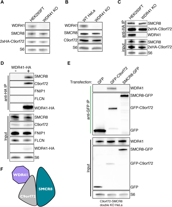
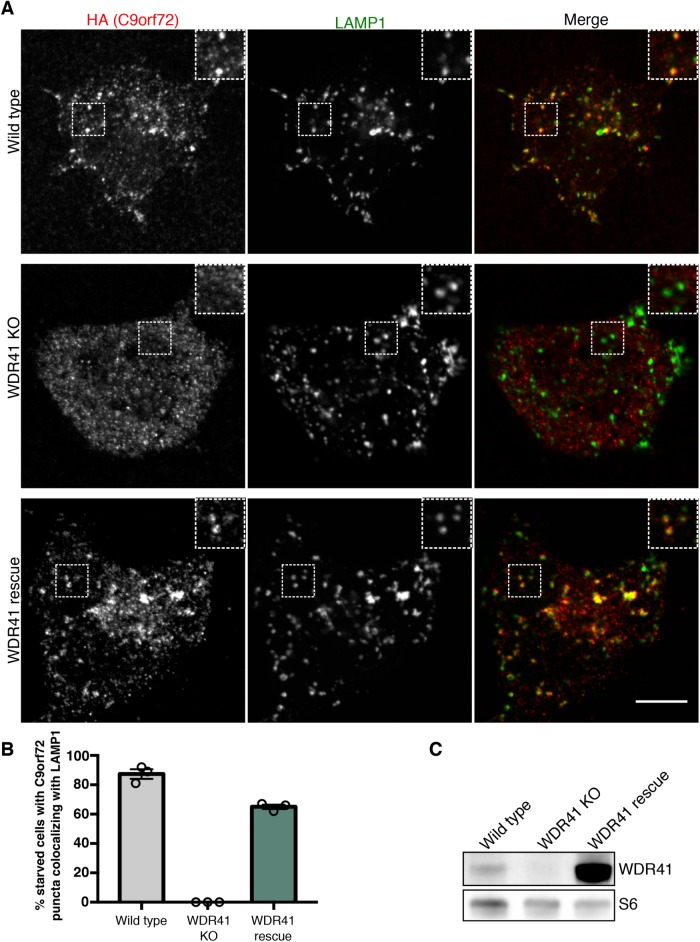
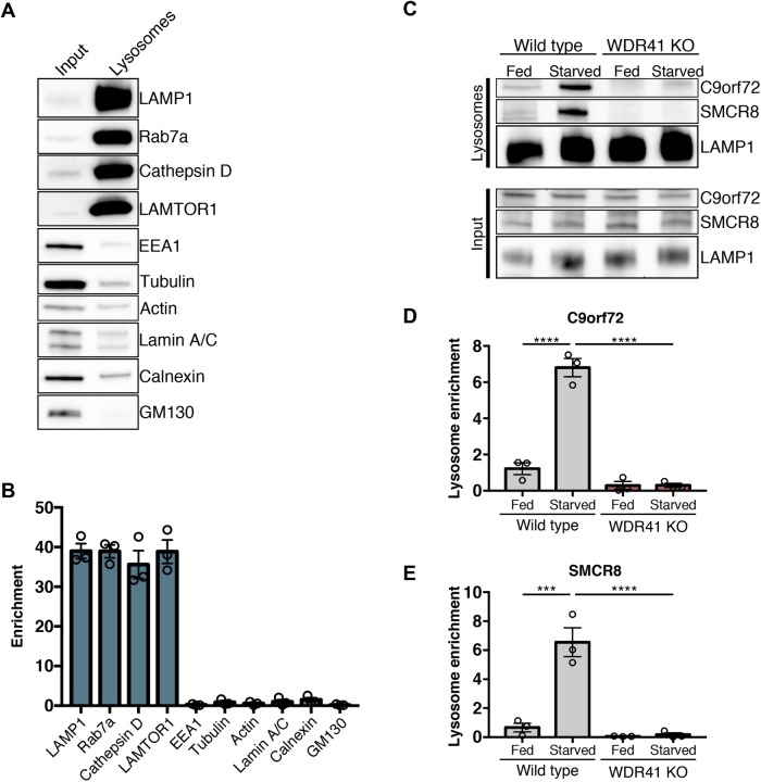
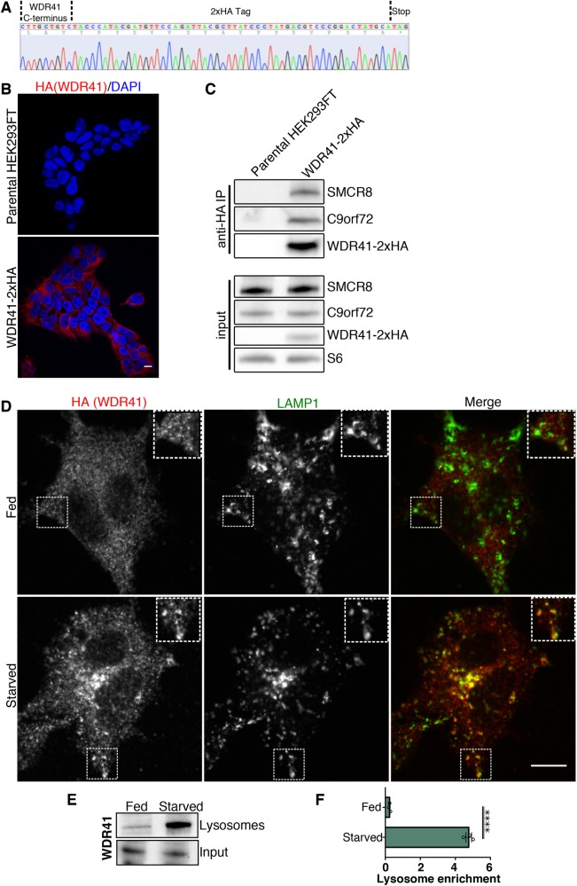
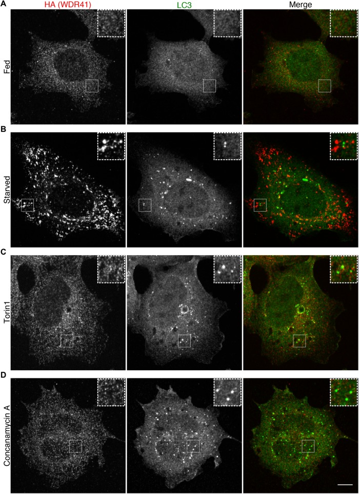
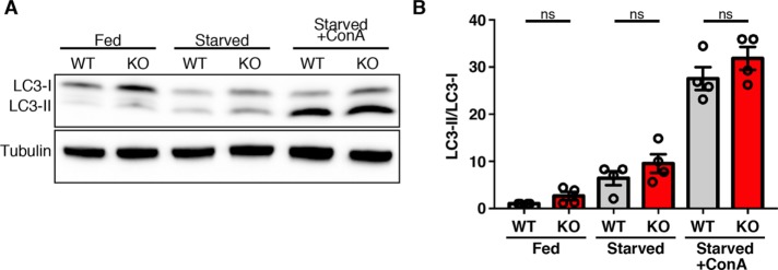
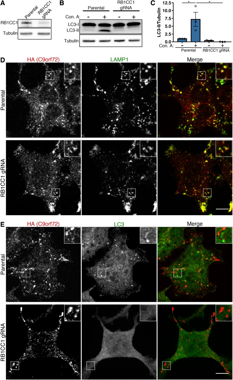
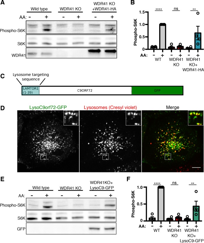
C9orf72 mutations are a major cause of amyotrophic lateral sclerosis and frontotemporal dementia. The C9orf72 protein undergoes regulated recruitment to lysosomes and has been broadly implicated in control of lysosome homeostasis. However, although evidence strongly supports an important function for C9orf72 at lysosomes, little is known about the lysosome recruitment mechanism. In this study, we identify an essential role for WDR41, a prominent C9orf72 interacting protein, in C9orf72 lysosome recruitment. Analysis of human WDR41 knockout cells revealed that WDR41 is required for localization of the protein complex containing C9orf72 and SMCR8 to lysosomes. Such lysosome localization increases in response to amino acid starvation but is not dependent on either mTORC1 inhibition or autophagy induction. Furthermore, WDR41 itself exhibits a parallel pattern of regulated association with lysosomes. This WDR41-dependent recruitment of C9orf72 to lysosomes is critical for the ability of lysosomes to support mTORC1 signaling as constitutive targeting of C9orf72 to lysosomes relieves the requirement for WDR41 in mTORC1 activation. Collectively, this study reveals an essential role for WDR41 in supporting the regulated binding of C9orf72 to lysosomes and solidifies the requirement for a larger C9orf72 containing protein complex in coordinating lysosomal responses to changes in amino acid availability.


Figures

FIGURE 1:
FIGURE 1:
WDR41 interacts with C9orf72 but is not required for the stability of C9orf72 and SMCR8. (A) Immunoblot analysis of WDR41 KO in HEK293FT cells that express 2xHA-tagged C9orf72 from the endogenous locus. (B) Immunoblot analysis of control and WDR41 KO HeLa cells. (C) Endogenously expressed 2x-HA C9orf72 was immunoprecipitated from the parental and WDR41 knockout cell line, followed by immunoblotting for the indicated proteins. (D) Anti-HA immunoprecipitation of control and WDR41-HA-transfected HeLa cells followed by immunoblotting for the indicated proteins. (E) C9orf72/SMCR8 double knockout cells were transfected with GFP, GFP-C9orf72, or SMCR8-GFP followed by anti-GFP immunoprecipitation and immunoblotting for the indicated proteins. (F) Immunoprecipitation results in E support a model wherein WDR41 associates with the C9orf72:SMCR8 complex primarily via an interaction with C9orf72.
FIGURE 2:
FIGURE 2:
WDR41 is required for the recruitment of C9orf72 to lysosomes. (A) Immunofluorescence images of C9orf72 localization (endogenously expressed 2xHA-C9orf72) in starved wild-type, WDR41 knockout, and WDR41 KO cells that were rescued by stable expression of a WDR41 transgene. Localization of C9orf72 to lysosomes (LAMP1 signal) is lost in WDR41 knockout cells. Scale bar, 10 μm. (B) For the indicated cell lines, the percentage of cells in starved conditions containing C9orf72 puncta that colocalize with LAMP1 are indicated. (C) Immunoblot analysis of WDR41 levels in wild-type, knockout, and rescue cell lines.
FIGURE 3:
FIGURE 3:
WDR41 is required for C9orf72 and SMCR8 enrichment on purified lysosomes. (A) Immunoblots of the indicated proteins in the total cell lysate (input) and in magnetically isolated lysosomes (lysosomes). Equal amounts of protein were loaded in each lane. LAMP1, Rab7a, Cathepsin D (mature form), and LAMTOR1 were used as markers of late endosomes/lysosomes; EEA, early endosomes; tubulin, actin, cytoskeleton; Lamin A/C, nucleus; Calnexin, ER; GM130, –Golgi. (B) Quantification of immunoblots for the indicated proteins expressed as a fold increase in the lysosome fraction relative to the input (mean ± SEM, n = 3). (C) Immunoblots of C9orf72, SMCR8, and LAMP1 in the total cell lysate (input) and in magnetically isolated lysosomes under fed and starved conditions in wild-type and WDR41 KO HeLa cells. Quantification of the levels of C9orf72 (D) and SMCR8 (E) on magnetically isolated lysosomes (mean ± SEM, n = 3, ****p < 0.0001, ***p ≤ 0.001 two-way analysis of variance [ANOVA] with Tukey’s multiple comparisons test).
FIGURE 4:
FIGURE 4:
WDR41 localizes to lysosomes in starved cells. (A) Sequencing result from the genomic DNA PCR product from HEK293FT cells that have a 2xHA epitope tag inserted at the endogenous WDR41 locus. The positions of the WDR41 C-terminal sequence, 2xHA epitope tag, and stop codon are indicated. (B) The specificity of the anti-HA immunofluorescence signal in WDR41-2xHA cells is supported by the absence of this signal in parental, non-gene-edited cells. Scale bar, 10 µm. (C) Anti-HA immunoprecipitations from WDR41-2xHA cells followed by immunoblotting for the indicated proteins. WDR41-2xHA retains the ability to interact with C9orf72 and SMCR8. Non-gene-edited HEK293FT cells served as a negative control. (D) Immunofluorescence images showing the localization of WDR41-2xHA (expressed from the endogenous locus) and LAMP1 in fed and starved (1.5-h serum and amino-acid-free) conditions. (E) Immunoblot analysis of WDR41 levels on magnetically-isolated lysosomes from HeLa cells in fed and starved conditions. (F) Quantification of WDR41 immunoblots expressed as a fold increase in the lysosome fraction relative to the input (mean ± SEM, n = 3, unpaired t test, ****p < 0.0001).
FIGURE 5:
FIGURE 5:
WDR41 is selectively recruited to lysosomes in response to starvation. Immunofluorescence images showing the localization of endogenously expressed WDR41-2xHA in (A) fed, (B) starved (1.5-h serum and amino-acid-free conditions), (C) torin1-treated (2 µM, 2 h), and (D) concanamycin A-treated (1 µM, 2 h). Scale bar, 10 µm.
FIGURE 6:
FIGURE 6:
LC3 lipidation and autophagic flux is unimpaired in WDR41 KO cells. (A) Immunoblot analysis of LC3 levels in wild-type and WDR41 knockout cells under fed, starved, and starved cells treated with the v-ATPase inhibitor concanamycin A (ConA). (B) Quantification of LC3-II/tubulin levels (mean ± SEM, n = 4, Sidak’s multiple comparisons test).
FIGURE 7:
FIGURE 7:
RB1CC1 and autophagy are not required for C9orf72 recruitment to lysosomes. (A) Immunoblot analysis of CRISPR-Cas9-mediated RB1CC1 depletion in the HEK293FT cell line that expresses 2xHA-C9orf72 from the endogenous locus. (B) Parental 2xHA-C9orf72 HEK293FT and RB1CC1-depleted cells were treated with and without concanamycin A (V-ATPase inhibitor) for 2 h and autophagic flux was assessed by immunoblotting. Although wild-type cells accumulate LC3-II when treated with concanamycin A, the RB1CC1-depleted cell line does not. (C) Quantification of the ratio of LC3-II to tubulin (mean ± SEM, n = 3, *p ≤ 0.05, two-way ANOVA with Tukey’s multiple comparisons test). (D) Immunofluorescence analysis of C9orf72 localization in starved wild-type and RB1CC1-depleted cells. Scale bar, 10 µm. (E) Immunofluorescence analysis of C9orf72 and LC3 in starved wild-type and RB1CC1-depleted cells. Starved wild-type cells have distinct C9orf72 and LC3 puncta (predominantly autophagosomes), while RB1CC1-depleted cells maintain C9orf72 puncta but lack LC3 puncta. Scale bar, 10 µm.
FIGURE 8:
FIGURE 8:
WDR41-dependent recruitment of C9orf72 to lysosomes is critical for the stimulation of mTORC1 activity by amino acids (A) Immunoblot analysis of phospho-S6 kinase (S6K-T389) levels during starvation (1.5 h) and subsequent amino acid (AA) refeeding (15 min) in WT, WDR41 knockout, and cells stably expressing WDR41-HA in the WDR41 knockout background. (B) Summary of S6 kinase phosphorylation levels normalized to total S6K levels; WT refeed normalized to 1 (mean ± SEM, n = 4, ****p < 0.0001, **p ≤ 0.01, ANOVA with Sidak’s multiple comparisons test). (C) Schematic diagram of the Lyso-C9orf72-GFP construct. The first 39 amino acids of LAMTOR1 were fused to the N-terminus of C9orf72 followed by a C-terminal GFP tag. (D) Live-cell imaging reveals the successful targeting of Lyso-C9orf72-GFP to lysosomes as illustrated by colocalization with cresyl violet, a fluorescent reporter of lysosome acidity. Scale bar = 10 μm. (E) Immunoblot analysis of phospho-S6K levels during starvation (1.5 h) and subsequent AA refeeding (15 min) in wild-type, WDR41 knockout, and cells stably expressing Lyso-C9orf72-GFP in the WDR41 knockout background. (F) Quantification of S6K phosphorylation levels normalized to total S6K levels (WT refeed normalized to 1; mean ± SEM, n = 4, ****p < 0.0001, **p ≤ 0.01, ANOVA with Sidak’s multiple comparisons test).

References

    1. Amick J, Ferguson SM. (2017). C9orf72: At the intersection of lysosome cell biology and neurodegenerative disease. Traffic , 267–276. - PMC - PubMed
    1. Amick J, Roczniak-Ferguson A, Ferguson SM. (2016). C9orf72 binds SMCR8, localizes to lysosomes, and regulates mTORC1 signaling. Mol Biol Cell , 3040–3051. - PMC - PubMed
    1. Baba M, Hong SB, Sharma N, Warren MB, Nickerson ML, Iwamatsu A, Esposito D, Gillette WK, Hopkins RF, 3rd, Hartley JL, et al (2006). Folliculin encoded by the BHD gene interacts with a binding protein, FNIP1, and AMPK, and is involved in AMPK and mTOR signaling. Proc Natl Acad Sci USA , 15552–15557. - PMC - PubMed
    1. Bajagic M, Archna A, Busing P, Scrima A. (2017). Structure of the WD40-domain of human ATG16L1. Protein Sci , 1828–1837. - PMC - PubMed
    1. Baker M, Mackenzie IR, Pickering-Brown SM, Gass J, Rademakers R, Lindholm C, Snowden J, Adamson J, Sadovnick AD, Rollinson S, et al (2006). Mutations in progranulin cause tau-negative frontotemporal dementia linked to chromosome 17. Nature , 916–919. - PubMed

Publication types