Review
. 2019 Jan;33(1):14-33.
doi: 10.1038/s41433-018-0158-4. Epub 2018 Jul 11.

Pachychoroid disease

Affiliations
Review

Pachychoroid disease

Chui Ming Gemmy Cheung et al. Eye (Lond). 2019 Jan.

Abstract

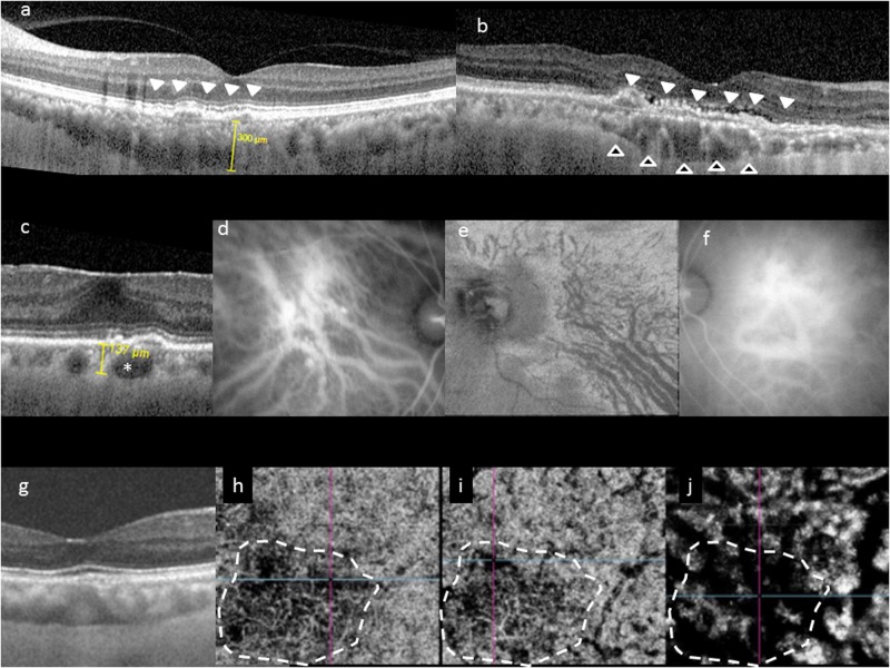
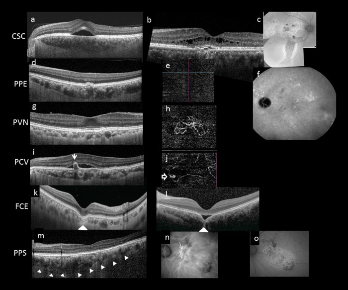
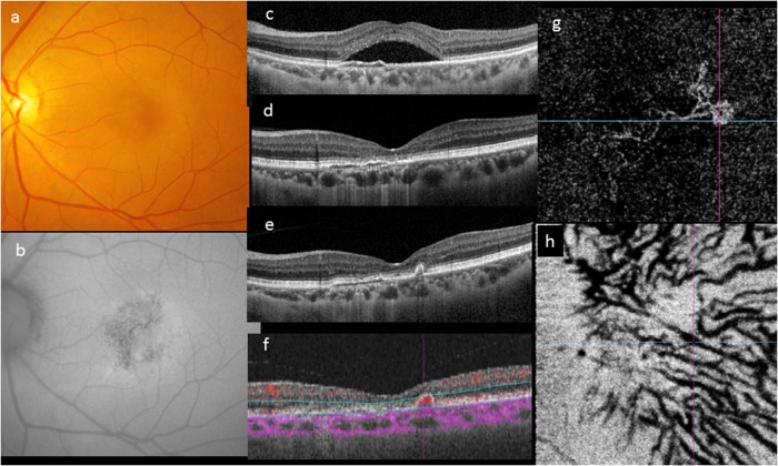
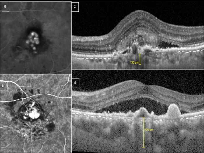
Pachychoroid is a relatively novel concept describing a phenotype characterized by attenuation of the choriocapillaris overlying dilated choroidal veins, and associated with progressive retinal pigment epithelium dysfunction and neovascularization. The emphasis in defining pachychoroid-related disorders has shifted away from simply an abnormally thick choroid (pachychoroid) toward a detailed morphological definition of a pathologic state (pachychoroid disease) with functional implications, which will be discussed in this review. Several clinical manifestations have been described to reside within the pachychoroid disease spectrum, including central serous chorioretinopathy, pachychoroid pigment epitheliopathy, pachychoroid neovasculopathy, polypoidal choroidal vasculopathy/aneurysmal type 1 neovascularization, focal choroidal excavation, peripapillary pachychoroid syndrome. These conditions all exhibit the characteristic choroidal alterations and are believed to represent different manifestations of a common pathogenic process. This review is based on both the current literature and the clinical experience of our individual authors, with an emphasis on the clinical and imaging features, management considerations, as well as current understanding of pathogenesis of these disorders within the context of the recent findings related to pachychoroid disease.

摘要: 肥厚型脉络膜疾病是近年来提出的新概念, 其描述了一类特征为脉络膜毛细血管层的静脉扩张, 并且与进行性视网膜色素上皮功能障碍和新生血管的形成有关的一类疾病。定义肥厚型脉络膜相关疾病的重点已经从简单的脉络膜厚度异常 (脉络膜肥厚) 转向影响功能的病理状态 (“肥厚型脉络膜疾病”) 的详细形态学定义, 这将在本综述中详细讨论。属于肥厚型脉络膜疾病谱的疾病, 包括中心性浆液性脉络膜视网膜病变, 肥厚型脉络膜色素上皮病变, 肥厚型脉络膜新生血管病变, 息肉状脉络膜血管病变/1型动脉瘤新生血管, 局灶性脉络膜凹陷, 视盘周围毛细脉络膜肥厚综合症。这些病症都表现出特征性的脉络膜改变, 并且被认为代表了常见致病过程的不同临床表现。 本综述基于目前已发表的文献和作者自身的临床经验以及最近在肥厚型脉络疾病的相关研究结果, 重点强调了临床表现、影像学特征、治疗要点以及目前对这些疾病发病机制的探讨。.


Conflict of interest statement

The authors declare that they have no conflict of interest.

Figures

Fig. 1
Fig. 1
Choroidal features common to pachychoroid phenotype. Typical choroidal features in the pachychoroid disease phenotype include diffuse or focal increase in choroidal thickness, which is typically associated with abnormally dilated Haller layer vessels (pachyvessels) and attenuation of the inner choroid (Sattler’s layer and choriocapillaris). Choroidal thickness can be readily evaluated in cross-sectional optical coherence tomography (OCT), which may show diffuse thickening and increased subfoveal choroidal thickness (a), or focal thickening (b, hollow arrowheads). In some eyes, an irregular elevation of the retinal pigment epithelium (RPE) can be seen to overlie these choroidal abnormalities (white arrowheads). Pachyvessels can be identified as a choroidal vessel with enlarged caliber (c, *) which can occupy almost the entire thickness of the choroid. Pachyvessels can also be seen as dilated submacular vessels which do not taper toward the posterior pole on ICGA (d) or on en face OCT (e). These pachyvessels may be distributed in a diffuse (d) or patchy manner (e). Pachyvessels usually exhibit choroidal vascular hyperpermeability with indocyanine green angiography (ICGA) (f) which may suggest choriocapillaris ischemia. Using OCT angiography (OCTA), the spatial correlation between choriocapillaris blood flow and pachyvessels can be evaluated in a depth-resolved manner. In gj, choroidal thickening and choriocapillaris attenuation seen in cross-sectional OCT (g) can be correlated with OCTA findings showing attenuation of flow signal (dash white outline) within the choriocapillaris (h) and inner choroid (i) which directly overlie areas with dilated outer choroidal vessels (j)
Fig. 2
Fig. 2
Multimodal imaging features of pachychoroid disease. Several clinical manifestations have been described to reside within the pachychoroid disease spectrum, including central serous chorioretinopathy (CSC) (ac), pachychoroid pigment epitheliopathy (PPE) (df), pachychoroid neovasculopathy (PVN) (g, h), polypoidal choroidal vasculopathy (PCV)/aneurysmal type 1 neovascularization (AT1) (i, j), focal choroidal excavation (FCE) (k, l), and peripapillary pachchoroid syndrome (PPS) (mo). Note that the choroidal features described in Fig. 1 can be seen in all cases in Fig. 2. In acute CSC (a), a solitary neurosensory detachment is commonly seen. In contrast, in chronic CSC (b), the neurosensory detachment tends to be shallower and broader. A shallow pigment epithelial detachment (PED), shallow subretinal fluid (SRF), and cystic intraretinal fluid can also be seen in this case. The RPE changes are best evaluated using fundus autofluorescence (FAF). In chronic CSC (c), multiple areas showing mixed hyperautofluorescence and hypoautofluorescence can be seen in the posterior pole. A downward gravitational tract of hypoautofluorescent can be seen. PPE is thought to be a forme fruste of CSC. On irregular RPE elevation overlying pachychoroid disease features without subretinal fluid can be seen in d. OCTA confirmed absence of neovascularization (e) and FAF showed milder granular autofluorescence disturbance than in c, without a gravitational tract. Cross-sectional OCT findings in PCN may be similar to those in chronic CSC and PPE, characterized by irregular RPE elevation with or without SRF. OCTA can readily detect the presence of neovascularization (h). Features of PCV/AT1 overlap significantly with PCN, with the additional feature of aneurysms. Although indocyanine green angiography (ICGA) is considered the gold standard for diagnosing PCV/AT1, common features on OCT include irregular RPE elevation overlying pachychoroid disease changes and narrow-peaked PEDs (arrow). In the corresponding OCTA (j), an aneurysm (hollow arrow) can be seen to arise from a branching vascular network. FCE is characterized by a localized area of choroidal excavation on OCT. In the conforming type (k), the photoreceptor tips are not separated from the underlying RPE, whereas in the non-conforming type (l) a hyporeflective space can be observed between the photoreceptor tips and RPE. Unusual hyperreflective choroidal tissue (arrowhead) can be seen to bridge the space between the bottom of the excavation and the outer choroidal boundary. Dilated choroidal vessel and thickened choroid can be seen on either side of the excavation. In PPS, the choroid is thicker on the nasal side to the fovea compared to the temporal side in the cross-sectional OCT (m). More dilated choroidal vessels (white arrows) are seen on the nasal side compared to the temporal side of the fovea on ICGA (n). Fundus autofluorescence illustrates mottled autofluorescence temporal to the disc and extending downwards (o)
Fig. 3
Fig. 3
Evolution of a case over 4 years. A 46-year-old woman first presented with a solitary neurosensory detachment and mottled fundus autofluorescence (ac). A shallow elevation of the RPE was noted. The SRF resolved spontaneously within 3 months and the eye remained unchanged for the following 4 years (d). During routine follow-up, development of a narrow-peaked PED without SRF was noted on OCT (e). OCTA showed a localized area of abnormal flow signal within the narrow-peaked PED (f). En face OCTA showed a neovascular network with aneurysms its temporal margin (g). The outline of pachyvessels can be seen as dark silhouettes in the OCTA segmented through Haller’s layer (h)
Fig. 4
Fig. 4
Choroidal features in polypoidal choroidal vasculopathy (PCV)/aneurysmal type 1 neovascularization (AT1). A bimodal distribution of choroidal thickness has been described in PCV/AT1. This figure shows two eyes with PCV/AT1 confirmed with indocyanine green angiography (ICGA) (a, b). Subfoveal choroidal thickness was 130 and 420 μm, respectively (c, d). However, in both cases, choriocapillaris/inner choroid (outline by dashed white line in preserved areas) appears to be compressed and attenuated by underlying outer choroidal vessels in the subfoveal area (c, d)

References

    1. Mrejen S, Spaide RF. Optical coherence tomography: imaging of the choroid and beyond. Surv Ophthalmol. 2013;58:387–429. - PubMed
    1. Yanagi Y, Ting DSW, Ng WY, et al. Choroidal vascular hyperpermeability as a predictor of treatment response for polypoidal choroidal vasculopathy. Retina. 2017 Jul 12. Epub ahead of print. - PubMed
    1. Guyer DR, Yannuzzi LA, Slakter JS, Sorenson JA, Hope-Ross M, Orlock DR. Digital indocyanine-green videoangiography of occult choroidal neovascularization. Ophthalmology. 1994;101:1727–35. - PubMed
    1. Dansingani KK, Balaratnasingam C, Naysan J, Freund KB. En face imaging of pachychoroid spectrum disorders with swept-source optical coherence tomography. Retina. 2016;36:499–516. - PubMed
    1. Ferrara D, Mohler KJ, Waheed N, et al. En face enhanced-depth swept-source optical coherence tomography features of chronic central serous chorioretinopathy. Ophthalmology. 2014;121:719–26. - PMC - PubMed

MeSH terms

LinkOut - more resources