. 2018 Jul;559(7714):410-414.
doi: 10.1038/s41586-018-0224-x. Epub 2018 Jun 6.

Mechanism of parkin activation by PINK1

Affiliations

Mechanism of parkin activation by PINK1

Christina Gladkova et al. Nature. 2018 Jul.

Abstract

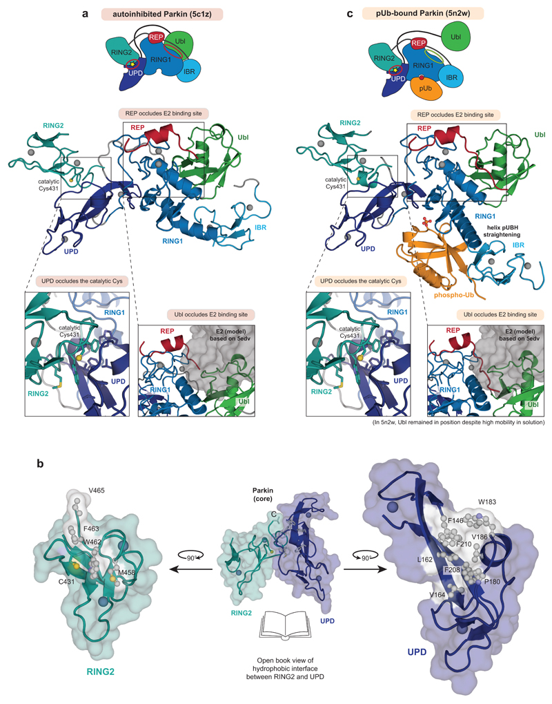
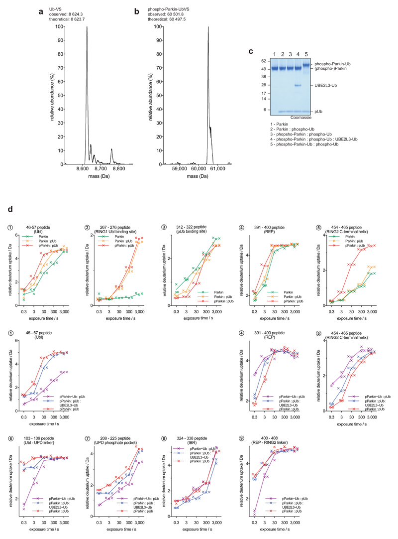
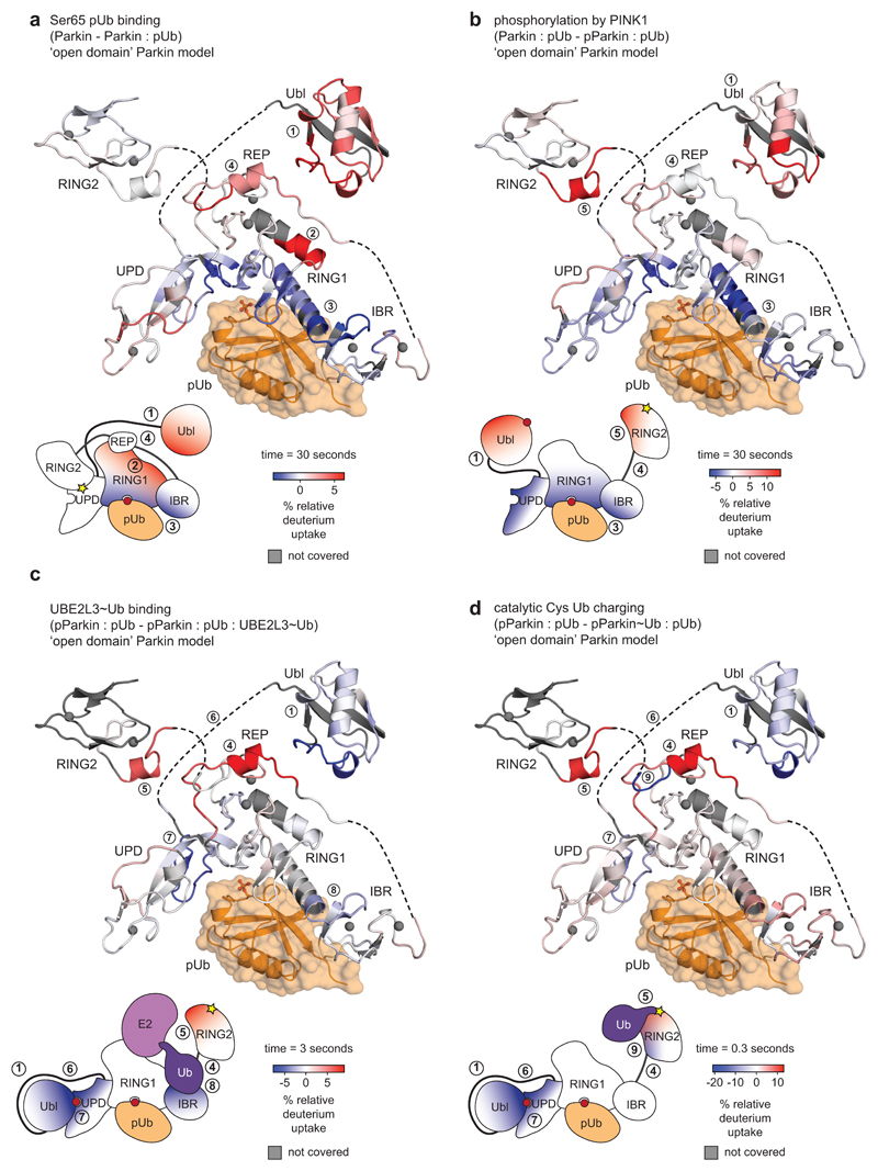
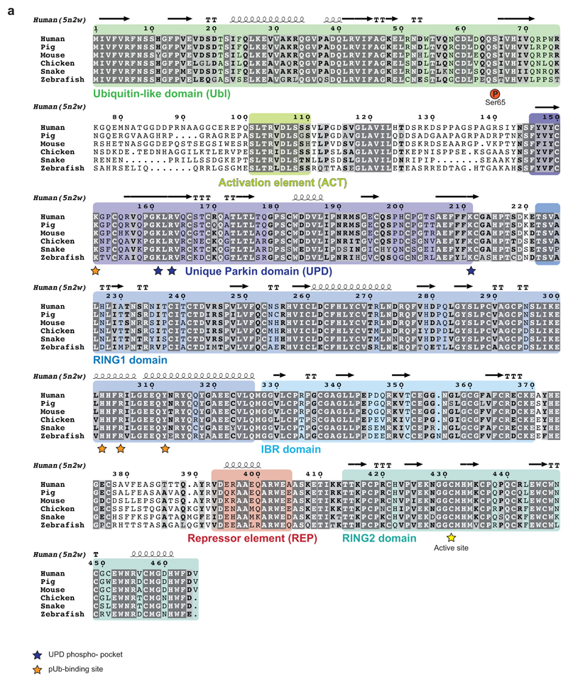
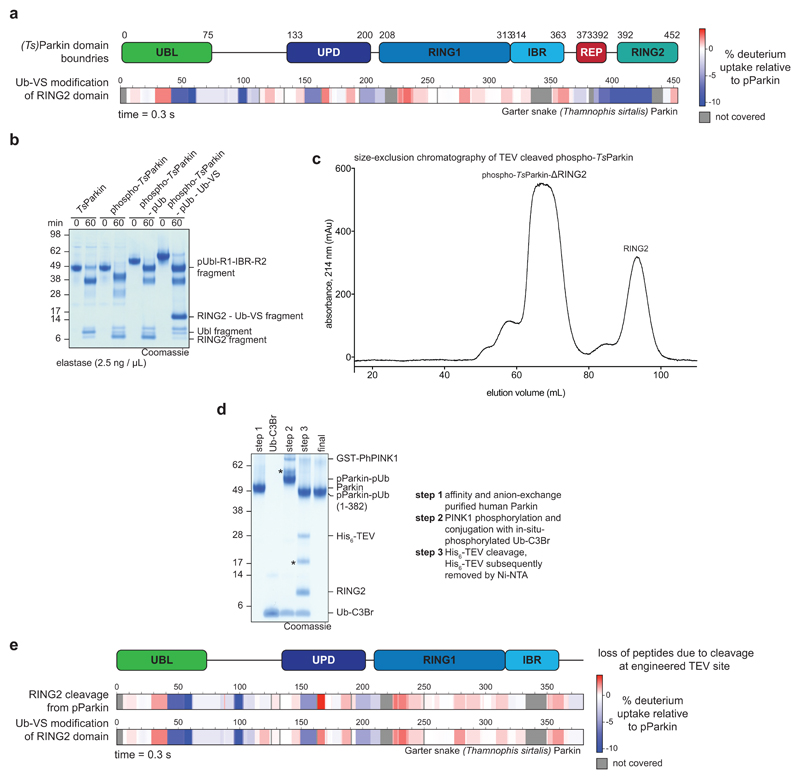
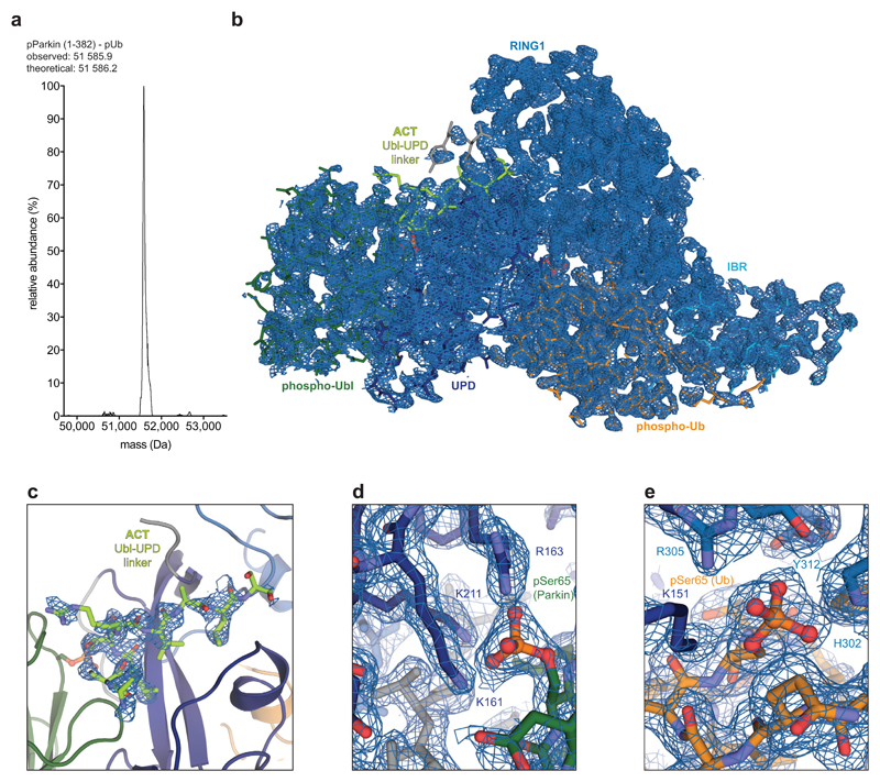
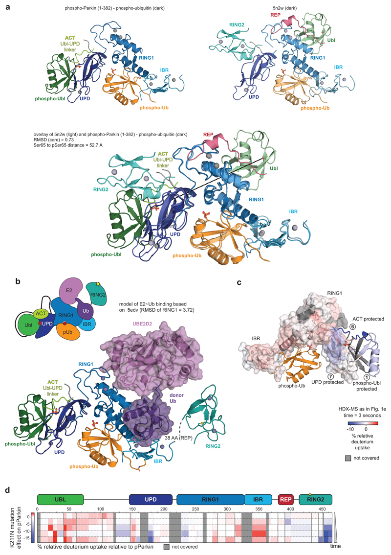
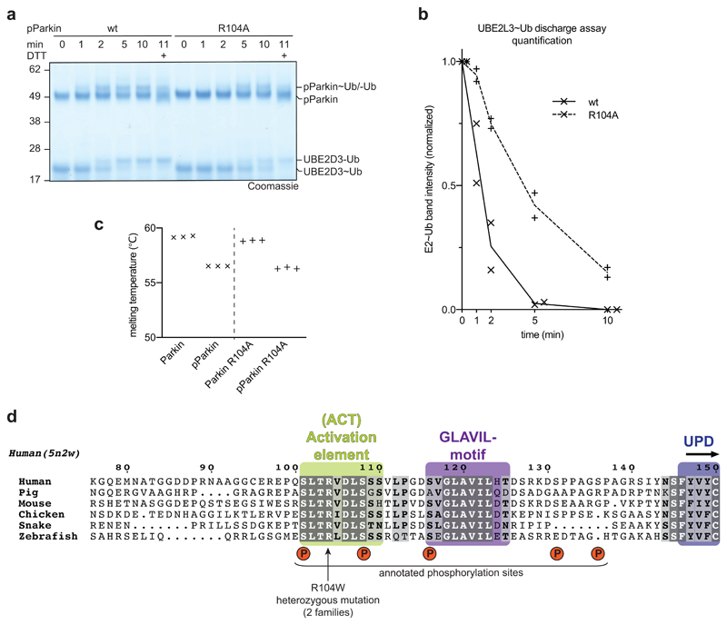
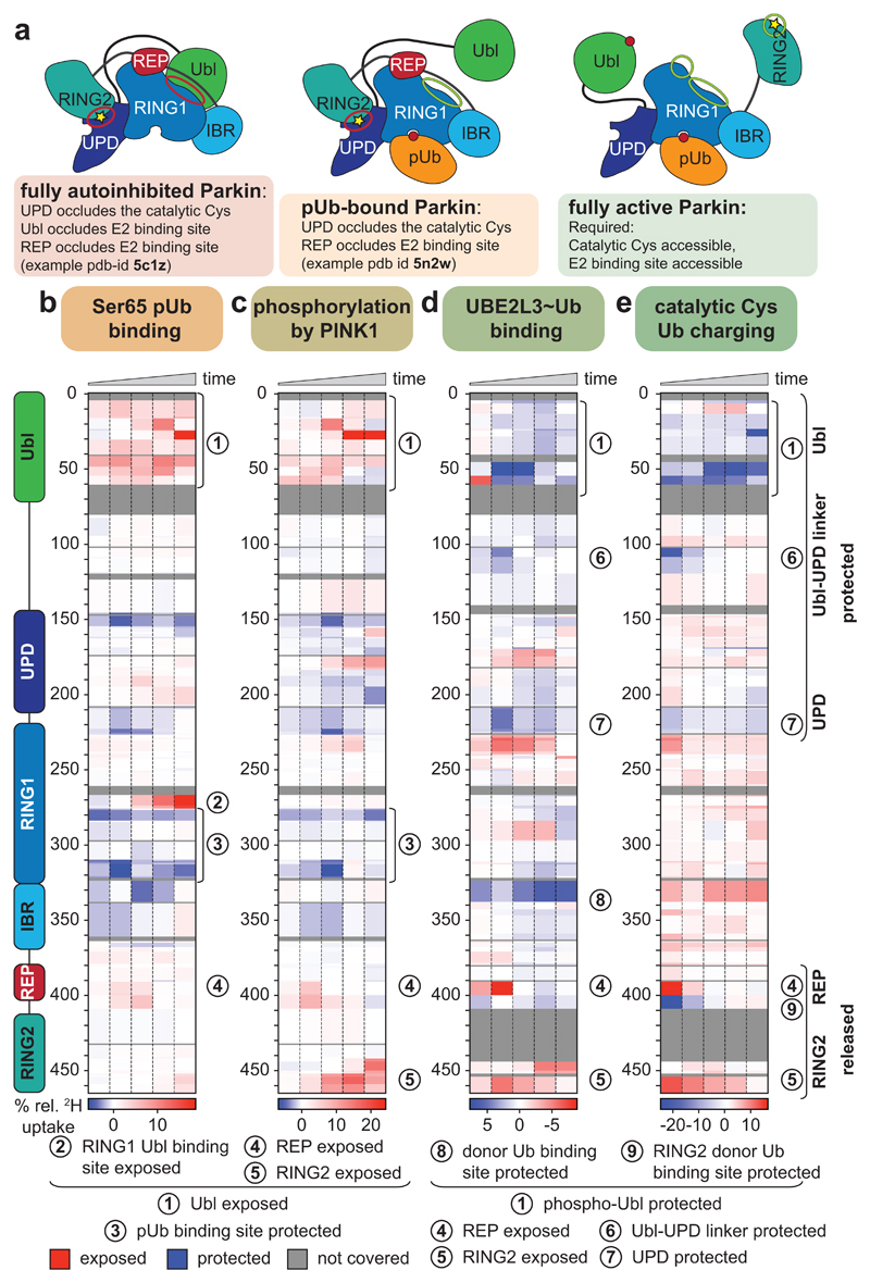
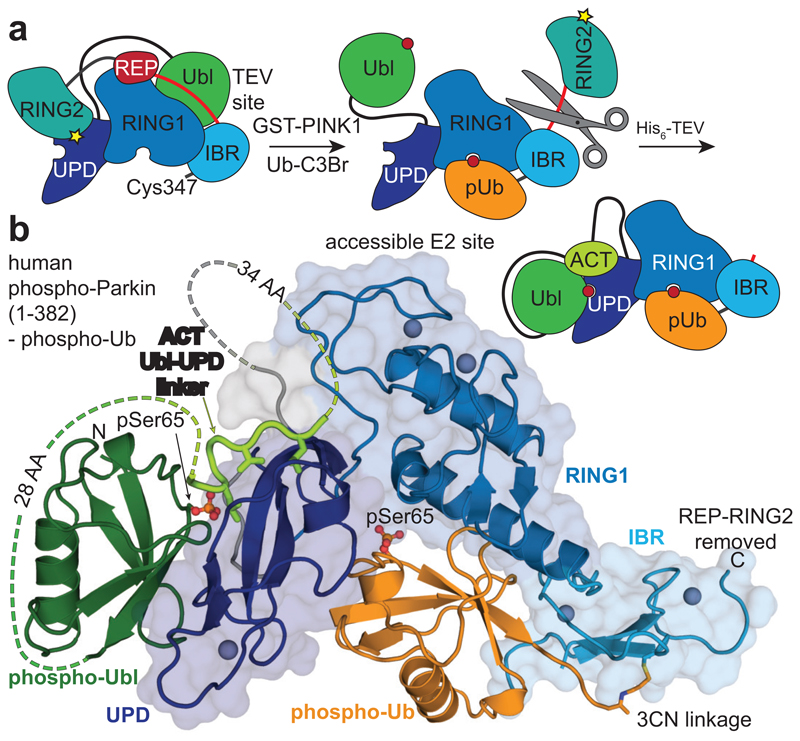
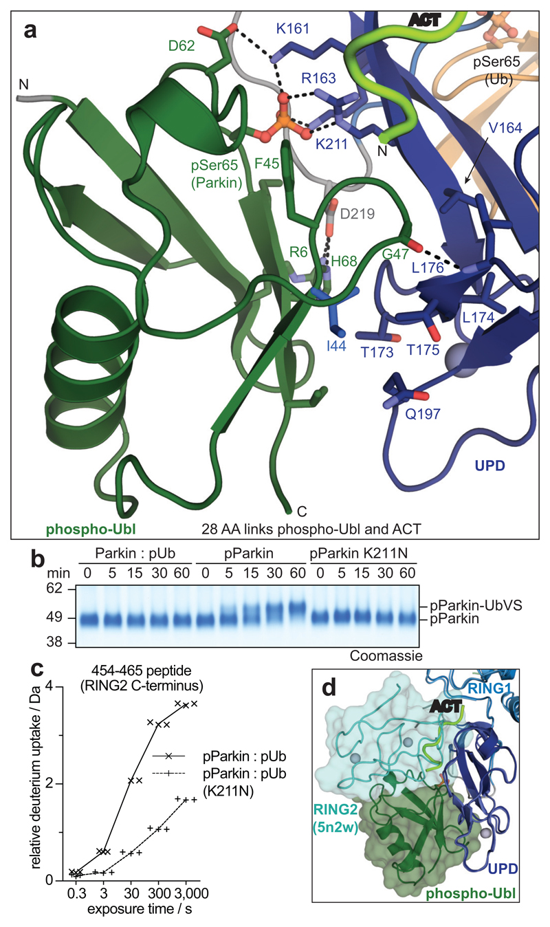
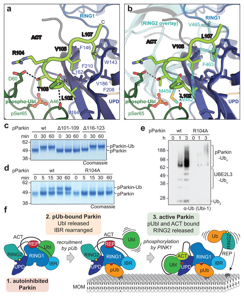
Mutations in the E3 ubiquitin ligase parkin (PARK2, also known as PRKN) and the protein kinase PINK1 (also known as PARK6) are linked to autosomal-recessive juvenile parkinsonism (AR-JP)1,2; at the cellular level, these mutations cause defects in mitophagy, the process that organizes the destruction of damaged mitochondria3,4. Parkin is autoinhibited, and requires activation by PINK1, which phosphorylates Ser65 in ubiquitin and in the parkin ubiquitin-like (Ubl) domain. Parkin binds phospho-ubiquitin, which enables efficient parkin phosphorylation; however, the enzyme remains autoinhibited with an inaccessible active site5,6. It is unclear how phosphorylation of parkin activates the molecule. Here we follow the activation of full-length human parkin by hydrogen-deuterium exchange mass spectrometry, and reveal large-scale domain rearrangement in the activation process, during which the phospho-Ubl rebinds to the parkin core and releases the catalytic RING2 domain. A 1.8 Å crystal structure of phosphorylated human parkin reveals the binding site of the phospho-Ubl on the unique parkin domain (UPD), involving a phosphate-binding pocket lined by AR-JP mutations. Notably, a conserved linker region between Ubl and the UPD acts as an activating element (ACT) that contributes to RING2 release by mimicking RING2 interactions on the UPD, explaining further AR-JP mutations. Our data show how autoinhibition in parkin is resolved, and suggest a mechanism for how parkin ubiquitinates its substrates via an untethered RING2 domain. These findings open new avenues for the design of parkin activators for clinical use.


Conflict of interest statement

Author information

The authors declare no competing interests.

Figures

Extended Data Figure 1
Extended Data Figure 1. Mechanisms of Parkin autoinhibition
a, Structure of autoinhibited, full-length human Parkin (pdb-id 5c1z, 19) shown schematically (top, as in Fig. 1a) and in cartoon representation in the same colours. Two insets show the UPD-RING2 interface (with Cys431 shows in ball-and-stick representation), and the blocked E2 binding site (with the E2 position, modelled according to 5edv shown as grey surface). Zn ions are shown as grey spheres. b, An ‘open-book’ view on the UPD-RING2 interface, with hydrophobic residues coloured white on each surface. c, Structure of phospho-ubiquitin bound to full-length Parkin (pdb-id 5n2w, 6) as in a. Phospho-ubiquitin binding leads to helix straightening, and IBR domain repositioning, which releases the Ubl domain for phosphorylation,. In the shown structures of unphosphorylated Parkin, the Ubl and REP (red) inhibit E2 binding, and the RING2-UPD interface is intact, with Cys431 being inaccessible. The Ubl-UPD linker was removed from both crystallised constructs,.
Extended Data Figure 2
Extended Data Figure 2. Samples preparation for HDX-MS and selected raw data.
a, Representative LC-MS spectrum of the prepared Ub-VS probe (see Online Methods). Experiment has been performed in duplicate. b, Representative LC-MS spectrum of Ub-VS-reacted phospho-Parkin. Experiment has been performed in duplicate. c, Samples used in HDX-MS analysis. In HDX-MS, non-covalent complexes with phospho-ubiquitin were used. Covalent complexes are indicated with a dash (‘-‘), thioester-based covalent complexes by a tilde (‘~’), and non-covalent complexes by a colon (‘:’). This is representative of at least three independent experiments, for gel source data, see Supplementary Fig. 1. d, Relative deuterium uptake (in Da) is shown for exemplary selected peptides across the Parkin molecule, over the timecourse of the experiment. Each point for the technical replicate experiments is shown. Data points were taken at identical time points, but are offset on the x-axis for clarity.
Extended Data Figure 3
Extended Data Figure 3. Graphical representation of HDX-MS data.
Data from HDX-MS experiments (Fig. 1b-e) was plotted onto a stylised ‘open domain’ model of Parkin, with identical colouring (blue, more protected from solvent exchange compared to previous state; red less protected from solvent exchange compared to previous state). Grey regions correspond to peptides that were not covered or could not be analysed due to modification. Schematic domain representations indicate an average change of the corresponding interfaces across all time points. White regions indicate no change. a, Parkin compared to Parkin:phospho-ubiquitin. b, Parkin:phospho-ubiquitin compared to phospho-Parkin:phospho-ubiquitin c, phospho-Parkin:phospho-ubiquitin compared to phospho-Parkin:phospho-ubiquitin in complex with UBE2L3-Ub isopeptide UBE2L3~Ub thioester mimetic (see Online Methods). This experiment confirmed a previously reported binding site for the E2-conjugated ubiquitin on the RBR, (8). d, phospho-Parkin:phospho-ubiquitin compared to Ub-VS-reacted phospho-Parkin:phospho-ubiquitin. Reaction with Ub-VS leads to modification of the catalytic Cys431 containing-peptide, generating non-identical peptides precluding comparison by HDX-MS. Low coverage of the RING2 domain can be explained by ubiquitin resistance to pepsin cleavage, leading to protection of the linked RING2 domain and subsequent peptide loss. To allow comparison, these peptides were also omitted form analysis of the UBE2L3 sample. In c and d, the structure representation is deceiving since REP and RING2 are highly mobile and are no longer bound to the Parkin core. Indeed, the high HD exchange in the REP sequence in active Parkin (see Fig. 1d, e, peptide (4) in Extended Data Fig. 2d) indicates an additional loss of secondary structure in this helical element when REP and RING2 are released.
Extended Data Figure 4
Extended Data Figure 4. A conserved linker between Ubl and UPD.
Sequence alignment of Parkin, with domains coloured corresponding to 5n2w as in Extended Data Fig. 1. Phosphate binding pockets are labelled. The linker region between Ubl and UPD (aa 76-143) contains two strings of highly conserved residues. Residues upstream and downstream of the conserved region are unconserved both in sequence and linker length. Thamnophis sirtalis (Ts, garter snake) Parkin shows the smallest number of residues in the linker (upstream, 25 aa in human Parkin, 18 aa in TsParkin; downstream, 18 aa in human Parkin, 11 aa in TsParkin). See also Extended Data Fig. 8d.
Extended Data Figure 5
a, HDX-MS experiment comparing phospho-TsParkin reacted with pUb-C3Br and phospho-TsParkin reacted with pUb-C3Br and Ub-VS with identical colouring (blue, more protected from solvent exchange; red less protected from solvent exchange; grey, not covered in all of the compared states, see Fig. 1). The experiment was performed in technical triplicate. The TsParkin profile is highly similar to the profile of human Parkin in an analogous state (Fig. 1e). Higher peptide resolution in this sample reveals protection of the RING2 interface by reacted Ub-VS, but importantly, the C-terminus of RING2 that binds to the UPD interface is surface exposed. Secondly, both phospho-Ubl and the Ubl-UPD linker are protected in activated Parkin. b, Limited proteolysis of TsParkin with Elastase, in different stages of activation. In unphosphorylated, autoinhibited TsParkin, the Ubl is cleaved off in the Ubl-UPD linker. In activated forms of TsParkin (phospho-TsParkin, phospho-TsParkin reacted with pUb-C3Br, phospho-TsParkin reacted with pUb-C3Br and Ub-VS), the RING2 is readily cleaved off, while the Ubl is not efficiently removed. this suggests that the Ubl-UPD linker is not accessible in activated forms of TsParkin. A representative gel from three independent experiments is shown. For gel source data, see Supplementary Fig. 1. c, A TEV cleavage site was introduced after the IBR domain, so that after activation by phospho-ubiquitin and Ubl-phosphorylation, the released RING2 domain can be removed. Once removed, RING2 is no longer stably associated with the remaining Parkin core. Shown is a gel filtration profile illustrating this point. A representative profile from three independent experiments is shown. d, SDS-PAGE analysis of sample preparation process (see Online methods). An asterisk (*) denotes ubiquitin probe (Ub-C3Br) reacted material that modifies the RING2 catalytic Cys, which explains the cleaved, probe-reacted RING2 band (* in step 3). A representative gel from three independent experiments is shown. For gel source data, see Supplementary Fig. 1. e, HDX-MS experiment on TsParkin, comparing phospho-TsParkin reacted with pUb-C3Br with either phospho-TsParkin reacted with pUb-C3Br and Ub-VS (bottom) or with RING2-TEV cleaved phospho-TsParkin reacted with pUb-C3Br (top), colouring as in a. Identical profiles were obtained, showing that RING2 removal has no effect on the activated core of Parkin. This further indicates that RING2 acts independently of the Parkin core upon full activation. Notably, in both comparisons, we observed concomitant protection of phospho-Ubl and the Ubl-UPD linker. The experiment was performed in technical triplicate.
Extended Data Figure 6
a, LC-MS spectrum of crystallised human phospho-Parkin (aa 1-382) bound to phospho-ubiquitin. This is representative of two independent experiments. b, Composite omit map (generated with simulated annealing) shown for the single complex in the asymmetric unit. 2|Fo|-|Fc| electron density is shown at 1σ. c, Electron density as in b for the Ubl-UPD linker. d, Electron density as in b for the Ser65 phospho-Ubl binding site on the UPD linker. e, Electron density as in b for the Ser65 phospho-Ub binding site. Since we are missing electron density for disordered regions in Ubl-ACT and ACT-UPD linkers, there is a remote possibility that the phospho-Ubl may interact in trans with a neighbouring Parkin molecule, which we cannot exclude.
Extended Data Figure 7
Extended Data Figure 7. The phospho-Ubl binding site on the UPD
a, Side by side view of phospho-Parkin-pUb (left) and Parkin-pUb (pdb-id 5n2w, , right), and superposition of both (below). The green Ubl domain changes position by >50 Å. b, E2~Ub from the structure of HOIP RBR domain in complex with UBE2D2~Ub, was modelled onto phospho-Parkin-pUb, by superposition of the RING1 domains of each complex. The E2-conjugated ubiquitin molecule in the ‘open’ conformation binds to the previously recognised cryptic ubiquitin binding interface on RING1/IBR . The contact points correlate with HDX-MS data (Fig. 1d, Extended Data Fig. 2, 3). c, HDX-MS data from Fig. 1e was plotted onto the phospho-Parkin-pUb structure with identical colouring (blue, more protected from solvent exchange; red less protected from solvent exchange; grey, not covered in all of the compared states, compare Fig. 1). Protected regions on UPD match the observed phospho-Ubl interface. d, HDX-MS experiments comparing a Parkin mutant with a mutation in the phospho-acceptor binding site on the UPD, (phospho-Parkin K211N:phospho-Ub) compared with phospho-Parkin:phospho-Ub, coloured as in c. The mutant is unable to protect the Ubl, and to release RING2 and REP. Experiments were done as technical triplicate.
Extended Data Figure 8
Extended Data Figure 8. A regulatory role of the Parkin Ubl-UPD linker.
a,b, E2 discharge assay resolved on a Coomassie stained SDS-PAGE gel (a) and quantified from band intensities (b) for phospho-Parkin and phospho-Parkin R104A. This is representative of at least two independent experiments, for gel source data, see Supplementary Fig. 1. The mutation in the ACT element leads to lower discharge activity, suggesting that the residue is required to dislodge RING2 from the Parkin core. c, Parkin R104A is equally stable as compared to wild-type Parkin, in the unphosphorylated or phosphorylated form. Thermal denaturation experiments were performed as technical triplicate. d, Sequence detail of the Ubl-UPD linker, which contains the here described ACT element. In the ACT element as bound to phospho-Parkin-pUb, the positions for two annotated (in PhosphoSitePlus) Parkin phosphorylation sites, Ser101 and Ser108, are resolved. Phosphorylation of Ser101 decreases Parkin activity, which is likely explained by phosphorylation preventing phospho-Ubl and/or linker binding to the UPD. It is hence highly likely that phosphorylation of Parkin on these residues provides additional layers of Parkin regulation that remain to be uncovered in future work. As an example, Parkin phosphorylation by PKA was recently reported to be a mechanism of Parkin inhibition in beige-to-white adipocyte transition, although phosphorylation sites remained unclear . Residues before the ACT element (aa 73-99), and after the ACT element, (aa 109-142) are disordered in our structure. The last ordered residue, Ser108, is tantalisingly close to the REP binding site as well as to the phospho-ubiquitin binding pocket, but disorder suggests that clear binding sites for other conserved linker residues, in particular for the Parkin GLAVIL motif, are not present. HDX-MS also does not reveal additional protection of the linker, even when the E2~Ub conjugate is bound, suggesting that the GLAVIL motif may not bind the E2 (Fig. 1, Extended Data Fig. 2, 3). On the other hand, there are at least three additional annotated phosphorylation sites, Ser116, Ser131 and Ser136,,,, suggesting that the second part of the linker may also be regulated. Phosphorylation on these residues could change its ability of the disordered parts of the linker to interact with Parkin in cis. For example, we would speculate that a phosphorylated Ser116 could e.g. reach the phosphate binding pocket occupied by phospho-Ser65 of ubiquitin. Alternatively, the remaining Ubl-UPD linker may be important for substrate recruitment, or involved in other, PINK1-independent mechanisms of Parkin activation.
Figure 1
Figure 1. Domain rearrangements in Parkin, resolved by HDX-MS.
a, Cartoon of Parkin activation. Left, Parkin is autoinhibited by several mechanisms (red circles)–. Middle, phospho-ubiquitin binding to Parkin releases the Ubl domain, but most mechanisms of autoinhibition remain,. Right, after Ubl phosphorylation, Parkin is fully active (green circles), but a structure of active Parkin has not been reported. Also see Extended Data Fig. 1. b-e, HDX-MS difference map with the shortest peptides covering any given region, coloured from blue (more protected from exchange compared to previous state) to red (more accessible to solvent exchange), peptides for grey coloured regions could not be analysed (see Extended Data Fig. 3d). The five rows per sample indicate different time lengths for HD exchange (0.3 s, 3 s, 30 s, 300 s and 3000 s). All experiments were performed with human full-length Parkin, as technical triplicates. See Extended Data Fig. 2 and 3 for raw data and structural mapping, respectively. b, Difference between Parkin and Parkin bound to phospho-ubiquitin. c, Difference between Parkin:phospho-ubiquitin and phospho-Parkin:phospho-ubiquitin. d, Difference between phospho-parkin:phospho-ubiquitin, and phospho-Parkin:phospho-ubiquitin in complex with a non-dischargeable UBE2L3-Ub complex (see Online Methods). e, Difference between phospho-Parkin:phospho-ubiquitin and phospho-Parkin:phospho-ubiquitin charged with Ub-VS (see Online Methods).
Figure 2
Figure 2. Structure of the phosphorylated Parkin core
a, Schematic for obtaining a crystallisable phosphorylated Parkin core. Scissors indicate the introduction of a tobacco etch virus (TEV) protease cleavage site after the IBR domain (aa 382). b, Crystal structure at 1.80 Å of the human phosphorylated Parkin core lacking RING2, bound to phospho-ubiquitin. Phosphorylated residues are shown in ball-and-stick representation. A cartoon representation akin Fig. 2a is shown to the right. Also see Extended Data Fig. 6 and Extended Data Table 1.
Figure 3
Figure 3. The Parkin UPD - phospho-Ubl interaction.
a, Structural detail of the binding site between Parkin phospho-Ubl (green) and UPD (blue). Key residues are shown, and phospho-Ser65 is highlighted. Grey spheres indicate Zn atoms, and hydrogen bonds are shown as dotted lines. b, Ub-VS probe reactivity of the RING2 catalytic Cys residue with Parkin:phospho-ubiquitin, phospho-Parkin, or phospho-Parkin K211N. The experiment was done in duplicate with identical results, for gel source data, see Supplementary Fig. 1. c, HDX-MS analysis of phospho-Parkin:phospho-ubiquitin in comparison to phospho-Parkin K211N:phospho-ubiquitin. The C-terminal peptide profiles are compared, see Extended Data Fig. 7d for overall data. The RING2 C-terminus remains solvent protected in the phospho-Parkin K211N background. Technical triplicates are shown for all time points. d, Superposition of Parkin-pUb (5n2w, 6), and phospho-Parkin-pUb showing relative positions of the RING2 (cyan surface) and phospho-Ubl (green surface), respectively, on the UPD domain.
Figure 4
Figure 4. An Activating element (ACT) in Parkin.
a, Structural detail of the ordered Activating element (ACT) within the Parkin phospho-Ubl-UPD linker. Three hydrophobic ACT residues bind the hydrophobic UPD groove, and polar ACT residues contact the phospho-Ubl. b, Superposition of the ACT with RING2 (5n2w, , semi-transparent) in the same orientation as in a. Hydrophobic ACT residues mimic RING2 interactions. c, Ub-VS charging assay of phospho-Parkin, and phospho-Parkin variants lacking the ACT (∆101-109) or the second conserved hydrophobic linker sequence (∆116-123). Experiments were performed in duplicate with identical results, for gel source data, see Supplementary Fig. 1. d, Ub-VS charging assay as in c, for phospho-Parkin wild-type (wt) or R104A mutant. Patients with Parkin R104W suffer from AR-JP. Experiments were performed in duplicate with identical results, for gel source data, see Supplementary Fig. 1. e, Activity of phospho-Parkin wild-type (wt) and R104A in vitro, with UBE2L3 as the E2 enzyme. The reaction was resolved by SDS-PAGE and Western blotted for ubiquitin. A representative gel of three independent experiments is shown. For source data, see Supplementary Fig. 1. f, Model of the sequential domain rearrangements required for full Parkin activation, extended from (also see Fig. 1a). In autoinhibited Parkin, the Ubl, REP and RING2 assume inhibitory positions. Phospho-ubiquitin binding induces MOM localisation, repositioning of the IBR domain and release of the Ubl domain. Phosphorylation of Parkin allows the phospho-Ubl domain and ACT element to bind to the UPD, to replace and release the RING2 and REP, enabling MOM protein ubiquitination.

References

    1. Corti O, Lesage S, Brice A. What genetics tells us about the causes and mechanisms of Parkinson's disease. Physiol Rev. 2011;91:1161–1218. - PubMed
    1. Pickrell AM, Youle RJ. The Roles of PINK1, Parkin, and Mitochondrial Fidelity in Parkinson's Disease. Neuron. 2015;85:257–273. - PMC - PubMed
    1. Harper JW, Ordureau A, Heo J-M. Building and decoding ubiquitin chains for mitophagy. Nat Rev Mol Cell Biol. 2018;19:93–108. - PubMed
    1. Pickles S, Vigié P, Youle RJ. Mitophagy and Quality Control Mechanisms in Mitochondrial Maintenance. Curr Biol. 2018;28:R170–R185. - PMC - PubMed
    1. Wauer T, Simicek M, Schubert AF, Komander D. Mechanism of phospho-ubiquitin-induced PARKIN activation. Nature. 2015;524:370–374. - PMC - PubMed

Publication types