

Directed differentiation of human induced pluripotent stem cells into mature kidney podocytes and establishment of a Glomerulus Chip
- PMID: 29995874
- PMCID: PMC6701189
- DOI: 10.1038/s41596-018-0007-8
Directed differentiation of human induced pluripotent stem cells into mature kidney podocytes and establishment of a Glomerulus Chip
Abstract
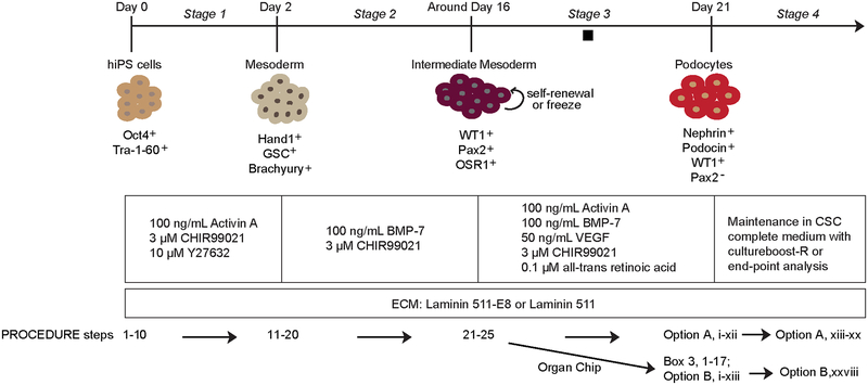
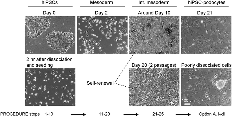
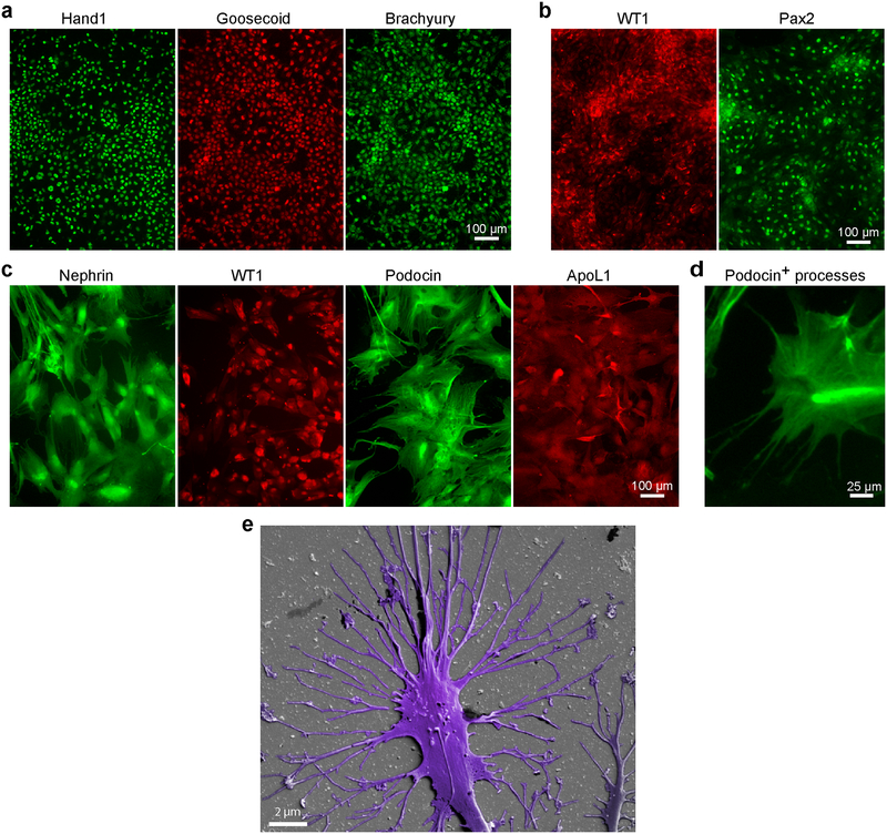
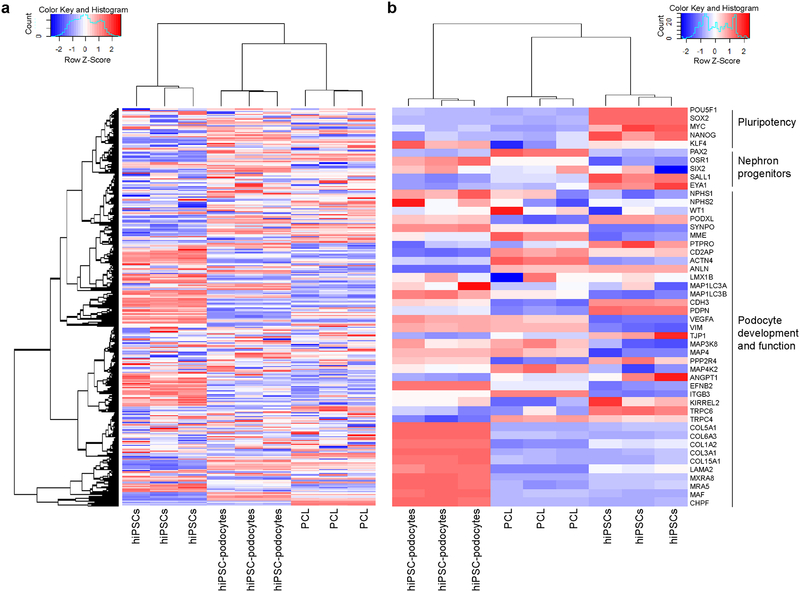
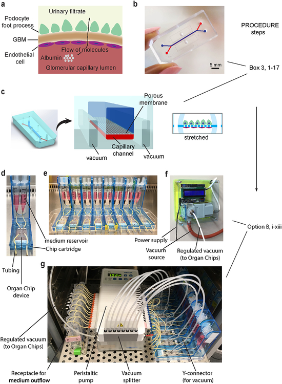
Protocols have been established to direct the differentiation of human induced pluripotent stem (iPS) cells into nephron progenitor cells and organoids containing many types of kidney cells, but it has been difficult to direct the differentiation of iPS cells to form specific types of mature human kidney cells with high yield. Here, we describe a detailed protocol for the directed differentiation of human iPS cells into mature, post-mitotic kidney glomerular podocytes with high (>90%) efficiency within 26 d and under chemically defined conditions, without genetic manipulations or subpopulation selection. We also describe how these iPS cell-derived podocytes may be induced to form within a microfluidic organ-on-a-chip (Organ Chip) culture device to build a human kidney Glomerulus Chip that mimics the structure and function of the kidney glomerular capillary wall in vitro within 35 d (starting with undifferentiated iPS cells). The podocyte differentiation protocol requires skills for culturing iPS cells, and the development of a Glomerulus Chip requires some experience with building and operating microfluidic cell culture systems. This method could be useful for applications in nephrotoxicity screening, therapeutic development, and regenerative medicine, as well as mechanistic study of kidney development and disease.
Conflict of interest statement
Competing Financial Interests
D.E.I. and S.M. are authors on a patent pending for methods for the generation of kidney glomerular podocytes from pluripotent stem cells (US patent application 14/950859). D.E.I. is a founder, holds equity and chairs the scientific advisory board at Emulate Inc.
Figures

Similar articles
-
Mature induced-pluripotent-stem-cell-derived human podocytes reconstitute kidney glomerular-capillary-wall function on a chip.Nat Biomed Eng. 2017;1:0069. doi: 10.1038/s41551-017-0069. Epub 2017 May 10. Nat Biomed Eng. 2017. PMID: 29038743 Free PMC article.
-
Human Induced Pluripotent Stem Cell-Derived Podocytes Mature into Vascularized Glomeruli upon Experimental Transplantation.J Am Soc Nephrol. 2016 Jun;27(6):1778-91. doi: 10.1681/ASN.2015010096. Epub 2015 Nov 19. J Am Soc Nephrol. 2016. PMID: 26586691 Free PMC article.
-
The directed differentiation of human iPS cells into kidney podocytes.PLoS One. 2012;7(9):e46453. doi: 10.1371/journal.pone.0046453. Epub 2012 Sep 28. PLoS One. 2012. PMID: 23029522 Free PMC article.
-
Induction of nephron progenitors and glomeruli from human pluripotent stem cells.Pediatr Nephrol. 2017 Feb;32(2):195-200. doi: 10.1007/s00467-016-3339-z. Epub 2016 Feb 11. Pediatr Nephrol. 2017. PMID: 26868670 Review.
-
Retinoids and glomerular regeneration.Semin Nephrol. 2014 Jul;34(4):429-36. doi: 10.1016/j.semnephrol.2014.06.009. Epub 2014 Jun 13. Semin Nephrol. 2014. PMID: 25217271 Review.
Cited by
-
Advanced in vitro Research Models to Study the Role of Endothelial Cells in Solid Organ Transplantation.Front Immunol. 2021 Feb 10;12:607953. doi: 10.3389/fimmu.2021.607953. eCollection 2021. Front Immunol. 2021. PMID: 33664744 Free PMC article. Review.
-
Activation of the Renin-Angiotensin System Disrupts the Cytoskeletal Architecture of Human Urine-Derived Podocytes.Cells. 2022 Mar 24;11(7):1095. doi: 10.3390/cells11071095. Cells. 2022. PMID: 35406662 Free PMC article.
-
Cells sorted off hiPSC-derived kidney organoids coupled with immortalized cells reliably model the proximal tubule.Commun Biol. 2023 May 4;6(1):483. doi: 10.1038/s42003-023-04862-7. Commun Biol. 2023. PMID: 37142732 Free PMC article.
-
Organoids as tools for fundamental discovery and translation-a Keystone Symposia report.Ann N Y Acad Sci. 2022 Dec;1518(1):196-208. doi: 10.1111/nyas.14874. Epub 2022 Sep 30. Ann N Y Acad Sci. 2022. PMID: 36177906 Free PMC article.
-
A Comprehensive Review of Organ-on-a-Chip Technology and Its Applications.Biosensors (Basel). 2024 May 1;14(5):225. doi: 10.3390/bios14050225. Biosensors (Basel). 2024. PMID: 38785699 Free PMC article. Review.
References
-
- Thomson J et al. Embryonic Stem Cell Lines Derived from Human Blastocysts. Science 282, 1145–1147 (1998). - PubMed
-
- Takahashi K et al. Induction of Pluripotent Stem Cells from Adult Human Fibroblasts by Defined Factors. Cell 131, 861–872 (2007). - PubMed
-
- Benam KH et al. Engineered in vitro disease models. Annu Rev Pathol 10, 195–262 (2015). - PubMed
Publication types
MeSH terms
Grants and funding
LinkOut - more resources
Full Text Sources
Other Literature Sources
