. 2018 Jul 11;13(7):e0199373.
doi: 10.1371/journal.pone.0199373. eCollection 2018.

Propofol prevents human umbilical vein endothelial cell injury from Ang II-induced apoptosis by activating the ACE2-(1-7)-Mas axis and eNOS phosphorylation

Affiliations

Propofol prevents human umbilical vein endothelial cell injury from Ang II-induced apoptosis by activating the ACE2-(1-7)-Mas axis and eNOS phosphorylation

Liangqing Zhang et al. PLoS One. .

Abstract

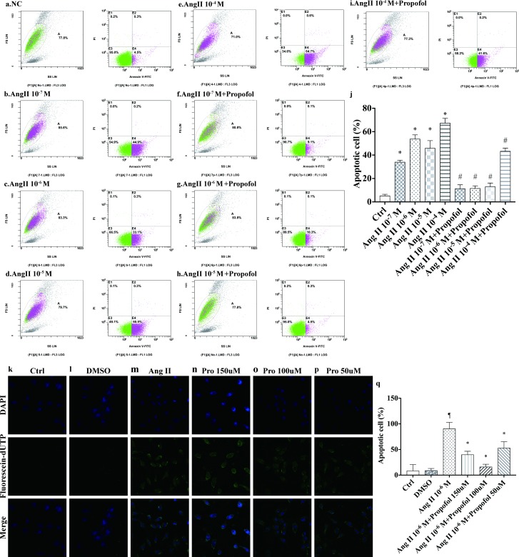
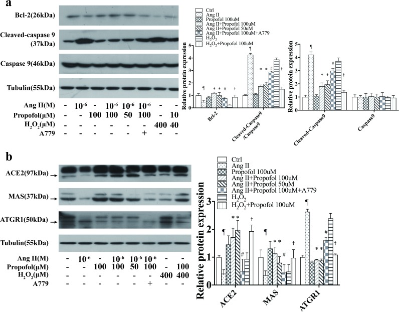
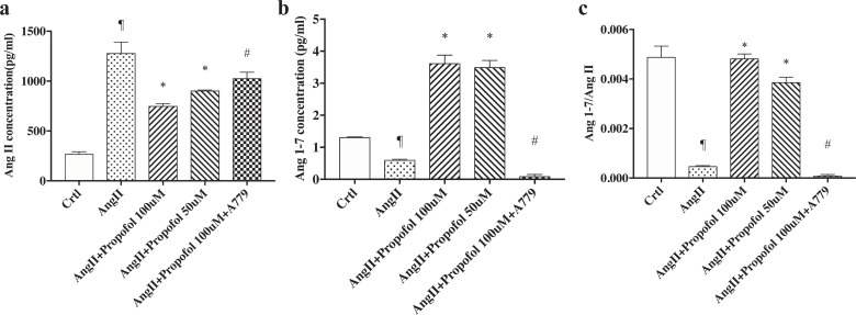
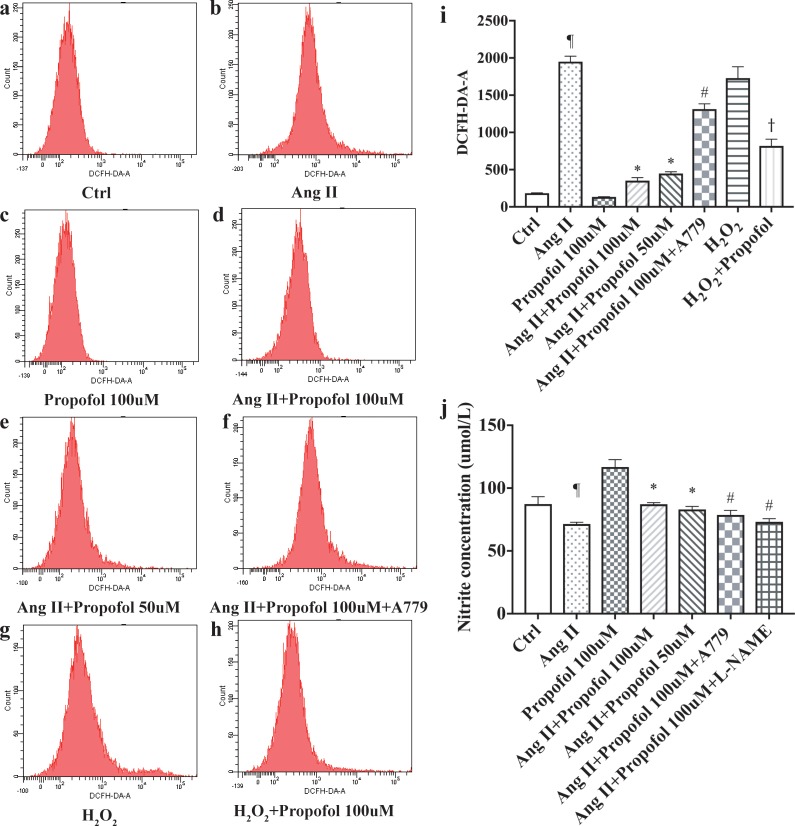
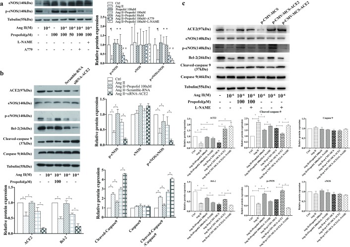
Angiotensin II (AngII), a vasoactive peptide that elevates arterial blood pressure and results in hypertension, has been reported to directly induce vascular endothelial cell apoptosis. Recent work has demonstrated that propofol pre-treatment attenuates angiotensin II-induced apoptosis in human coronary artery endothelial cells. However, the underlying mechanism remains largely unknown. Here, we investigated human umbilical vein endothelial cells (HUVECs) subjected to angiotensin II-induced apoptosis in the presence or absence of propofol treatment and found that angiotensin II-induced apoptosis was attenuated by propofol in a dose-dependent manner. Furthermore, ELISA assays demonstrated that the ratio of angiotensin (1-7) (Ang (1-7)) to Ang II was increased after propofol treatment. We examined the expression of ACE2, Ang (1-7) and Mas and found that the ACE2-Ang (1-7)-Mas axis was up-regulated by propofol, while ACE2 overexpression increased phosphorylated endothelial nitric oxide synthase (phosphorylated eNOS) expression and siACE2 resulted in the repression of endothelial nitric oxide synthase (eNOS) phosphorylation. In conclusion, our study revealed that propofol can inhibit endothelial cell apoptosis induced by Ang II by activating the ACE2-Ang (1-7)-Mas axis and further up-regulating the expression and phosphorylation of eNOS.


Conflict of interest statement

The authors have declared that no competing interests exist.

Figures

Fig 1
Fig 1. Propofol inhibits AngII-induced apoptosis in HUVECs.
(a-j) Detection of apoptosis with Annexin V-FITC and propidium iodide staining. Cells were preconditioned with increasing concentrations of AngII (10−7–10-4M) for 24 h and then treated with or without propofol for 4 h. Each group of Annexin V- and propidium iodide-stained cells was measured by flow cytometry. The histogram represents the percentage of apoptotic cells, and the data represent the mean±s.d. of three independent experiments. (n = 3) *P<0.01 versus the control group and # P<0.05 versus corresponding concentration AngII injury groups (k-q). AngII-pre-treated HUVECs were treated with different concentrations (0–150μmol/L) of propofol for 4 h. Cell apoptosis was evaluated via TUNEL assay. Data are from n = 3 independent experiments. ¶versus the DMSO group (P<0.01) and *versus the AngII injury group (P<0.01).
Fig 2
Fig 2. Propofol inhibits AngII-induced apoptosis by up-regulating the ACE2-Ang (1–7)-Mas axis.
(a) Western blotting was performed to evaluate the expression of the apoptosis-related proteins caspase-9 and Bcl-2. The expression of cleaved caspase-9 decreased and that of Bcl-2 was significantly increased after propofol treatment for 4 h compared with the control group. After A779 administration, the expression of cleaved caspase-9 was down-regulated and Bcl-2 levels increased. (b) ACE2 and Mas protein levels were up-regulated; however, ATGR1 protein level was decreased in the propofol group compared with the control group. A779 decreased the protein levels of ACE2 and Mas while up-regulated ATGR1 after A779 treatment. The data are representative of six independent experiments, *P<0.01 versus the AngII -treated group, # P<0.01 versus the AngII+propofol 100μM group, ¶P<0.01 versus the control group, and †P<0.01 versus the H2O2 -treated group.
Fig 3
Fig 3. Propofol up-regulates the expression of Ang (1–7), as quantified by ELISA.
HUVECs cultured in 96-well plates were treated with AngII for 24 h, followed by treatment with propofol with or without A779 for 4 h. Data are shown as the mean±s.d. from n = 3 independent experiments. (a) The level of Ang (1–7) was quantified by ELISA; *P<0.01 versus the AngII group, and #P<0.05 versus the control group. (b) The level of AngII was quantified by ELISA using a commercial kit. # P<0.05 versus the AngII group. (c) The ratio of Ang (1–7) to AngII expression was calculated and shown as (c). *P<0.01 versus the AngII -treated group, # P<0.05 versus the AngII+propofol 100μM group, and ¶P<0.01 versus the control group.
Fig 4
Fig 4. Propofol inhibited Ang II-mediated oxidative damage.
(a–h) ROS assay showed that treatment with propofol reduced Ang II-stimulated ROS formation. (i–j) NO assay showed that Addition of propofol exhibited higher NO production. The results are shown as the mean ± SD (n = 3). ¶ p<0.05 compared to the Ctrl group; * p<0.05 compared to the Ang II group; # p<0.05 compared to the Propofol 100uM group; and † P<0.05 compared to H2O2 group.
Fig 5
Fig 5. Propofol prevents AngII-induced apoptosis by regulating ACE2 and phosphorylated eNOS.
(a) A representative Western blot of eNOS activation in AngII-pre-treated HUVECs among normal (Control), propofol-treated and with or without A779 and L-NAME addition after propofol treatment for 4 h. Following the process described in the Materials and Methods, equal amounts of total protein from each cell sample were subjected to Western blotting using specific antibodies against eNOS and phosphorylated eNOS. β-Tubulin was used to confirm equal protein loading. The results are shown as the mean ± SD (n = 3). ¶ p<0.05 compared to the Ctrl group; * p<0.05 compared to the Ang II group; # p<0.05 compared to the Propofol 100uM group. (b) Expression of phosphorylated eNOS in HUVECs in AngII-treated medium, with or without propofol-treatment. HUVECs transfected with siRNA-ACE2 and scramble-RNA were harvested 48h after transfection. Data are representative of n = 3 independent WB experiments; *P<0.01. (c) Expression of phosphorylated eNOS in HUVECs in AngII-treated medium, propofol-treated and with or without L-NAME addition after propofol treatment for 4 h. HUVECs transfected with an ACE2-overexpression plasmid were harvested 48h after transfection. Data are representative of n = 3 independent WB experiments; *P<0.01.

References

    1. Zicha J, Kunes J. Ontogenetic aspects of hypertension development: analysis in the rat. Physiol Rev. 1999;79(4):1227–82. Epub 1999/10/03. doi: 10.1152/physrev.1999.79.4.1227 . - DOI - PubMed
    1. Davignon J, Ganz P. Role of endothelial dysfunction in atherosclerosis. Circulation. 2004;109(23 Suppl 1):Iii27–32. Epub 2004/06/17. doi: 10.1161/01.CIR.0000131515.03336.f8 . - DOI - PubMed
    1. Bader M, Ganten D. Update on tissue renin-angiotensin systems. J Mol Med (Berl). 2008;86(6):615–21. Epub 2008/04/17. doi: 10.1007/s00109-008-0336-0 . - DOI - PubMed
    1. Bader M. Tissue renin-angiotensin-aldosterone systems: Targets for pharmacological therapy. Annu Rev Pharmacol Toxicol. 2010;50:439–65. Epub 2010/01/09. doi: 10.1146/annurev.pharmtox.010909.105610 . - DOI - PubMed
    1. Wang B, Shravah J, Luo H, Raedschelders K, Chen DD, Ansley DM. Propofol protects against hydrogen peroxide-induced injury in cardiac H9c2 cells via Akt activation and Bcl-2 up-regulation. Biochem Biophys Res Commun. 2009;389(1):105–11. Epub 2009/08/26. doi: 10.1016/j.bbrc.2009.08.097 ; PubMed Central PMCID: PMCPMC3631547. - DOI - PMC - PubMed

Publication types

MeSH terms

LinkOut - more resources