. 2018 Jul 10;24(2):271-277.
doi: 10.1016/j.celrep.2018.06.037.

A Neural Circuit Underlying the Generation of Hot Flushes

Affiliations

A Neural Circuit Underlying the Generation of Hot Flushes

Stephanie L Padilla et al. Cell Rep. .

Abstract

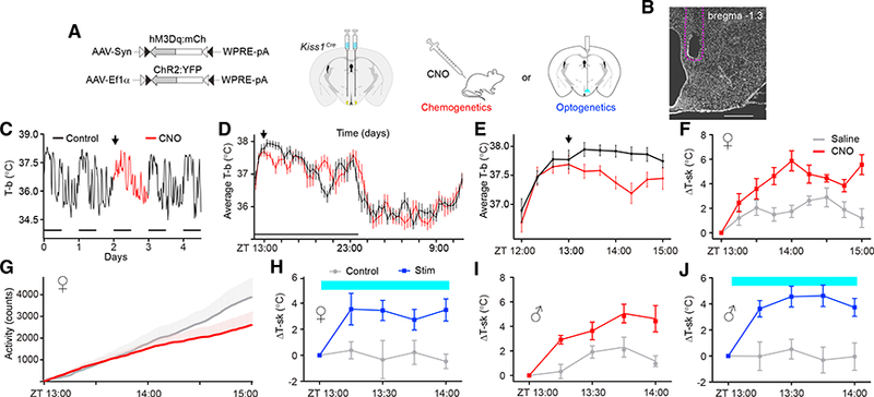
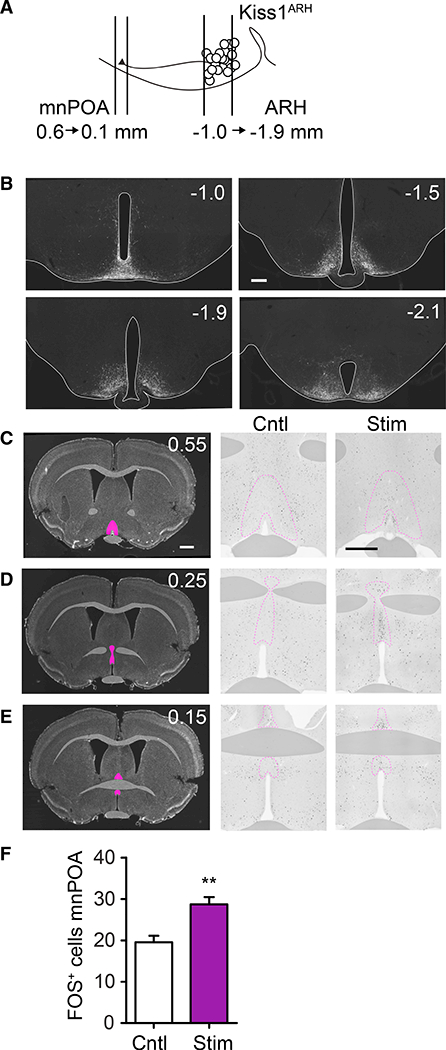
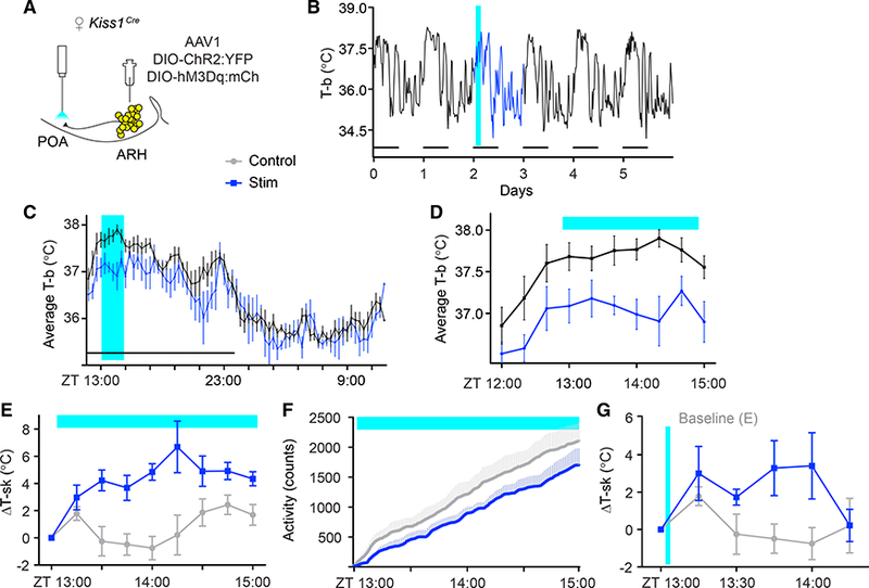
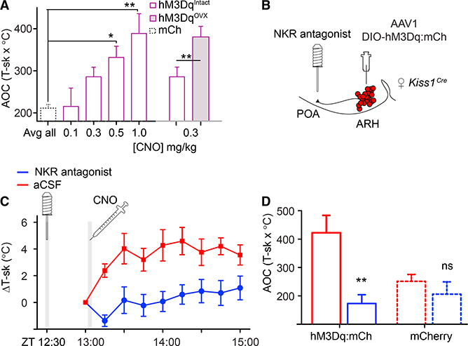
Hot flushes are a sudden feeling of warmth commonly associated with the decline of gonadal hormones at menopause. Neurons in the arcuate nucleus of the hypothalamus that express kisspeptin and neurokinin B (Kiss1ARH neurons) are candidates for mediating hot flushes because they are negatively regulated by sex hormones. We used a combination of genetic and viral technologies in mice to demonstrate that artificial activation of Kiss1ARH neurons evokes a heat-dissipation response resulting in vasodilation (flushing) and a corresponding reduction of core-body temperature in both females and males. This response is sensitized by ovariectomy. Brief activation of Kiss1ARH axon terminals in the preoptic area of the hypothalamus recapitulates this response, while pharmacological blockade of neurokinin B (NkB) receptors in the same brain region abolishes it. We conclude that transient activation of Kiss1ARH neurons following sex-hormone withdrawal contributes to the occurrence of hot flushes via NkB release in the rostral preoptic area.

Keywords: chemogenetics; estrogen; hot flashes; kisspeptin; menopause; neurokinin B; optogenetics; temperature regulation.


Conflict of interest statement

DECLARATION OF INTERESTS

The authors declare no competing interests.

Figures

Figure 1.
Figure 1.. Artificial Activation of Kiss1ARH Neurons Is Sufficient to Drive Heat Dissipation in Both Female and Male Mice
(A) Schematic representation of the viral vectors and injection targets. A unilateral fiber-optic ferrule was implanted above the ARH. (B) DAPI staining of a coronal brain section indicates the fiber-optic terminal in the ARH. Scale bar, 500 μm. (C—E) Telemetry recordings of core body temperature (T-b) show that Kiss1ARH neuron stimulation (CNO, 1 mg/kg) was sufficient to transiently decrease T-b. (C) 5-day T-b recording of a single mouse, with CNO injected on day 2 at ZT= 13:00 (arrow). The animals were housed on a 12-hr:12-hr lightdark cycle (horizontal bars); Zeitgeber time (ZT) = 12:00 refers to the onset of the dark. (D) Average T-b comparing baseline versus CNO treatment on a scale of 24 hr. (E) Average T-b comparing baseline vs. CNO-treatment on a scale of 2 hr; n = 9; F(1, 144) = 7.50, p = 0.0146. (F) Thermal imaging of tail-skin temperature (T-sk), saline versus CNO-treated females; n = 9; F(1, 128) = 17.07, p < 0.00001. (G) Telemetry recordings of locomotor activity; n = 9; F(1,2178) = 46.48, p < 0.00001. (H) Optogenetic stimulation (2 Hz) of Kiss1ARH neurons was sufficient to increase T-sk in females; n = 6; F(1,40) = 11.23, p < 0.00001. (I) T-sk in saline versus CNO-treated males; n = 7; F(1,48) = 26.35, p < 0.00001. (J) Optogenetic stimulation (2 Hz) of Kiss1ARH neurons was sufficient to increase T-sk in males; n = 9; F(1,64) (male) = 12.97, p = 0.0024. F statistics represent the main effect of treatment by two-way repeated measures (RMs)-ANOVA. Error bars represent ± SEM. Crossover design with randomized starts.
Figure 2.
Figure 2.. Activation of Kiss1ARH Neurons Induces Fos Expression in the Rostral POA
(A) Schematic diagram of Kiss1ARH projections to the POA (numbers indicate distance from Bregma in mm). (B) Serial, coronal sections of hM3Dq:mCherry-transduced Kiss1Cre neurons with immunohistochemistry for mCherry. (C-E) Representative Fos staining in POA from rostral to caudal (top to bottom). 0.55 (C), 0.25 (D), and 0.15 (E) from Bregma. Left panels: DAPI-stained coronal sections, with pink highlights indicating the POA. Middle and right panels: Fos immunoreactivity in the POA comparing control versus stimulated mice, respectively. (F) There were more Fos-positive cells in the rostral POA of stimulated mice; for Cntl, M = 19.57, SD = 7.56; for Stim, M = 28.73, SD = 13.56; t(83) = 3.06; **p = 0.003. All scale bars, 500 μm.
Figure 3.
Figure 3.. Optogenetic Stimulation of Kiss1ARH Axons in the Rostral POA Promotes a Heat-Dissipation Response in Female Mice
(A) Schematic representation of the targeted viral injection into the ARH and fiber-optic placement 0.5 mm above the POA. (B-D) Optogenetic stimulation (2 Hz) of Kiss1ARH neuron fibers in the POA was sufficient to transiently decrease T-b. Within-subject comparison of unstimulated baseline (black trace) vs. stimulated (blue trace). (B) 6-day T-b recording of a single mouse, subjected to blue-light stimulation from ZT = 13:00–15:00 on day 2 (blue). (C) Average T-b comparing baseline versus stimulation on a scale of 24 hr. (D) Average T-b comparing baseline vs. stimulation on a scale of 2 hr; n = 5; F(1,72) = 9.589, p = 0.0147. (E) T-sk in control versus terminal-stimulated females; n = 5; F(1,64) = 17.83, p = 0.0029. (F) Telemetry recording of locomotor activity, n = 5, F(1,960) (activity) = 185.8, p < 0.00001. (G) T-sk following a brief (2-min, 2 Hz) stimulation; n = 5; F(1,48) = 9.41, p = 0.0035. F statistics represent the main effect of treatment by two-way RM-ANOVA. Error bars represent ± SEM. Crossover design with randomized starts.
Figure 4.
Figure 4.. Evoked Hot Flushes Are Sensitive to Ovarian Estrogens and Are Blocked by Neurokinin Receptor Antagonists Delivered to the POA
(A) Dose response of T-sk to CNO. CNO was injected at ZT = 13:00, and T-sk was monitored for 2 hr; data are presented as the average area under the curve (AOC; hours x °C). mCherry-expressing controls were treated with CNO (0.05 to 1 mg/kg) and averaged. One-way ANOVA: for mCh, n = 8; for hM3Dq, n = 6; **p = 0.0006, asterisks indicate Bonferroni post-test results. Following ovariectomy, females were significantly more sensitive to 0.3 mg/kg CNO. Paired t test, n = 6. Intact: M = 285.6, SD = 22.74; ovari- ectomized, M = 393.1, SD = 29.31. t(5) = 2.605; *p = 0.0480. (B) Schematic representation of targeted viral injection into the ARH and placement of infusion cannula in the POA. (C) Infusion of the cocktail of antagonists was sufficient to block CNO-induced flushing. 30 min prior to recording, either saline or antagonists were infused into the POA of h M3Dq-expressi ng females. T-sk was recorded every 15 min starting immediately prior to CNO delivery at ZT = 13:00. Two-way RM-ANOVA, n = 6; F(1, 45) = 84.05, p < 0.00001. (D) T-sk from ZT = 13:00–15:00 represented as AOC. hM3Dq data were extracted from (B) and compared to mCherry-expressing controls that were treated in the same manner. Two-way RM-ANOVA main effect of antagonists, n = 6 or 8 per group (hM3Dq or mCh); F(1, 12) = 16.49, **p = 0.0016; Bonferroni post-tests compare artificial cerebral spinal fluid (ACSF) with antagonists for paired animals in each group. ns, not significant. Experiments were conducted using a crossover design with randomized start times. All error bars represent SEM.

References

    1. Casper RF, Yen SS, and Wilkes MM (1979). Menopausal flushes: a neuroendocrine link with pulsatile luteninizing hormone secreation. Science 205, 823–825. - PubMed
    1. Clarkson J, Han SY, Piet R, McLennan T, Kane GM, Ng J, Porteous RW, Kim JS, Colledge WH, Iremonger KJ, and Herbison AE (2017). Definition of the hypothalamic GnRH pulse generator in mice. Proc. Natl. Acad. Sci. USA 114, E10216–E10223. - PMC - PubMed
    1. d’Anglemont de Tassigny, X., Fagg LA, Dixon JP, Day K, Leitch HG, Hendrick AG, Zahn D, Franceschini I, Caraty A, Carlton MB, et al. (2007). Hypogonadotropic hypogonadism in mice lacking a functional Kiss1 gene. Proc. Natl. Acad. Sci. USA 104, 10714–10719. - PMC - PubMed
    1. Dacks PA, Krajewski SJ, and Rance NE (2011). Activation of neurokinin 3 receptors in the median preoptic nucleus decreases core temperature in the rat. Endocrinology 152, 4894–4905. - PMC - PubMed
    1. de Croft S, Boehm U, and Herbison AE (2013). Neurokinin B activates arcuate kisspeptin neurons through multiple tachykinin receptors in the male mouse. Endocrinology 154, 2750–2760. - PubMed

Publication types