. 2018 Jul 10;6(3):41.
doi: 10.3390/vaccines6030041.

Respiratory Tract Deposition and Distribution Pattern of Microparticles in Mice Using Different Pulmonary Delivery Techniques

Affiliations

Respiratory Tract Deposition and Distribution Pattern of Microparticles in Mice Using Different Pulmonary Delivery Techniques

Nitesh K Kunda et al. Vaccines (Basel). .

Abstract

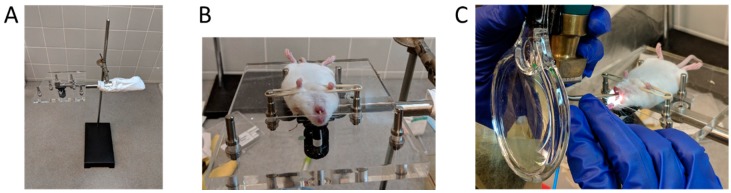
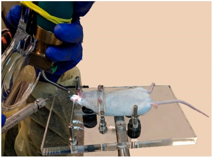
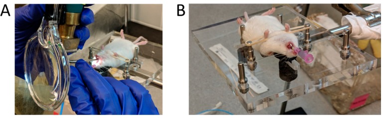
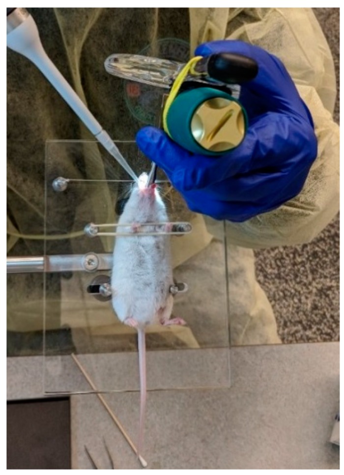
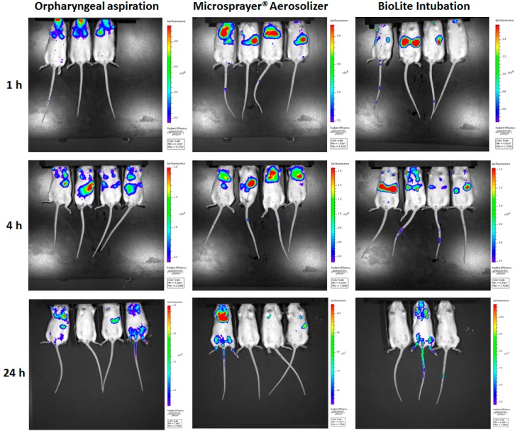
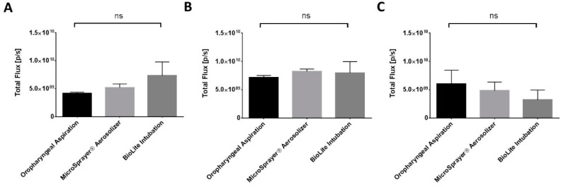
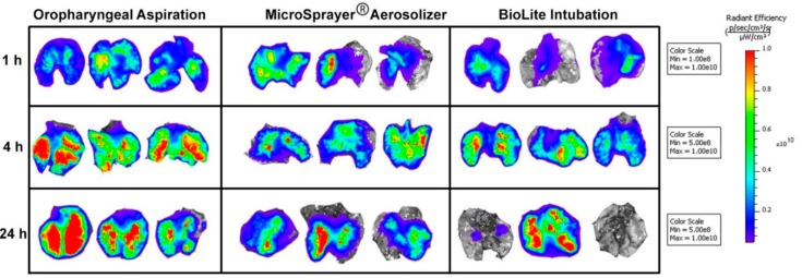
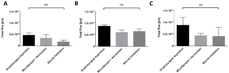
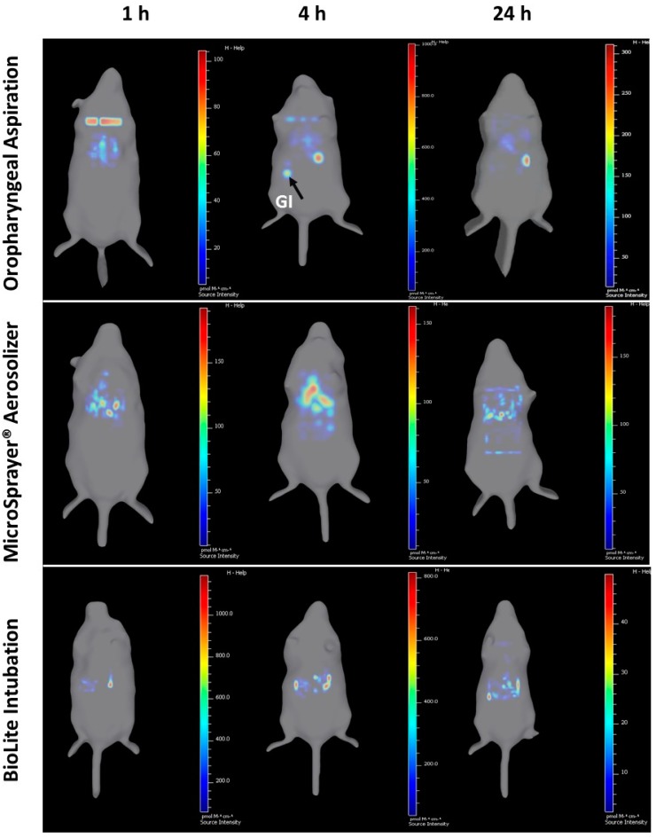
Pulmonary delivery of drugs and vaccines is an established route of administration, with particulate-based carriers becoming an attractive strategy to enhance the benefits of pulmonary therapeutic delivery. Despite the increasing number of publications using the pulmonary route of delivery, the lack of effective and uniform administration techniques in preclinical models generally results in poor translational success. In this study, we used the IVIS Spectrum small-animal in vivo imaging system to compare the respiratory tract deposition and distribution pattern of a microsphere suspension (5 µm) in mice after 1, 4, and 24 h when delivered by oropharyngeal aspiration, the Microsprayer® Aerosolizer, and the BioLite Intubation System, three-widely reported preclinical inhalation techniques. We saw no significant differences in microsphere deposition in whole body images and excised lungs (at 1, 4, and 24 h); however, the three-dimensional (3D) images showed more localized deposition in the lungs with the MicroSprayer® and BioLite delivery techniques. Further, oropharyngeal aspiration (at 1 h) showed microsphere deposition in the oral cavity, in contrast to the MicroSprayer® and BioLite systems. The studies shown here will allow researchers to choose the appropriate pulmonary delivery method in animal models based on their study requirements.

Keywords: BioLite intubation; IVIS; MicroSprayer® Aerosolizer; microparticles; oropharyngeal aspiration; pulmonary delivery; respiratory tract deposition; vaccines.


Conflict of interest statement

The authors declare no conflict of interest.

Figures

Figure 1
Figure 1
Steps for mouse intubation. (A) Set-up of the intubation platform; (B) Anesthetized mouse placed on its back and secured on the intubation platform; and (C) Visualization of the trachea using a small animal laryngoscope, with an attached 3X magnifying glass, to facilitate administration of the test agent.
Figure 2
Figure 2
Mouse intubation using the MicroSprayer® Aerosolizer (Penn-Century, Wyndmoor, PA, USA). The microsprayer is inserted into the trachea of the mouse with the help of a small animal laryngoscope.
Figure 3
Figure 3
Mouse intubation using the BioLite Intubation System (Braintree Scientific Ltd., Braintree, MA, USA). (A) Visualization of the trachea using a small animal laryngoscope and a fiber-optic stylet. (B) Cannula inserted into the trachea of the mouse, ready for attaching the syringe and administration of the test agent.
Figure 4
Figure 4
Administration of fluorescent microparticles by the ‘oropharyngeal aspiration’ method. The tracheal opening was visualized with the help of a small animal laryngoscope.
Figure 5
Figure 5
Whole animal images of mice acquired using IVIS Spectrum at 1, 4, and 24 h after pulmonary administration of near-infrared fluorescent Degradex® poly(d,l-lactide-co-glycolide) (PLGA) microspheres using oropharyngeal aspiration, the MicroSprayer® Aerosolizer, and the BioLite Intubation System.
Figure 6
Figure 6
Total fluorescence flux (ρ/s) (oral cavity, trachea, and the lungs) in mice at (A) 1 h, (B) 4 h, and (C) 24 h after pulmonary administration of near-infrared fluorescent Degradex® poly(d,l-lactide-co-glycolide) (PLGA) microspheres using oropharyngeal aspiration, the MicroSprayer® Aerosolizer, and the BioLite Intubation System (n = 3–4, mean ± SEM, one-way ANOVA, Tukey’s multiple comparison test).
Figure 7
Figure 7
Images of excised lungs of mice acquired using IVIS Spectrum at 1, 4, and 24 h after pulmonary administration of near-infrared fluorescent Degradex® poly(d,l-lactide-co-glycolide) (PLGA) microspheres using oropharyngeal aspiration, the MicroSprayer® Aerosolizer, and the BioLite Intubation System.
Figure 8
Figure 8
Total flux (ρ/s) of fluorescence in excised lungs of mice at (A) 1 h, (B) 4 h, and (C) 24 h after administration of near-infrared fluorescent Degradex® poly(d,l-lactide-co-glycolide) (PLGA) microspheres using oropharyngeal aspiration, the MicroSprayer® Aerosolizer, and the BioLite Intubation System (n = 3–4, mean ± SEM, one-way ANOVA, Tukey’s multiple comparison test).
Figure 9
Figure 9
Three-dimensional (3D) longitudinal imaging of mouse at 1, 4, and 24 h using IVIS Spectrum after administration of near-infrared fluorescent Degradex® poly(d,l-lactide-co-glycolide) (PLGA) microspheres using oropharyngeal aspiration, the MicroSprayer® Aerosolizer, and the BioLite Intubation System (3D video available as Supplementary Video S1). Gastrointestinal (GI) deposition using the oropharyngeal aspiration technique is shown with an arrow.

Similar articles

Cited by

References

    1. Kunda N.K., Somavarapu S., Gordon S.B., Hutcheon G.A., Saleem I.Y. Nanocarriers Targeting Dendritic Cells for Pulmonary Vaccine Delivery. Pharm. Res. 2013;30:325–341. doi: 10.1007/s11095-012-0891-5. - DOI - PubMed
    1. Eleftheriadis G.K., Akrivou M., Bouropoulos N., Tsibouklis J., Vizirianakis I.S., Fatouros D.G. Polymer–Lipid Microparticles for Pulmonary Delivery. Langmuir. 2018;34:3438–3448. doi: 10.1021/acs.langmuir.7b03645. - DOI - PubMed
    1. Price D.N., Stromberg L.R., Kunda N.K., Muttil P. In vivo pulmonary delivery and magnetic-targeting of dry powder nano-in-microparticles. Mol. Pharm. 2017;14:4741–4750. doi: 10.1021/acs.molpharmaceut.7b00532. - DOI - PMC - PubMed
    1. Cauley L.S., Lefrançois L. Guarding the perimeter: Protection of the mucosa by tissue-resident memory T cells. Mucosal Immunol. 2013;6:14–23. doi: 10.1038/mi.2012.96. - DOI - PMC - PubMed
    1. Walrath J., Zukowski L., Krywiak A., Silver R.F. Resident Th1-like effector memory cells in pulmonary recall responses to Mycobacterium tuberculosis. Am. J. Respir. Cell Mol. Biol. 2005;33:48–55. doi: 10.1165/rcmb.2005-0060OC. - DOI - PMC - PubMed

LinkOut - more resources