

The invariant arginine within the chromatin-binding motif regulates both nucleolar localization and chromatin binding of Foamy virus Gag
- PMID: 29996845
- PMCID: PMC6042332
- DOI: 10.1186/s12977-018-0428-z
The invariant arginine within the chromatin-binding motif regulates both nucleolar localization and chromatin binding of Foamy virus Gag
Abstract
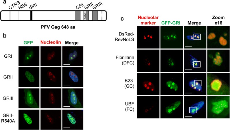
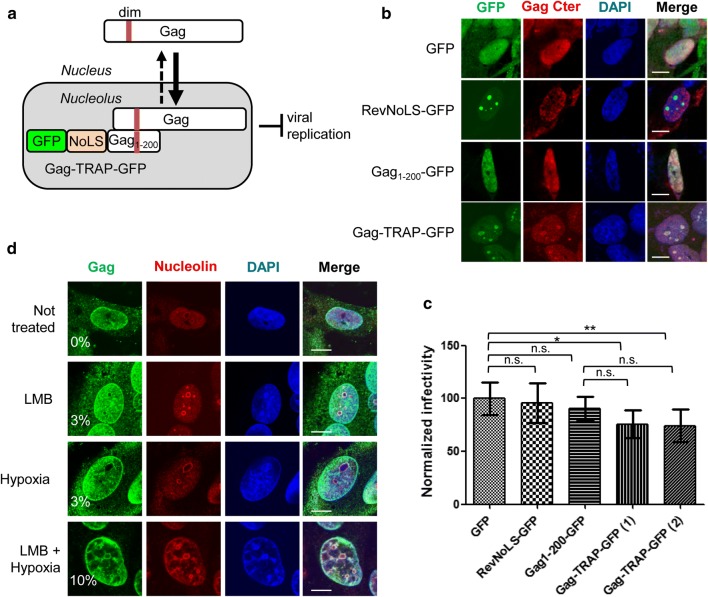
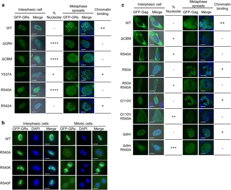
Background: Nuclear localization of Gag is a property shared by many retroviruses and retrotransposons. The importance of this stage for retroviral replication is still unknown, but studies on the Rous Sarcoma virus indicate that Gag might select the viral RNA genome for packaging in the nucleus. In the case of Foamy viruses, genome encapsidation is mediated by Gag C-terminal domain (CTD), which harbors three clusters of glycine and arginine residues named GR boxes (GRI-III). In this study we investigated how PFV Gag subnuclear distribution might be regulated.
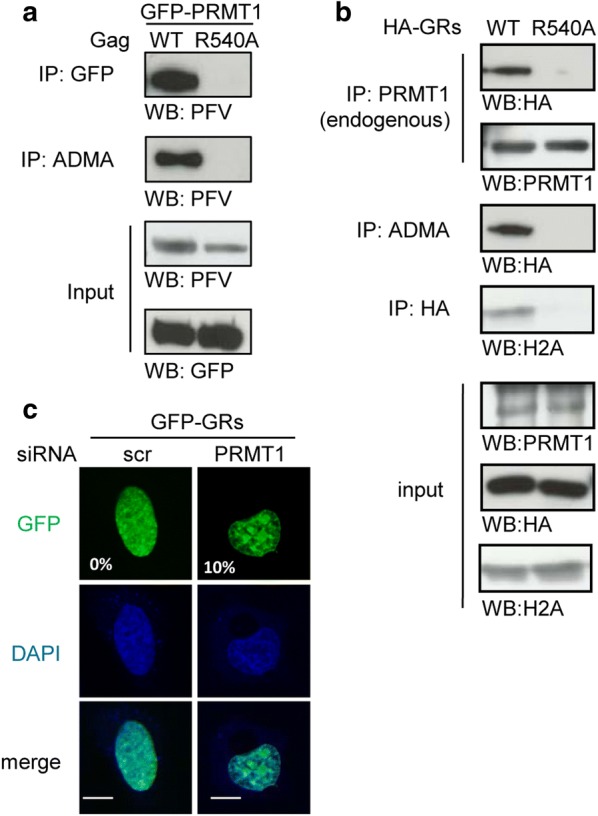
Results: We show that the isolated GRI and GRIII boxes act as nucleolar localization signals. In contrast, both the entire Gag CTD and the isolated GRII box, which contains the chromatin-binding motif, target the nucleolus exclusively upon mutation of the evolutionary conserved arginine residue at position 540 (R540), which is a key determinant of FV Gag chromatin tethering. We also provide evidence that Gag localizes in the nucleolus during FV replication and uncovered that the viral protein interacts with and is methylated by Protein Arginine Methyltransferase 1 (PRMT1) in a manner that depends on the R540 residue. Finally, we show that PRMT1 depletion by RNA interference induces the concentration of Gag C-terminus in nucleoli.
Conclusion: Altogether, our findings suggest that methylation by PRMT1 might finely tune the subnuclear distribution of Gag depending on the stage of the FV replication cycle. The role of this step for viral replication remains an open question.
Keywords: Chromatin-binding; Foamy virus; Gag; Methylation; Nuclear trafficking; Nucleolus; PRMT; Post-translational modification.
Figures
Similar articles
-
The cooperative function of arginine residues in the Prototype Foamy Virus Gag C-terminus mediates viral and cellular RNA encapsidation.Retrovirology. 2014 Oct 8;11:87. doi: 10.1186/s12977-014-0087-7. Retrovirology. 2014. PMID: 25292281 Free PMC article.
-
Prototype foamy virus gag nuclear localization: a novel pathway among retroviruses.J Virol. 2011 Sep;85(18):9276-85. doi: 10.1128/JVI.00663-11. Epub 2011 Jun 29. J Virol. 2011. PMID: 21715475 Free PMC article.
-
The carboxyl terminus of the human foamy virus Gag protein contains separable nucleic acid binding and nuclear transport domains.J Virol. 1996 Dec;70(12):8255-62. doi: 10.1128/JVI.70.12.8255-8262.1996. J Virol. 1996. PMID: 8970944 Free PMC article.
-
The foamy virus Gag proteins: what makes them different?Viruses. 2013 Mar 26;5(4):1023-41. doi: 10.3390/v5041023. Viruses. 2013. PMID: 23531622 Free PMC article. Review.
-
Particle assembly and genome packaging.Curr Top Microbiol Immunol. 2003;277:89-110. doi: 10.1007/978-3-642-55701-9_4. Curr Top Microbiol Immunol. 2003. PMID: 12908769 Review.
Cited by
-
The First Co-Opted Endogenous Foamy Viruses and the Evolutionary History of Reptilian Foamy Viruses.Viruses. 2019 Jul 12;11(7):641. doi: 10.3390/v11070641. Viruses. 2019. PMID: 31336856 Free PMC article.
-
Post-Translational Modifications of Retroviral HIV-1 Gag Precursors: An Overview of Their Biological Role.Int J Mol Sci. 2021 Mar 11;22(6):2871. doi: 10.3390/ijms22062871. Int J Mol Sci. 2021. PMID: 33799890 Free PMC article. Review.
-
Visualizing Association of the Retroviral Gag Protein with Unspliced Viral RNA in the Nucleus.mBio. 2020 Apr 7;11(2):e00524-20. doi: 10.1128/mBio.00524-20. mBio. 2020. PMID: 32265329 Free PMC article.
-
The Unique, the Known, and the Unknown of Spumaretrovirus Assembly.Viruses. 2021 Jan 13;13(1):105. doi: 10.3390/v13010105. Viruses. 2021. PMID: 33451128 Free PMC article. Review.
-
Arginine methylation of SARS-Cov-2 nucleocapsid protein regulates RNA binding, its ability to suppress stress granule formation, and viral replication.J Biol Chem. 2021 Jul;297(1):100821. doi: 10.1016/j.jbc.2021.100821. Epub 2021 May 23. J Biol Chem. 2021. PMID: 34029587 Free PMC article.
References
-
- Heneine W, Schweizer M, Sandstrom P, Folks T. Human infection with foamy viruses. Curr Top Microbiol Immunol. 2003;277:181–196. - PubMed
Publication types
MeSH terms
Substances
Grants and funding
LinkOut - more resources
Full Text Sources
Other Literature Sources
