

Chiral polymer modified nanoparticles selectively induce autophagy of cancer cells for tumor ablation
- PMID: 29996877
- PMCID: PMC6040058
- DOI: 10.1186/s12951-018-0383-9
Chiral polymer modified nanoparticles selectively induce autophagy of cancer cells for tumor ablation
Abstract
Background: Autophagy regulation through exogenous materials has aroused intensive attention to develop treatment protocols according to diverse human diseases. However, to the best of our knowledge, few examples have been reported to selectively control autophagy process and ultimately achieve efficient therapeutic potential.
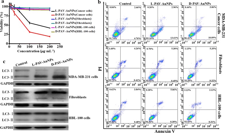
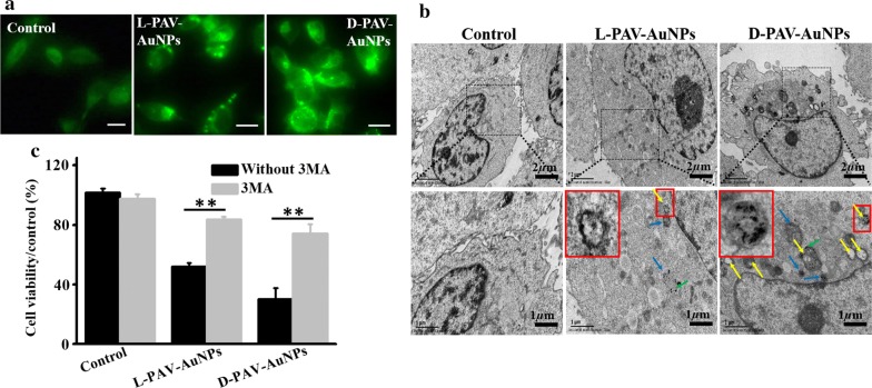
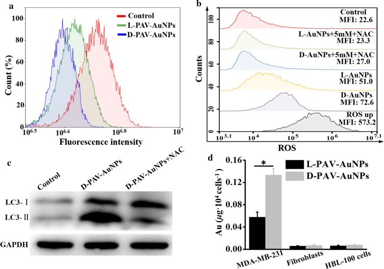
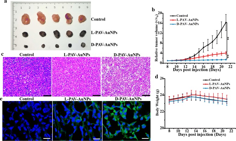
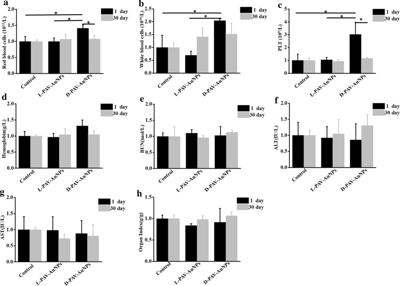
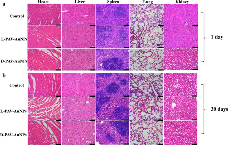
Results: In this study, monolayers of poly (acryloyl-L, D and racemic valine) (L-PAV-AuNPs, D-PAV-AuNPs and L/D-PAV-AuNPs) chiral molecules were anchored on the surfaces of gold nanoparticles (PAV-AuNPs), and the subsequent chirality-selective effects on autophagy activation were thoroughly studied. The cytotoxicity induced by PAV-AuNPs towards MDA-MB-231 cells (Breast cancer cells) was achieved mainly through autophagy and showed chirality-dependent, with D-PAV-AuNPs exhibiting high autophagy-inducing activity in vitro and in vivo. In contrast, the PAV-AuNPs exhibited autophagy inactivation for normal cells, e.g., 3T3 fibroblasts and HBL-100 cells. The chirality-selective autophagy activation effect in MDA-MB-231 cells was likely attributed to the chirality-variant ROS generation, cellular uptake and their continuous autophagy stimulus. Furthermore, the intratumoral injection of D-PAV-AuNPs could largely suppress the tumor growth but exhibit negligible toxicity in vivo.
Conclusions: As the first exploration on stereospecific NPs for autophagy induction, this work not only substantiates that chiral polymer coated NPs can selective induce autophagy-specific in cancer cells and achieve a high tumor eradication efficiency in vivo, but also opens up a new direction in discovering unprecedented stereospecific nanoagents for autophagy-associated tumor treatment.
Keywords: Autophagy; Chiral polymer; Cytotoxicity; Nanomaterials; Tumor ablation.
Figures


Similar articles
-
Chiral nanomaterials for tumor therapy: autophagy, apoptosis, and photothermal ablation.J Nanobiotechnology. 2021 Jul 22;19(1):220. doi: 10.1186/s12951-021-00965-7. J Nanobiotechnology. 2021. PMID: 34294083 Free PMC article. Review.
-
Cytotoxicity of gold nanoparticles with different structures and surface-anchored chiral polymers.Acta Biomater. 2017 Apr 15;53:610-618. doi: 10.1016/j.actbio.2017.01.082. Epub 2017 Feb 15. Acta Biomater. 2017. PMID: 28213095
-
Surface-anchored poly(acryloyl-L(D)-valine) with enhanced chirality-selective effect on cellular uptake of gold nanoparticles.Sci Rep. 2016 Aug 17;6:31595. doi: 10.1038/srep31595. Sci Rep. 2016. PMID: 27531648 Free PMC article.
-
Influence of Albumin Configuration by the Chiral Polymer-Grafted Gold Nanoparticles.Langmuir. 2016 Jun 7;32(22):5608-16. doi: 10.1021/acs.langmuir.6b01447. Epub 2016 May 26. Langmuir. 2016. PMID: 27181989
-
Polymer decorated gold nanoparticles in nanomedicine conjugates.Adv Colloid Interface Sci. 2017 Nov;249:386-399. doi: 10.1016/j.cis.2017.01.007. Epub 2017 Feb 15. Adv Colloid Interface Sci. 2017. PMID: 28259207 Review.
Cited by
-
Structure-Based Varieties of Polymeric Nanocarriers and Influences of Their Physicochemical Properties on Drug Delivery Profiles.Adv Sci (Weinh). 2022 Apr;9(10):e2105373. doi: 10.1002/advs.202105373. Epub 2022 Feb 3. Adv Sci (Weinh). 2022. PMID: 35112798 Free PMC article. Review.
-
Chiral nanomaterials for tumor therapy: autophagy, apoptosis, and photothermal ablation.J Nanobiotechnology. 2021 Jul 22;19(1):220. doi: 10.1186/s12951-021-00965-7. J Nanobiotechnology. 2021. PMID: 34294083 Free PMC article. Review.
-
Differential Interactions of Chiral Nanocapsules with DNA.Int J Mol Sci. 2021 Jan 8;22(2):584. doi: 10.3390/ijms22020584. Int J Mol Sci. 2021. PMID: 33430158 Free PMC article.
-
Nanomedicine Approaches for Autophagy Modulation in Cancer Therapy.Small Sci. 2025 Apr 11;5(6):2400607. doi: 10.1002/smsc.202400607. eCollection 2025 Jun. Small Sci. 2025. PMID: 40529859 Free PMC article.
-
Targeting autophagy using metallic nanoparticles: a promising strategy for cancer treatment.Cell Mol Life Sci. 2019 Apr;76(7):1215-1242. doi: 10.1007/s00018-018-2973-y. Epub 2018 Nov 27. Cell Mol Life Sci. 2019. PMID: 30483817 Free PMC article. Review.
References
-
- Badve S, Dabbs DJ, Schnitt SJ, Baehner F, Decker T, Eusebi V, Fox S, Ichihara S, Jacquemier J, Lakhani SR, et al. Basal-like and triple-negative breast cancers: a critical review with an emphasis on the implications for pathologists and oncologists. Mod Pathol. 2011;24:157–167. doi: 10.1038/modpathol.2010.200. - DOI - PubMed
MeSH terms
Substances
Grants and funding
LinkOut - more resources
Full Text Sources
Other Literature Sources
Research Materials
Miscellaneous
