. 2018 Jul 11;20(1):74.
doi: 10.1186/s13058-018-0996-9.

Estrogen promotes estrogen receptor negative BRCA1-deficient tumor initiation and progression

Affiliations

Estrogen promotes estrogen receptor negative BRCA1-deficient tumor initiation and progression

Chuying Wang et al. Breast Cancer Res. .

Abstract

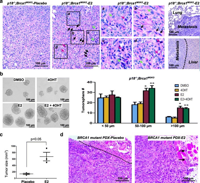
Background: Estrogen promotes breast cancer development and progression mainly through estrogen receptor (ER). However, blockage of estrogen production or action prevents development of and suppresses progression of ER-negative breast cancers. How estrogen promotes ER-negative breast cancer development and progression is poorly understood. We previously discovered that deletion of cell cycle inhibitors p16Ink4a (p16) or p18Ink4c (p18) is required for development of Brca1-deficient basal-like mammary tumors, and that mice lacking p18 develop luminal-type mammary tumors.

Methods: A genetic model system with three mouse strains, one that develops ER-positive mammary tumors (p18 single deletion) and the others that develop ER-negative tumors (p16;Brca1 and p18;Brca1 compound deletion), human BRCA1 mutant breast cancer patient-derived xenografts, and human BRCA1-deficient and BRCA1-proficient breast cancer cells were used to determine the role of estrogen in activating epithelial-mesenchymal transition (EMT), stimulating cell proliferation, and promoting ER-negative mammary tumor initiation and metastasis.

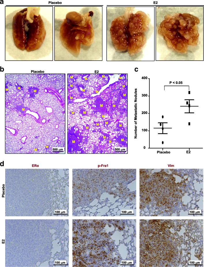
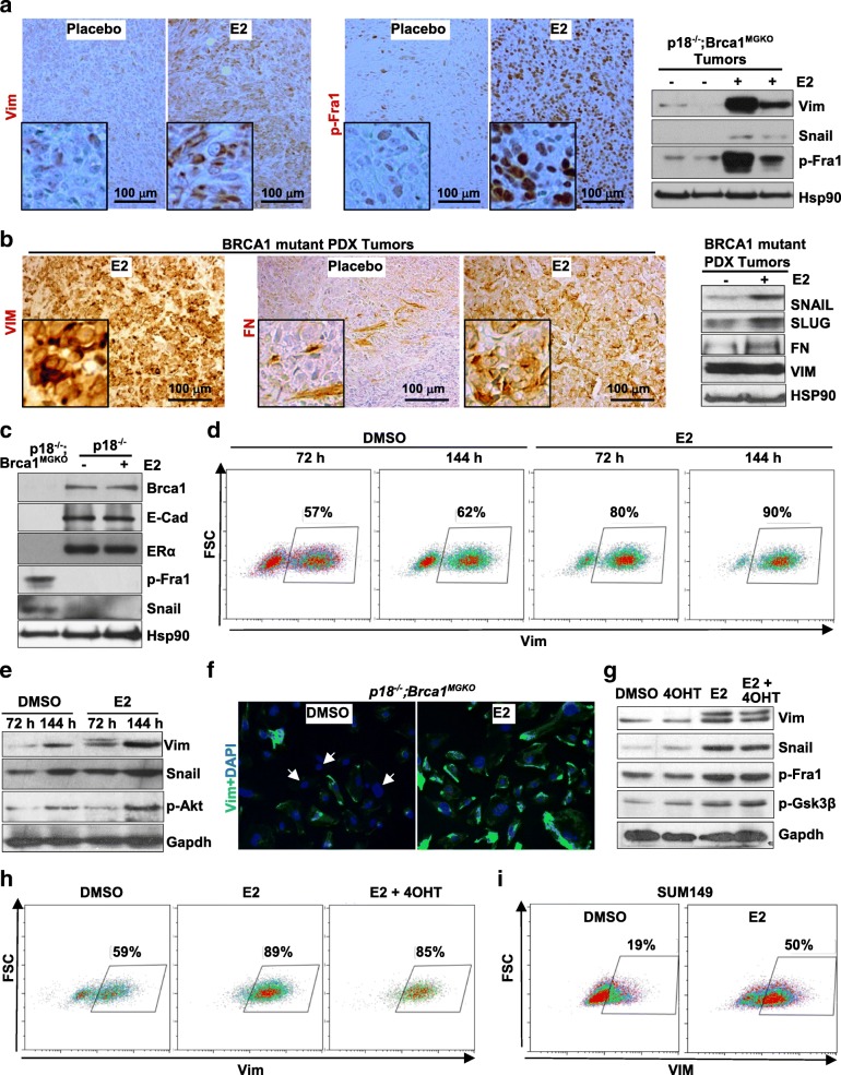
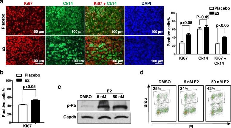
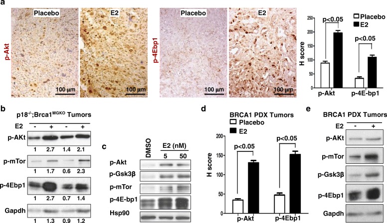
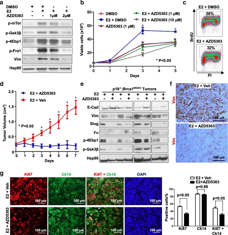
Results: Estrogen stimulated the proliferation and tumor-initiating potential of both ER-positive Brca1-proficient and ER-negative Brca1-deficient tumor cells. Estrogen activated EMT in a subset of Brca1-deficient mammary tumor cells that maintained epithelial features, and enhanced the number of cancer stem cells, promoting tumor progression and metastasis. Estrogen activated EMT independent of ER in Brca1-deficient, but not Brca1-proficient, tumor cells. Estrogen activated the AKT pathway in BRCA1-deficient tumor cells independent of ER, and pharmaceutical inhibition of AKT activity suppressed EMT and cell proliferation preventing BRCA1 deficient tumor progression.

Conclusions: This study reveals for the first time that estrogen promotes BRCA1-deficient tumor initiation and progression by stimulation of cell proliferation and activation of EMT, which are dependent on AKT activation and independent of ER.

Keywords: BRCA1; Cancer stem cells; EMT; Estrogen; Estrogen receptor.


Conflict of interest statement

Ethics approval

The Institutional Animal Care and Use Committee at the University of Miami approved all animal procedures.

Consent for publication

Not applicable.

Competing interests

The authors declare that they have no competing interests.

Publisher’s Note

Springer Nature remains neutral with regard to jurisdictional claims in published maps and institutional affiliations.

Figures

Fig. 1
Fig. 1
Estrogen promotes Brca1-deficient mammary tumor initiation and progression. a Representative H.E staining of regenerated p18−/−;Brca1MGKO mammary tumors treated with or without E2. Note that the metastasized tumors in lung and liver and the highly heterogeneous tumor cell types in E2-treated mammary tumors. Boxed areas, a, b, c, and d, are enlarged to show the four different group of cells in E2-treated tumors. Spindle cells (black arrows), cells with high nuclear-cytoplasm ratio (green arrows), and mitotic cells (red arrows) are indicated. b p18−/−;Brca1MGKO mammary tumor cells were analyzed by tumorsphere formation assay in the presence of E2, 4OHT, dimethyl sulfoxide (DMSO), or E2 + 4OHT. The number of spheres large than 30 μm was quantified; *p < 0.05 between the E2-treated group and DMSO-treated group; **p < 0.05 between the group treated with E2 + 4OHT and the 4OHT-treated group. c BRCA1 mutant patient-derived xenograft (PDX) tumors were transplanted into the mammary fat pads of NSG mice along with E2 pellets or placebo pellets under the skin. Tumor size was measured and plotted after 4 weeks of transplantation. Data are represented as mean ± SD for tumors in each group (n = 3). d Representative H.E staining of BRCA1 mutant PDX tumors treated with or without E2. Note the clear boundary between placebo-treated tumor tissue and surrounding tumor-free tissue (left), and the E2-treated tumor front (indicated by arrows) invading the surrounding muscle tissue (right)
Fig. 2
Fig. 2
Estrogen promotes Brca1-deficient mammary tumor metastasis. a-d We inoculated 1 × 106 metastatic p18−/−;Brca1MGKO tumor (donor B) cells into the mammary fat pads of 4-week-old female NSG mice in which either E2 or placebo Beeswax pellet was implanted subcutaneously. When newly generated tumors reached maximum size (1.3 cm3) allowed by the IACUC in 3–6 weeks, or the mice became moribund, lungs were examined for gross appearance (a), H.E. staining (b), and quantification of the number of metastatic nodules (c). M, metastatic nodules. Data in (c) are mean ± SD for the numbers of metastatic nodules detected in all lobes of the lungs in each group (n = 4). d Representative immunohistochemical analysis of lung metastasis with antibodies against estrogen receptor (ER)α, p-fos-related antigen 1 (Fra1), and vimentin (Vim)
Fig. 3
Fig. 3
Estrogen activates epithelial-mesenchymal transition in Brca1-deficient and estrogen receptor (ER)-negative tumor cells. a, b p18−/−;Brca1MGKO (a) and BRCA1 mutant patient-derived xenograft (PDX) (b) tumors treated with E2 or placebo were analyzed by immunohistochemical staining and western blot. Samples in (a) were derived from four different tumors developed in four individual mice. c The expression of genes indicated in the regenerated p18−/− tumors treated with E2 and primary p18−/− tumors was determined by western blot; p18−/−;Brca1MGKO tumor was used as a control. d, e p18−/−;Brca1MGKO tumor cells were treated with 50 nM E2 and dimethyl sulfoxide (DMSO) for 72 h and 144 h, respectively, and analyzed by fluorescence-activated cell sorting (FACS) (d) and western blot (e). The percentages of vimentin (Vim)-positive cells in (d) are indicated. f p18−/−; Brca1MGKO tumor cells were treated with 50 nM E2 or DMSO for 120 h and stained with anti-Vim. g, h p18−/−;Brca1MGKO tumor cells were treated with DMSO or 50 nM E2 in the presence or absence of 5 μM 4OHT for 72 h and analyzed by western blot (g) and FACS (h). i SUM149 breast cancer cells were treated with DMSO or 50 nM for 72 h and analyzed by FACS. The percentages of VIM-positive cells are indicated
Fig. 4
Fig. 4
Estrogen stimulates proliferation of Brca1-deficient tumor cells. a, b Expression of Ki67 and Ck14 in p18−/−;Brca1MGKO (a) and BRCA1 mutant patient-derived xenograft (b) tumors treated with E2 or placebo was analyzed by immunostaining. The percentages of Ki67+ and/or Ck14+ cells were calculated from 4',6-diamidino-2-phenylindole (DAPI)+ tumor cells and quantitated in four randomly selected fields for each section of a tumor, and the results represent the mean ± SD of three individual tumors per group. c, d p18−/−;Brca1MGKO tumor cells were treated with DMSO or E2 for 24 h. The expression of p-RB was determined by western blot (c) and the bromodeoxyuridine (Brdu) incorporation was determined by fluorescence-activated cell sorting (FACS) (d). The percentages of Brdu-positive cells in (d) are indicated
Fig. 5
Fig. 5
Estrogen activates the Akt pathway in Brca1-deficient mammary tumors. a, b Representative p18−/−;Brca1MGKO tumors treated with E2 or placebo were analyzed by immunohistochemical staining (IHC) (a) and western blot (b). H-scores for p-Akt and p-4E-bp1 in a were calculated in four randomly selected fields for each section of a tumor, and the results represent the mean ± SD of three individual tumors per group. c p18−/−;Brca1MGKO tumor cells were treated with DMSO or E2, and the expression of p-Akt, p-mTor, p-Gsk3β and p-4E-bp1 was analyzed by western blot. d BRCA1 mutant patient-derived xenograft (PDX) tumors treated with E2 or placebo were analyzed by IHC. H-scores for p-Akt and p-4E-bp1 were calculated in four randomly selected fields for each section of a tumor, and the results represent the mean ± SD of three individual tumors per group. e Representative BRCA1 mutant PDX tumors treated with E2 or placebo were analyzed by western blot
Fig. 6
Fig. 6
Pharmaceutical inhibition of Akt suppresses epithelial-mesenchymal transition and cell proliferation preventing Brca1-deficient tumor progression. a p18−/−;Brca1MGKO tumor cells were treated with dimethyl sulfoxide (DMSO) or 5 nM E2 in the presence or absence of different dosage of AZD5363 for 2 h, the expression of p-mTor, p-4E-bp1, p-Gsk3β, p-Fra1 and vimentin (Vim) were analyzed by western blot. b p18−/−; Brca1MGKO tumor cells were treated with DMSO or 5 nM E2 in the presence or absence of different dosage of AZD5363, and the numbers of viable cells were determined on day 1, day 3, and day 5. *p < 0.05 between E2-treated and E2 + AZD5363-treated groups at the time points (Student t test). Data are represented as mean ± SD (n = 4). c p18−/−;Brca1MGKO tumor cells were treated with 5 nM E2 with or without 1 μM AZD5363 for 24 h, and bromodeoxyuridine (Brdu) incorporation was then determined by fluorescence-activated cell sorting (FACS). d-g We transplanted 1 × 106 p18−/−; Brca1MGKO tumor cells into the mammary fat pads of NSG mice along with E2 pellet under the skin, and tumors were allowed to reach ~ 250 mm3 in size. Mice were then treated with AZD5363 at 150 mg/kg body weight or vehicle daily by oral gavage. The tumor size was determined and plotted (d). Data in d are represented as mean ± SD of four tumors in each group. *p < 0.05 between two groups at each time point (Student t test). p18−/−; Brca1MGKO tumors treated with AZD5363 or vehicle for 7 days (d) were analyzed by western blot (e), immunohistochemical staining (f), and immunofluorescent staining (g). Samples in e were derived from eight different tumors developed in eight individual mice. The percentages of Ki67+ and/or Ck14+ cells (g) were quantitated in four randomly selected fields for each section of a tumor, and the results represent the mean ± SD of three individual tumors per group

Similar articles

Cited by

References

    1. Yue W, Yager JD, Wang JP, Jupe ER, Santen RJ. Estrogen receptor-dependent and independent mechanisms of breast cancer carcinogenesis. Steroids. 2013;78:161–170. doi: 10.1016/j.steroids.2012.11.001. - DOI - PubMed
    1. Dall GV, Britt KL. Estrogen effects on the mammary gland in early and late life and breast cancer risk. Front Oncol. 2017;7:110. doi: 10.3389/fonc.2017.00110. - DOI - PMC - PubMed
    1. Swain SM. Tamoxifen for patients with estrogen receptor-negative breast cancer. J Clin Oncol. 2001;19:93S–97S. - PubMed
    1. Rebbeck TR, Kauff ND, Domchek SM. Meta-analysis of risk reduction estimates associated with risk-reducing salpingo-oophorectomy in BRCA1 or BRCA2 mutation carriers. J Natl Cancer Inst. 2009;101:80–87. doi: 10.1093/jnci/djn442. - DOI - PMC - PubMed
    1. Sun Y, Wang Y, Fan C, Gao P, Wang X, Wei G, Wei J. Estrogen promotes stemness and invasiveness of ER-positive breast cancer cells through Gli1 activation. Mol Cancer. 2014;13:137. doi: 10.1186/1476-4598-13-137. - DOI - PMC - PubMed

Publication types

MeSH terms

LinkOut - more resources