. 2018 Jul 11;20(1):139.
doi: 10.1186/s13075-018-1631-y.

Methods for high-dimensional analysis of cells dissociated from cryopreserved synovial tissue

Collaborators, Affiliations

Methods for high-dimensional analysis of cells dissociated from cryopreserved synovial tissue

Laura T Donlin et al. Arthritis Res Ther. .

Abstract

Background: Detailed molecular analyses of cells from rheumatoid arthritis (RA) synovium hold promise in identifying cellular phenotypes that drive tissue pathology and joint damage. The Accelerating Medicines Partnership RA/SLE Network aims to deconstruct autoimmune pathology by examining cells within target tissues through multiple high-dimensional assays. Robust standardized protocols need to be developed before cellular phenotypes at a single cell level can be effectively compared across patient samples.

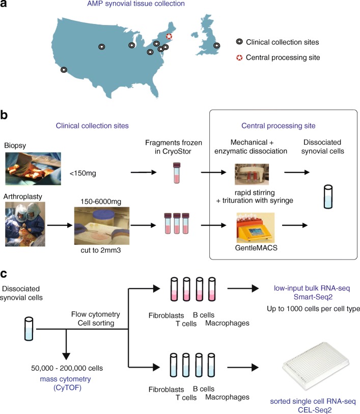
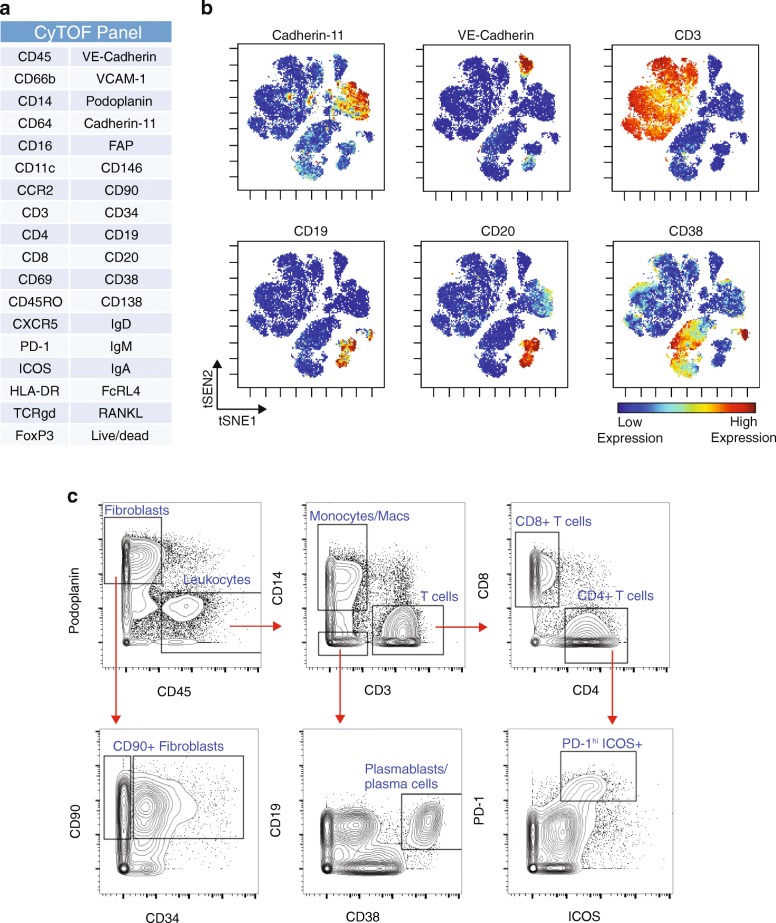
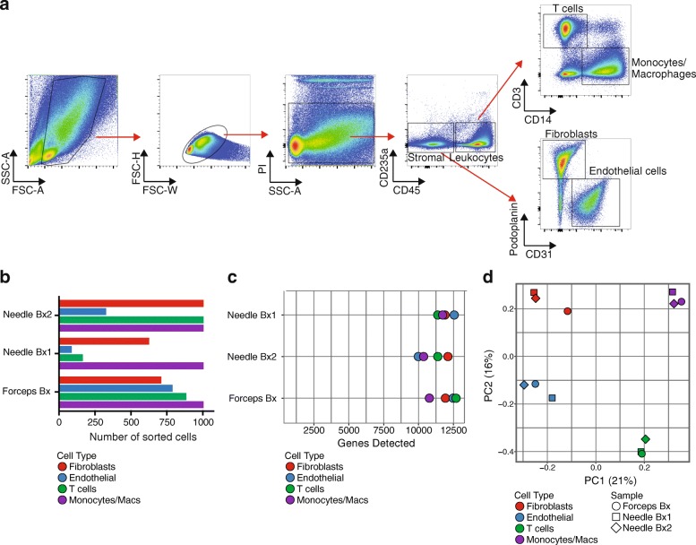
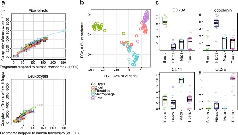
Methods: Multiple clinical sites collected cryopreserved synovial tissue fragments from arthroplasty and synovial biopsy in a 10% DMSO solution. Mechanical and enzymatic dissociation parameters were optimized for viable cell extraction and surface protein preservation for cell sorting and mass cytometry, as well as for reproducibility in RNA sequencing (RNA-seq). Cryopreserved synovial samples were collectively analyzed at a central processing site by a custom-designed and validated 35-marker mass cytometry panel. In parallel, each sample was flow sorted into fibroblast, T-cell, B-cell, and macrophage suspensions for bulk population RNA-seq and plate-based single-cell CEL-Seq2 RNA-seq.

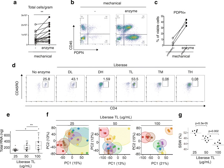
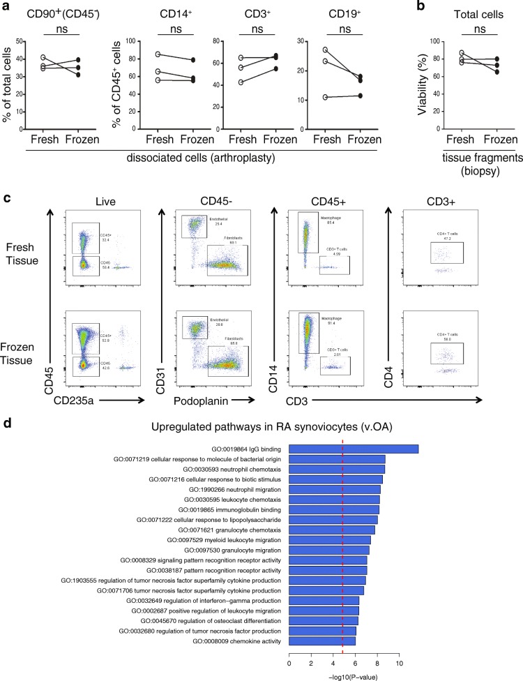
Results: Upon dissociation, cryopreserved synovial tissue fragments yielded a high frequency of viable cells, comparable to samples undergoing immediate processing. Optimization of synovial tissue dissociation across six clinical collection sites with ~ 30 arthroplasty and ~ 20 biopsy samples yielded a consensus digestion protocol using 100 μg/ml of Liberase™ TL enzyme preparation. This protocol yielded immune and stromal cell lineages with preserved surface markers and minimized variability across replicate RNA-seq transcriptomes. Mass cytometry analysis of cells from cryopreserved synovium distinguished diverse fibroblast phenotypes, distinct populations of memory B cells and antibody-secreting cells, and multiple CD4+ and CD8+ T-cell activation states. Bulk RNA-seq of sorted cell populations demonstrated robust separation of synovial lymphocytes, fibroblasts, and macrophages. Single-cell RNA-seq produced transcriptomes of over 1000 genes/cell, including transcripts encoding characteristic lineage markers identified.

Conclusions: We have established a robust protocol to acquire viable cells from cryopreserved synovial tissue with intact transcriptomes and cell surface phenotypes. A centralized pipeline to generate multiple high-dimensional analyses of synovial tissue samples collected across a collaborative network was developed. Integrated analysis of such datasets from large patient cohorts may help define molecular heterogeneity within RA pathology and identify new therapeutic targets and biomarkers.

Keywords: Accelerating Medicines Partnership; Arthroplasty; CyTOF; Mass cytometry; RNA sequencing; Rheumatoid arthritis; Synovial biopsy; Synovial tissue.


Conflict of interest statement

Ethics approval and consent to participate

The study received institutional review board approval at each site. Sites performing research synovial biopsies included informed consent specifically for this procedure. Where samples were used for transcriptome analysis, informed consent included consent for genetic analysis and deposition of AMP project data into public NIH databases.

Competing interests

The authors declare that they have no competing interests.

Publisher’s Note

Springer Nature remains neutral with regard to jurisdictional claims in published maps and institutional affiliations.

Figures

Fig. 1
Fig. 1
AMP consortium pipeline for high-dimensional synovial tissue assays. a Numerous Clinical Collection Sites contributed cryopreserved synovial samples to the Central Processing Site. b At the Clinical Collection Sites, synovial tissue was excised during research-oriented biopsies or routine removal in arthroplasty surgeries. Tissues and dissociated synovial cells cryopreserved for centralized processing. c Synovial tissue mechanically disaggregated via magnetic bar stirring in heated water bath for smaller biopsy fragments or gentleMACS dissociation for larger arthroplasty specimens. Tissue dissociation also involved incubation with protein and nucleic acid degrading enzymes at 37 °C. After filtration of debris, sample aliquots processed in parallel for mass cytometry (CyTOF) analysis and FACS-based sorting of four cell types for single-cell and bulk RNA sequencing (RNA-seq). AMP Accelerating Medicines Partnership
Fig. 2
Fig. 2
High synovial cell yield, preserved surface markers, and reproducible transcriptomic results with mechanical and enzymatic disaggregation protocol. a Total cell counts per gram from synovial tissue mechanically disaggregated with or without Liberase™ TL proteolytic enzyme treatment. n = 16, paired t test. b, c Flow cytometry detection and quantification of stromal cells (CD45PDPN+) upon mechanical disaggregation with or without Liberase™ TL proteolytic enzyme treatment. d Flow cytometry of dissociated synovial cells treated with panel of proteolytic Liberase™ TL formulations. Cells gated for viability and CD3 T-cell receptor subunit. Plots representative of n = 4 biologic replicates. e Total RNA yield from synovial tissue dissociated with three concentrations of Liberase™ TL. Two or three technical replicates from nine tissues used for each enzyme concentration. ANOVA with Tukey's comparison, *p<0.05 and **p<0.01. f Principal component analyses on RNA-seq gene expression results for samples from (e), whereby each technical replicate of same sample is represented by a dot of same color while variation between replicates is indicated by size of encircling cloud of same color. g Sum of squares within (SSW) replicates from same donor divided by total sum of squares, using gene expression profiles from (e) and (f). Horizontal lines indicate SSW values using all replicates. Individual dots are SSW values computed after leave-one-out strategy where replicates from one donor are left out and SSW is computed with remaining samples. Student’s t test used to test whether bootstrapped SSW values differ significantly. DH Dispase-High, DL Dispase-Low, PC principal component, PDPN podoplanin, TH Thermolysin-High, TL Thermolysin-Low, TM Thermolysin-Medium
Fig. 3
Fig. 3
Cryopreservation of synovial fragments retains stromal and hematopoietic cell viability and RA synovial gene expression patterns. a Frequency of synovial cell subpopulations from arthroplasty samples dissociated from freshly isolated or cryopreserved aliquots. b Cell viability frequency assayed by flow cytometry for synovial biopsy samples dissociated from fresh or cryopreserved tissue fragments. c Flow cytometry analysis of cells dissociated from synovial tissue either immediately or after cryopreservation. d Top 20 GO terms enriched for genes upregulated in RA patient disaggregated synovial cells in comparison to patients with OA. N = 10 RA and 10 OA samples with 2–8 replicates per donor. ns no significant difference, OA osteoarthritis, RA rheumatoid arthritis
Fig. 4
Fig. 4
Mass cytometry analysis of dissociated synovial tissue reveals synovial cell heterogeneity via surface marker detection. a Antibody marker panel for synovial cell mass cytometry analysis. b viSNE visualization of synovial cells analyzed by mass cytometry from cryopreserved synovial biopsy sample obtained from knee of an RA patient. Each dot is a cell. Color reflects level of expression of marker indicated at top of plot. c Biaxial contour plots of same mass cytometry data shown in (b), showing serial gating of cell subpopulations. Representative of at least six synovial samples
Fig. 5
Fig. 5
Low-input RNA-seq distinguishes cell populations sorted from dissociated RA synovial cells. a Flow cytometric detection of distinct cell populations obtained from cryopreserved RA synovial biopsy. b Number of cells of each targeted cell populations sorted from three different RA patient synovial biopsy (Bx) samples (two obtained by needle biopsy and one by portal and forceps biopsy from knee joint) for low-input RNA-seq. c Number of genes detected by low-input RNA-seq for each cell population sorted from same biopsies as (b). d Principal components analysis of sorted cell populations from three RA synovial biopsy samples based on 5006 genes with greatest variance. Color indicates sorted cell type, while shape indicates individual donor. FSC forward scatter, PC principal component, PI propidium iodide, SSC side scatter
Fig. 6
Fig. 6
Single-cell RNA-seq transcriptomic analysis distinguishes different cell types sorted from cryopreserved RA synovium. a Saturation curves indicating complexity of transcriptomes obtained from single cells (fibroblasts or leukocytes) sorted from cryopreserved RA knee arthroplasty tissue. At least five read fragments required for gene to be considered “detected”. b Principal components analysis of single cells from cryopreserved RA synovium, colored by cell type as determined by flow cytometry at time of cell sorting. Top 648 most variable expressed genes (F statistic) used, centered and scaled. PC scores for PC1 (x axis) and PC2 (y axis). c Expression (log2(TPM + 1)) of selected lineage-characteristic genes in sorted cell populations from cryopreserved RA arthroplasty sample. A total of 96 cells (24 for each type) filtered down to 69 by eliminating low-quality cells (those with more than 10% of reads in well mapped to ERCC) and presumed doublets (those with greater than 2.8 million reads). Representative of two synovial samples. PC principal component

References

    1. Gravallese EM, Manning C, Tsay A, Naito A, Pan C, Amento E, Goldring SR. Synovial tissue in rheumatoid arthritis is a source of osteoclast differentiation factor. Arthritis Rheum. 2000;43(2):250–258. doi: 10.1002/1529-0131(200002)43:2<250::AID-ANR3>3.0.CO;2-P. - DOI - PubMed
    1. Meednu N, Zhang H, Owen T, Sun W, Wang V, Cistrone C, Rangel-Moreno J, Xing L, Anolik JH. Production of RANKL by memory B cells: a link between B cells and bone Erosion in rheumatoid arthritis. Arthritis Rheumatol. 2016;68(4):805–816. doi: 10.1002/art.39489. - DOI - PMC - PubMed
    1. McInnes IB, Schett G. The pathogenesis of rheumatoid arthritis. N Engl J Med. 2012;365(23):2205–2219. doi: 10.1056/NEJMra1004965. - DOI - PubMed
    1. Ai R, Hammaker D, Boyle DL, Morgan R, Walsh AM, Fan S, Firestein GS, Wang W. Joint-specific DNA methylation and transcriptome signatures in rheumatoid arthritis identify distinct pathogenic processes. Nat Commun. 2016;7:11849. doi: 10.1038/ncomms11849. - DOI - PMC - PubMed
    1. Rao DA, Gurish MF, Marshall JL, Slowikowski K, Fonseka CY, Liu Y, Donlin LT, Henderson LA, Wei K, Mizoguchi F, et al. Pathologically expanded peripheral T helper cell subset drives B cells in rheumatoid arthritis. Nature. 2017;542(7639):110–114. doi: 10.1038/nature20810. - DOI - PMC - PubMed

Publication types

MeSH terms