. 2018 Jul 11;3(4):e00164-18.
doi: 10.1128/mSphere.00164-18.

Regulation of Trypanosoma brucei Acetyl Coenzyme A Carboxylase by Environmental Lipids

Affiliations

Regulation of Trypanosoma brucei Acetyl Coenzyme A Carboxylase by Environmental Lipids

Sunayan S Ray et al. mSphere. .

Abstract

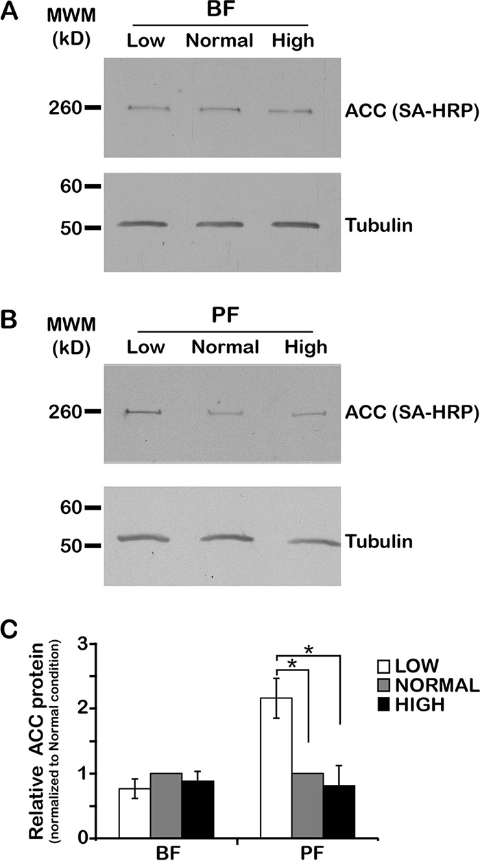
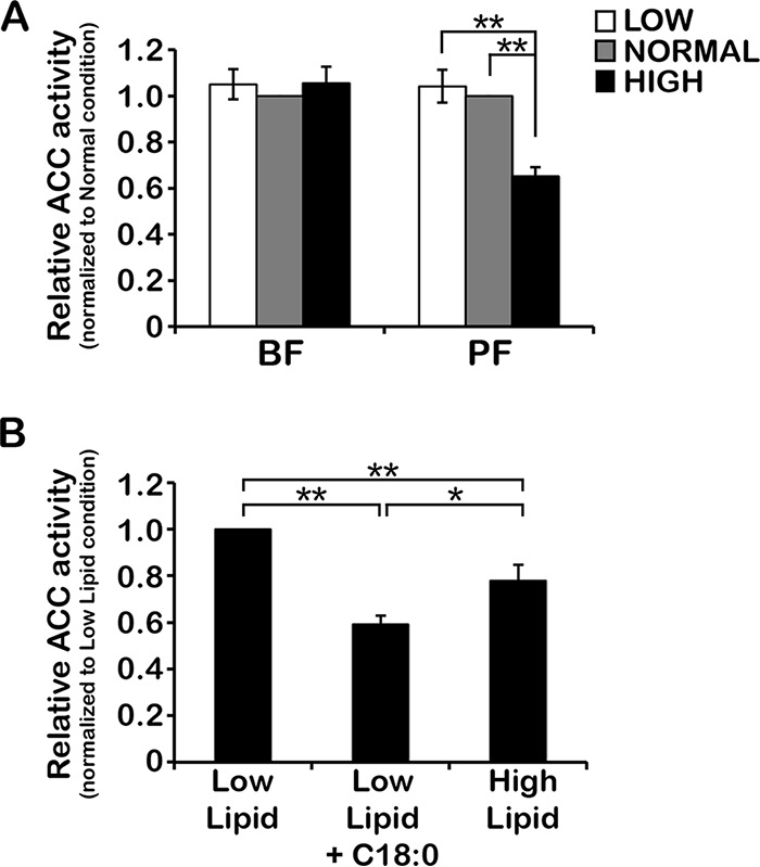
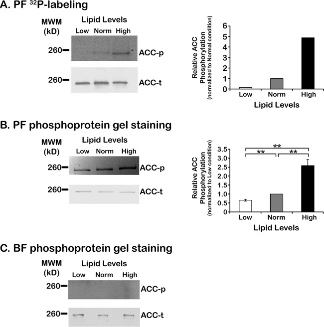
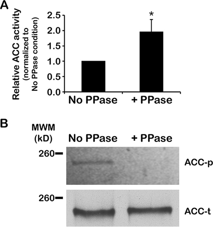
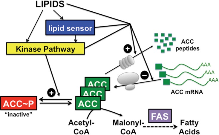
To satisfy its fatty acid needs, the extracellular eukaryotic parasite Trypanosoma brucei relies on two mechanisms: uptake of fatty acids from the host and de novo synthesis. We hypothesized that T. brucei modulates fatty acid synthesis in response to environmental lipid availability. The first committed step in fatty acid synthesis is catalyzed by acetyl coenzyme A (acetyl-CoA) carboxylase (ACC) and serves as a key regulatory point in other organisms. To test our hypothesis, T. brucei mammalian bloodstream and insect procyclic forms were grown in low-, normal-, or high-lipid media and the effect on T. brucei ACC (TbACC) mRNA, protein, and enzymatic activity was examined. In bloodstream form T. brucei, media lipids had no effect on TbACC expression or activity. In procyclic form T. brucei, we detected no change in TbACC mRNA levels but observed 2.7-fold-lower TbACC protein levels and 37% lower TbACC activity in high-lipid media than in low-lipid media. Supplementation of low-lipid media with the fatty acid stearate mimicked the effect of high lipid levels on TbACC activity. In procyclic forms, TbACC phosphorylation also increased 3.9-fold in high-lipid media compared to low-lipid media. Phosphatase treatment of TbACC increased activity, confirming that phosphorylation represented an inhibitory modification. Together, these results demonstrate a procyclic-form-specific environmental lipid response pathway that regulates TbACC posttranscriptionally, through changes in protein expression and phosphorylation. We propose that this environmental response pathway enables procyclic-form T. brucei to monitor the host lipid supply and downregulate fatty acid synthesis when host lipids are abundant and upregulate fatty acid synthesis when host lipids become scarce.IMPORTANCETrypanosoma brucei is a eukaryotic parasite that causes African sleeping sickness. T. brucei is transmitted by the blood-sucking tsetse fly. In order to adapt to its two very different hosts, T. brucei must sense the host environment and alter its metabolism to maximize utilization of host resources and minimize expenditure of its own resources. One key nutrient class is represented by fatty acids, which the parasite can either take from the host or make themselves. Our work describes a novel environmental regulatory pathway for fatty acid synthesis where the parasite turns off fatty acid synthesis when environmental lipids are abundant and turns on synthesis when the lipid supply is scarce. This pathway was observed in the tsetse midgut form but not the mammalian bloodstream form. However, pharmacological activation of this pathway in the bloodstream form to turn fatty acid synthesis off may be a promising new avenue for sleeping sickness drug discovery.

Keywords: Trypanosoma brucei; acetyl-CoA carboxylase; enzyme regulation; fatty acids; lipids; parasitology; protein phosphorylation.


Figures

FIG 1
FIG 1
Environmental lipids affect TbACC protein levels in PF but not BF. (A and B) BF (A) and PF (B) wild-type (WT) cells were grown in low-, normal-, and high-lipid media to the mid-logarithmic (mid-log) phase (~3 days). Lysates prepared in the presence of a phosphatase inhibitor cocktail were resolved by the use of SDS-PAGE (10 µg total protein/lane) and transferred to nitrocellulose. TbACC was detected by SA-HRP blotting (A and B, top panels), and the same blots were reprobed for tubulin as a loading control (bottom panels). Representative blots from three independent experiments are shown. MWM, molecular weight marker. (C) Densitometric quantification of the results from the three independent experiments described in the panel A and B legends. The TbACC signal was normalized to the tubulin loading control. Means ± standard errors of the means (SEM) are shown. *, P ≤ 0.05 (two-tailed Student’s t test).
FIG 2
FIG 2
Environmental lipids affect TbACC activity in PF but not BF. (A) BF and PF WT cells were grown under low-, normal-, and high-lipid conditions to the mid-log phase (~3 days). Lysates prepared in the presence of phosphatase inhibitor cocktail (equal total levels of protein, 2 to 10 µg) were assayed for TbACC activity by measuring incorporation of [14C]NaHCO3 into the acid-resistant malonyl-CoA product in the presence of ATP and acetyl-CoA. Values were first normalized to the no-ATP negative control before averaging was performed. Average values were then expressed relative to that of normal-lipid media. Means ± SEM of results from three independent experiments performed in pentuplicate are shown. **, P ≤ 0.005 (two-tailed Student’s t test). (B) PF WT cells grown under low-lipid conditions to the mid-log phase (~2 days) were subdivided into three cultures and grown for an additional 24 h, with one culture maintained in low-lipid media (Low Lipid), one supplemented with a final concentration of 20% FBS (High Lipid), and one supplemented with 35 µM stearate fatty acid (Low Lipid + C18:0). Hypotonic lysates were prepared and assayed for TbACC activity as described above. Values were normalized to the no-ATP control before averaging was performed, and averaged values are expressed relative to the low-lipid condition. Means ± SEM of results from three independent experiments are shown. *, P ≤ 0.05; **, P ≤ 0.005 (two-tailed Student’s t test).
FIG 3
FIG 3
Effect of environmental lipids on PF TbACC phosphorylation. (A) PF TbACC-myc cells were grown in normal-lipid media to the mid-log phase (~2 days). Cells were harvested and then incubated with 1 to 2 mCi [32P]orthophosphate in low (Low)-, normal (Norm)-, and high (High)-lipid phosphate-free media for 16 h. TbACC-myc immunoprecipitates were resolved by the use of SDS-PAGE, transferred to nitrocellulose, and assessed by autoradiography (top left panel). An identically loaded blot was prepared in parallel and probed for total TbACC by SA-HRP blotting (bottom left panel). Densitometric analysis of the blot is shown in the right panel. TbACC-myc phosphorylation values were normalized to total TbACC loaded levels. This experiment was performed once. (B) PF TbACC-myc cells were grown in low-, normal-, and high-lipid media to the mid-log phase (~3 days). TbACC-myc immunoprecipitates were resolved by the use of 10% SDS-PAGE, and phosphorylated TbACC-myc was detected by phosphoprotein gel staining and imaging under UV (upper left panel, ACC-p). Identically loaded gels prepared in parallel were transferred to nitrocellulose and probed for total TbACC by SA-HRP blotting (lower left panel, ACC-t). A representative gel and blot from three independent experiments are shown. Densitometric analysis of three independent experiments is shown in the right panel. PF TbACC-myc phosphorylation values (ACC-p) were normalized to total TbACC-myc loaded levels (ACC-t). Means ± SEM are shown. **, P ≤ 0.005 (two-tailed Student’s t test). (C) BF TbACC-myc cells were grown in low-, normal-, and high-lipid media to the mid-log phase (~3 days). TbACC-myc immunoprecipitates were resolved by the use of SDS-PAGE and assessed for phosphorylated TbACC (ACC-p) by phosphoprotein gel staining (top panel) and total TbACC (ACC-t) by SA-HRP blotting (bottom panel) as described for panel B. No densitometry data are shown due to a lack of phosphorylated TbACC.
FIG 4
FIG 4
Phosphorylation of TbACC reduces activity. (A) PF TbACC-myc cells were grown in normal media to the mid-log phase (~3 days). TbACC-myc was immunoprecipitated from lysates and directly treated on-bead with 400 U of Lambda phosphatase (+PPase) or was subjected to mock treatment as a control (No PPase). Phosphatase- and mock-treated TbACC-myc was directly assayed on-bead for ACC activity. Values were first normalized to the no-ATP negative control before averaging was performed. Average values were then expressed relative to that of normal-lipid media. Means ± SEM of results from three independent experiments are shown. *, P ≤ 0.05 (two-tailed Student’s t test). (B) To confirm dephosphorylation, results of phosphatase- and mock-treated TbACC-myc pulldown experiments were resolved by the use of SDS-PAGE and assessed for phosphorylation by phosphoprotein gel staining (upper panel, ACC-p). An identically loaded gel was prepared in parallel, transferred to nitrocellulose, and probed for total TbACC by SA-HRP blotting (lower panel, ACC-t). The image was digitally processed to enable better visualization of the bands (see Materials and Methods). This experiment was performed once.
FIG 5
FIG 5
Current model for how environmental lipids modulate TbACC. Environmental lipids activate a kinase signaling pathway that leads to the phosphorylation and inactivation of TbACC. Environmental lipids also increase TbACC protein expression through repression of TbACC mRNA translation and/or stimulation of protein degradation. Lipids may regulate TbACC activity and expression directly or through the action of a lipid sensor. In addition, lipids may coordinately control TbACC protein levels and activity via the same kinase signaling pathway, or TbACC protein and activity may be regulated by distinct mechanisms. FAS, fatty acid synthesis.

References

    1. Roheim PS, Carey M, Forte T, Vega GL. 1979. Apolipoproteins in human cerebrospinal fluid. Proc Natl Acad Sci U S A 76:4646–4649. doi:10.1073/pnas.76.9.4646. - DOI - PMC - PubMed
    1. Trindade S, Rijo-Ferreira F, Carvalho T, Pinto-Neves D, Guegan F, Aresta-Branco F, Bento F, Young SA, Pinto A, Van Den Abbeele J, Ribeiro RM, Dias S, Smith TK, Figueiredo LM. 2016. Trypanosoma brucei parasites occupy and functionally adapt to the adipose tissue in mice. Cell Host Microbe 19:837–848. doi:10.1016/j.chom.2016.05.002. - DOI - PMC - PubMed
    1. Bringaud F, Rivière L, Coustou V. 2006. Energy metabolism of trypanosomatids: adaptation to available carbon sources. Mol Biochem Parasitol 149:1–9. doi:10.1016/j.molbiopara.2006.03.017. - DOI - PubMed
    1. Dyer NA, Rose C, Ejeh NO, Acosta-Serrano A. 2013. Flying tryps: survival and maturation of trypanosomes in tsetse flies. Trends Parasitol 29:188–196. doi:10.1016/j.pt.2013.02.003. - DOI - PubMed
    1. Field MC, Lumb JH, Adung’a VO, Jones NG, Engstler M. 2009. Macromolecular trafficking and immune evasion in African trypanosomes. Int Rev Cell Mol Biol 278:1–67. doi:10.1016/S1937-6448(09)78001-3. - DOI - PubMed

Publication types

MeSH terms

LinkOut - more resources