Polymerization pathway of mammalian nonmuscle myosin 2s
- PMID: 29997172
- PMCID: PMC6065031
- DOI: 10.1073/pnas.1808800115
Polymerization pathway of mammalian nonmuscle myosin 2s
Abstract
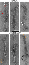
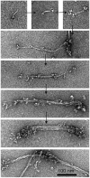
The three mammalian nonmuscle myosin 2 (NM2) monomers, like all class 2 myosin monomers, are hexamers of two identical heavy (long) chains and two pairs of light (short) chains bound to the heavy chains. The heavy chains have an N-terminal globular motor domain (head) with actin-activated ATPase activity, a lever arm (neck) to which the two light chains bind, and a coiled-coil helical tail. Monomers polymerize into bipolar filaments, with globular heads at each end separated by a bare zone, by antiparallel association of their coiled-coil tails. NM2 filaments are highly dynamic in situ, frequently disassembling and reassembling at different locations within the cell where they are essential for multiple biological functions. Therefore, it is important to understand the mechanisms of filament polymerization and depolymerization. Monomers can exist in two states: folded and unfolded. It has been thought that unfolded monomers form antiparallel dimers that assemble into bipolar filaments. We now show that polymerization in vitro proceeds from folded monomers to folded antiparallel dimers to folded antiparallel tetramers that unfold forming antiparallel bipolar tetramers. Folded dimers and tetramers then associate with the unfolded tetramer and unfold, forming a mature bipolar filament consisting of multiple unfolded tetramers with an entwined bare zone. We also demonstrate that depolymerization is essentially the reverse of the polymerization process. These results will advance our understanding of NM2 filament dynamics in situ.
Keywords: filaments; nonmuscle myosin 2; polymerization.
Conflict of interest statement
The authors declare no conflict of interest.
Figures


Similar articles
-
Muscle myosins form folded monomers, dimers, and tetramers during filament polymerization in vitro.Proc Natl Acad Sci U S A. 2020 Jul 7;117(27):15666-15672. doi: 10.1073/pnas.2001892117. Epub 2020 Jun 22. Proc Natl Acad Sci U S A. 2020. PMID: 32571956 Free PMC article.
-
Effect of ATP and regulatory light-chain phosphorylation on the polymerization of mammalian nonmuscle myosin II.Proc Natl Acad Sci U S A. 2017 Aug 8;114(32):E6516-E6525. doi: 10.1073/pnas.1702375114. Epub 2017 Jul 24. Proc Natl Acad Sci U S A. 2017. PMID: 28739905 Free PMC article.
-
Suggesting Dictyostelium as a Model for Disease-Related Protein Studies through Myosin II Polymerization Pathway.Cells. 2024 Jan 31;13(3):263. doi: 10.3390/cells13030263. Cells. 2024. PMID: 38334655 Free PMC article.
-
The heavy chain has its day: regulation of myosin-II assembly.Bioarchitecture. 2013 Jul-Aug;3(4):77-85. doi: 10.4161/bioa.26133. Bioarchitecture. 2013. PMID: 24002531 Free PMC article. Review.
-
The regulation of myosin II in Dictyostelium.Eur J Cell Biol. 2006 Sep;85(9-10):969-79. doi: 10.1016/j.ejcb.2006.04.004. Epub 2006 Jun 30. Eur J Cell Biol. 2006. PMID: 16814425 Review.
Cited by
-
Myosin 2 - A general contractor for the cytoskeleton.Curr Opin Cell Biol. 2025 Jun;94:102522. doi: 10.1016/j.ceb.2025.102522. Epub 2025 May 3. Curr Opin Cell Biol. 2025. PMID: 40319507 Review.
-
Filament evanescence of myosin II and smooth muscle function.J Gen Physiol. 2021 Mar 1;153(3):e202012781. doi: 10.1085/jgp.202012781. J Gen Physiol. 2021. PMID: 33606000 Free PMC article. Review.
-
Antibodies in cerebral cavernous malformations react with cytoskeleton autoantigens in the lesional milieu.J Autoimmun. 2020 Sep;113:102469. doi: 10.1016/j.jaut.2020.102469. Epub 2020 Apr 30. J Autoimmun. 2020. PMID: 32362501 Free PMC article.
-
Comparative Analysis of the Roles of Non-muscle Myosin-IIs in Cytokinesis in Budding Yeast, Fission Yeast, and Mammalian Cells.Front Cell Dev Biol. 2020 Nov 19;8:593400. doi: 10.3389/fcell.2020.593400. eCollection 2020. Front Cell Dev Biol. 2020. PMID: 33330476 Free PMC article. Review.
-
Drebrin is Required for Myosin-facilitated Actin Cytoskeletal Remodeling during Pulmonary Alveolar Development.Am J Respir Cell Mol Biol. 2024 Apr;70(4):308-321. doi: 10.1165/rcmb.2023-0229OC. Am J Respir Cell Mol Biol. 2024. PMID: 38271699 Free PMC article.
References
-
- Sellers JR. Myosins: A diverse superfamily. Biochim Biophys Acta. 2000;1496:3–22. - PubMed
Publication types
MeSH terms
Substances
LinkOut - more resources
Full Text Sources
Other Literature Sources

