. 2018 Jul 24;115(30):7783-7788.
doi: 10.1073/pnas.1722056115. Epub 2018 Jul 11.

CD52 glycan binds the proinflammatory B box of HMGB1 to engage the Siglec-10 receptor and suppress human T cell function

Affiliations

CD52 glycan binds the proinflammatory B box of HMGB1 to engage the Siglec-10 receptor and suppress human T cell function

Esther Bandala-Sanchez et al. Proc Natl Acad Sci U S A. .

Erratum in

Abstract

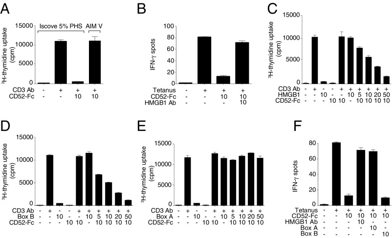
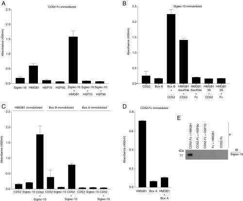
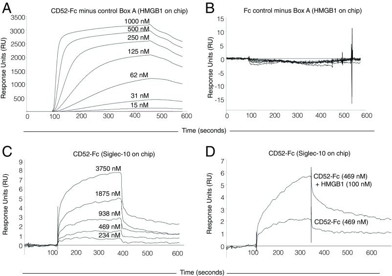
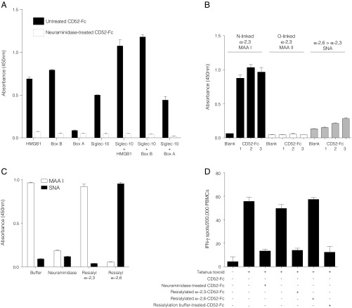
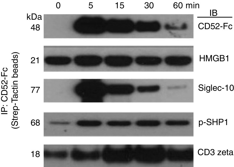
CD52, a glycophosphatidylinositol (GPI)-anchored glycoprotein, is released in a soluble form following T cell activation and binds to the Siglec (sialic acid-binding Ig-like lectin)-10 receptor on T cells to suppress their function. We show that binding of CD52-Fc to Siglec-10 and T cell suppression requires the damage-associated molecular pattern (DAMP) protein, high-mobility group box 1 (HMGB1). CD52-Fc bound specifically to the proinflammatory Box B domain of HMGB1, and this in turn promoted binding of the CD52 N-linked glycan, in α-2,3 sialic acid linkage with galactose, to Siglec-10. Suppression of T cell function was blocked by anti-HMGB1 antibody or the antiinflammatory Box A domain of HMGB1. CD52-Fc induced tyrosine phosphorylation of Siglec-10 and was recovered from T cells complexed with HMGB1 and Siglec-10 in association with SHP1 phosphatase and the T cell receptor (TCR). Thus, soluble CD52 exerts a concerted immunosuppressive effect by first sequestering HMGB1 to nullify its proinflammatory Box B, followed by binding to the inhibitory Siglec-10 receptor, triggering recruitment of SHP1 to the intracellular immunoreceptor tyrosine-based inhibitory motif of Siglec-10 and its interaction with the TCR. This mechanism may contribute to immune-inflammatory homeostasis in pathophysiologic states and underscores the potential of soluble CD52 as a therapeutic agent.

Keywords: CD52; HMGB1; Siglec-10; T cell; sialoglycan.


Conflict of interest statement

The authors declare no conflict of interest.

Figures

Fig. 1.
Fig. 1.
T cell suppression by soluble CD52 requires HMGB1 Box B and is antagonized by HMGB1 Box A. (A) Proliferation of human PBMCs incubated in serum-containing IP5 medium (Iscove’s modified Dulbecco’s medium with 5% PHS) or serum-free medium (AIM V) for 48 h alone or with plate-bound anti-CD3 antibody (1 μg/mL) and CD52-Fc. (B) IFN-γ production measured by ELISpot from human PBMCs (1 × 10⁵) incubated in serum-containing medium with no antigen or tetanus toxoid (10 Lfu/mL) in the presence of CD52-Fc and neutralizing antibody to HMGB1. (C) Proliferation of human PBMCs incubated in serum-free medium alone or with plate-bound anti-CD3 antibody (1 μg/mL), CD52-Fc and with increasing concentrations of disulfide-HMGB1, or HMGB1 Box B (D), or HMGB1 Box A (E). Proliferation was measured as [3H] thymidine uptake. (F) IFN-γ production measured by ELISpot from human PBMCs (1 × 10⁵) incubated in serum-containing medium in the presence of HMGB1 Box A or HMGB1 Box B recombinant proteins and neutralizing antibody to HMGB1. Concentrations of recombinant proteins in every figure are in μg/mL. Serum-free medium was used in experiments in A, C, D, and E.
Fig. 2.
Fig. 2.
HMGB1 promotes binding of soluble CD52-Fc to Siglec-10. Binding of CD52-Fc to recombinant HMGB1 and Siglec-10-Fc is shown. Proteins immobilized in flat bottom 96-well microtiter plate wells were as follows: CD52-Fc (at 10 μg/mL) (A and D), Siglec-10-Fc (at 10 μg/mL) (B), HMGB1, HMGB1 Box B, and HMGB1 Box A (each at 20 μg/mL) (C). After blocking the plate with binding buffer, different proteins as indicated were added (50 μL per well, 10 μg/mL) to triplicate wells, and binding was detected as described in SI Appendix, SI Materials and Methods. Results are mean ± SD of three separate experiments. (E) CD52-Fc or Fc (each 20 μg/mL) and Siglec-10-Fc (10 μg/mL) were incubated separately or together with HMGB1 or HSPs 70 or 90 (each 20 μg/mL) overnight at 4 °C. CD52-Fc (or Fc) was then precipitated (P) with Strep-Tactin beads and the pellet fractionated by SDS/PAGE and immunoblotted (IB) with anti-Siglec-10 antibody.
Fig. 3.
Fig. 3.
Surface plasmon resonance quantifies binding of CD52-Fc to HMGB1. Binding of CD52-Fc (A) or Fc control protein to HMGB1 (B), CD52-Fc to Siglec-10-Fc (C), and CD52-Fc with or without HMGB1 to Siglec-10-Fc (D). HMGB1 (100 µg/mL) or Siglec-10-Fc (25 µg/mL) were immobilized on CM5 dextran sensor chips. As a control for nonspecific binding, recombinant Box A was used in A and B and recombinant Fc in C and D. Data are representative of three experiments. Further details are provided in SI Appendix, SI Materials and Methods.
Fig. 4.
Fig. 4.
CD52-Fc glycan sialic acid is required for binding to HMGB1 and Siglec-10. (A) Binding of CD52-Fc to HMGB1, Box A, Box B, and Siglec-10 pretreated with Clostridium perfringens type V neuraminidase or with vehicle alone to plate-bound proteins as indicated. (B) Binding of untreated CD52-Fc to lectins (MAA I, MAA II, and SNA) immobilized on a microtiter plate, showing that CD52-Fc binds MAA I, a lectin that recognizes sialic acid in α-2,3 linkage with galactose. (C) Resialylation of neuraminidase-treated CD52-Fc with either α-2,3 (CstII) or α-2,6 (PdST6Ga1I) sialyltransferases, confirmed by binding to MAA I lectin (α-2,3 linkage) or SNA lectin (α-2,6 linkage). (D) ELISpot assay demonstrating functional activity of CD52-Fc reconstituted with sialic acid in α-2,3 linkage with galactose. Data are mean ± SEM of three independent experiments.
Fig. 5.
Fig. 5.
CD52–HMGB1–Siglec-10 trimolecular complex interacts with the T cell receptor. Flow-sorted CD3+CD4+ T cells were incubated with anti-CD3/CD28 antibody beads for the indicated times in the presence of CD52-Fc (40 µg/mL) at 37 °C. The reaction was stopped with ice-cold PBS containing protease and phosphatase inhibitors. Cells were washed in cold PBS with inhibitors and solubilized in 1% digitonin buffer. CD52-Fc was precipitated (IP) with Strep-Tactin beads, fractionated in SDS/PAGE under reducing conditions, and immunoblotted (IB) with antibodies to CD52, HMGB1, Siglec-10, p-SHP1, or TCR CD3 zeta. Coomassie Brilliant Blue protein staining was similar in all lanes.

References

    1. Xia MQ, Tone M, Packman L, Hale G, Waldmann H. Characterization of the CAMPATH-1 (CDw52) antigen: Biochemical analysis and cDNA cloning reveal an unusually small peptide backbone. Eur J Immunol. 1991;21:1677–1684. - PubMed
    1. Hale G. CD52 (CAMPATH1) J Biol Regul Homeost Agents. 2001;15:386–391. - PubMed
    1. Kirchhoff C, Schröter S. New insights into the origin, structure and role of CD52: A major component of the mammalian sperm glycocalyx. Cells Tissues Organs. 2001;168:93–104. - PubMed
    1. Ito K, Hasegawa A, Komori S, Koyama K. Biochemical property and immunogenicity of mouse male reproductive tract CD52 (mrt-CD52) J Reprod Immunol. 2007;75:32–39. - PubMed
    1. Bandala-Sanchez E, et al. T cell regulation mediated by interaction of soluble CD52 with the inhibitory receptor Siglec-10. Nat Immunol. 2013;14:741–748. - PubMed

Publication types