. 2018 Jul 12;3(13):e95319.
doi: 10.1172/jci.insight.95319.

Elderly human hematopoietic progenitor cells express cellular senescence markers and are more susceptible to pyroptosis

Affiliations

Elderly human hematopoietic progenitor cells express cellular senescence markers and are more susceptible to pyroptosis

Tinhinane Fali et al. JCI Insight. .

Abstract

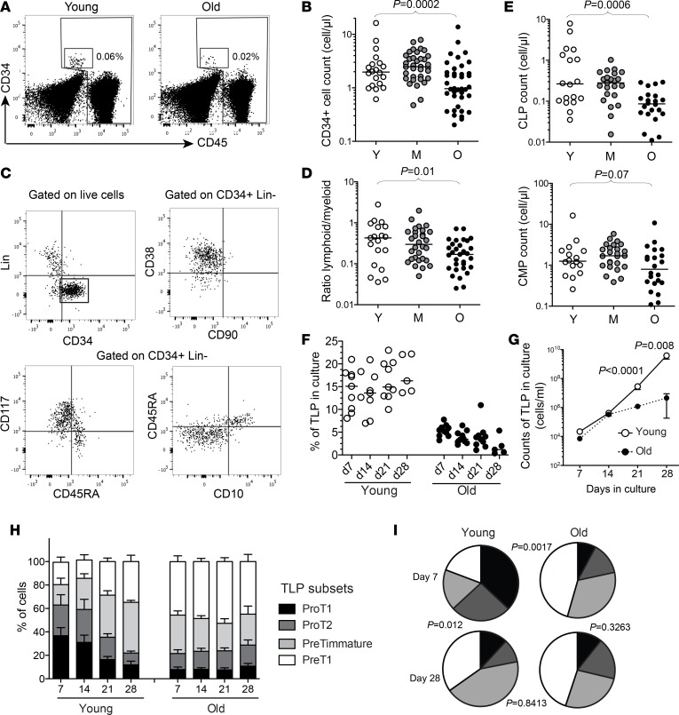
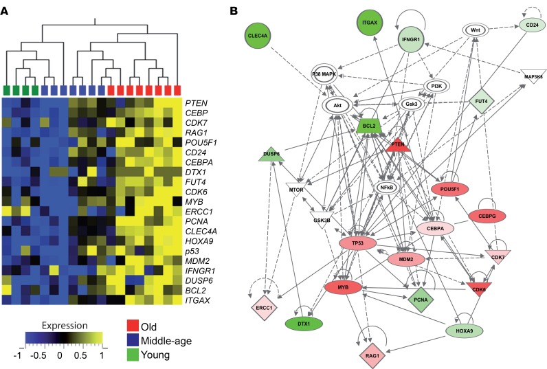
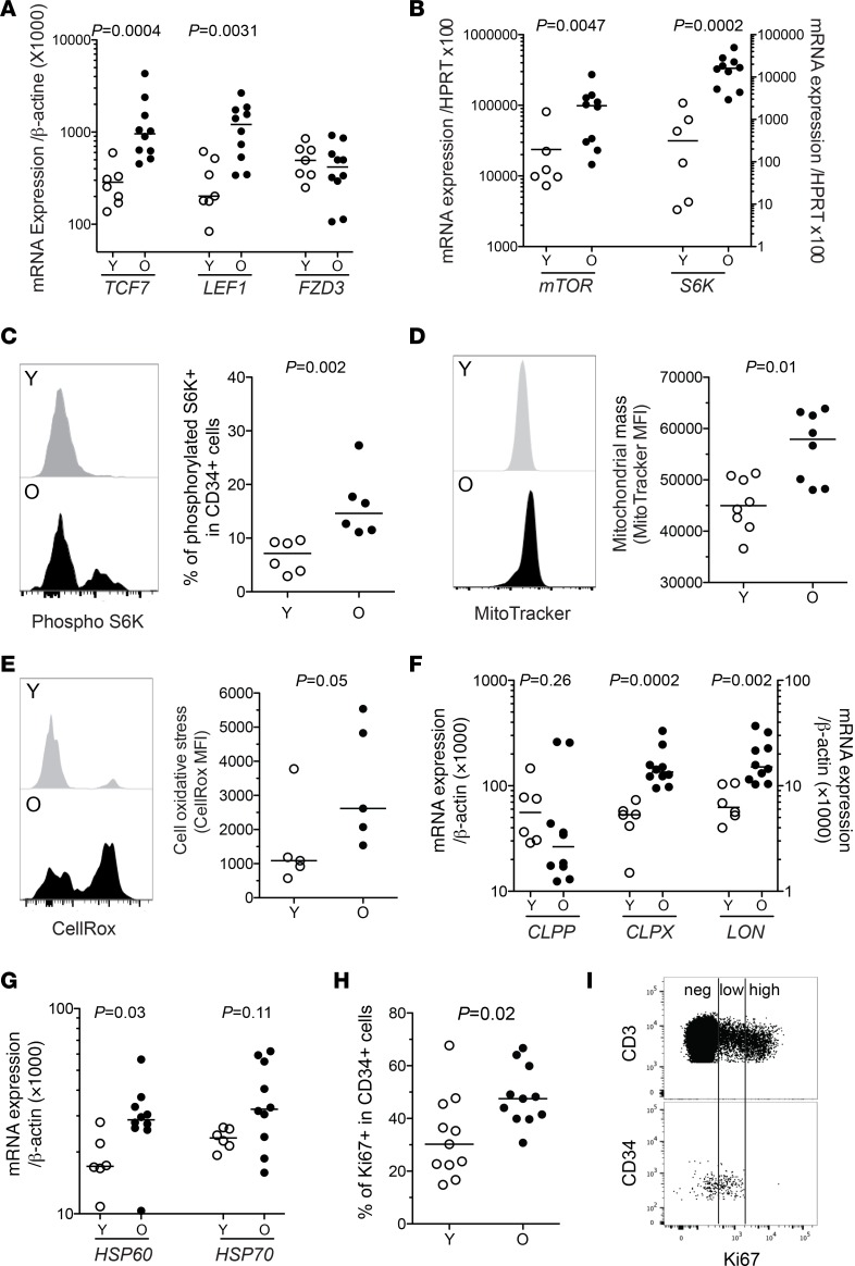
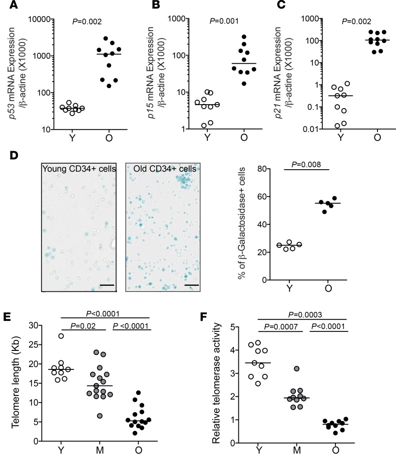
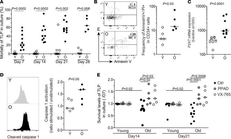
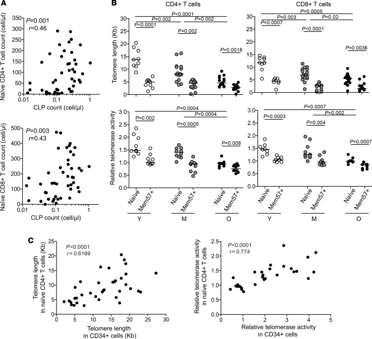
The maintenance of effective immunity over time is dependent on the capacity of hematopoietic stem cells (HSCs) to sustain the pool of immunocompetent mature cells. Decline of immune competence with old age may stem from HSC defects, including reduced self-renewal potential and impaired lymphopoiesis, as suggested in murine models. To obtain further insights into aging-related alteration of hematopoiesis, we performed a comprehensive study of blood hematopoietic progenitor cells (HPCs) from older humans. In the elderly, HPCs present active oxidative phosphorylation and are pressed to enter cell cycling. However, p53-p21 and p15 cell senescence pathways, associated with telomerase activity deficiency, strong telomere attrition, and oxidative stress, are engaged, thus limiting cell cycling. Moreover, survival of old HPCs is impacted by pyroptosis, an inflammatory form of programmed cell death. Lastly, telomerase activity deficiency and telomere length attrition of old HPCs may be passed on to progeny cells such as naive T lymphocytes, further highlighting the poor hematopoietic potential of the elderly. This pre-senescent profile is characteristic of the multiple intrinsic and extrinsic factors affecting HPCs in elderly individuals and represents a major obstacle in terms of immune reconstitution and efficacy with advanced age.

Keywords: Aging; Cellular senescence; Hematopoietic stem cells; Human stem cells; Stem cells.


Conflict of interest statement

Conflict of interest: The authors have declared that no conflict of interest exists.

Figures

Figure 1
Figure 1. Altered lymphopoietic capacity of circulating CD34+ hematopoietic progenitors from elderly humans.
(A) Representative examples of CD34 and CD45 staining to identify HPCs within the CD45lo population. (B) Absolute counts of CD34+CD45loLin cells in young (Y, n = 20), middle-aged (M, n = 35), or old (O, n = 40) healthy adults. (C) Representative staining for CD38, CD90, CD117, CD45RA, and CD10 on bead-enriched CD34+ cells from PBMCs of a healthy adult. (D) Ratio of common lymphoid progenitors (CLPs, CD38+CD117CD45RA+CD10+) versus common myeloid progenitors (CMPs, CD38+CD117+CD45RACD10) within CD34+ cells from PBMCs in young, middle-aged, or old healthy adults. (E) Frequency of CLPs or CMPs in the blood of young, middle-aged, or old healthy adults. (F) Frequency of TLPs upon in vitro differentiation of FACS-isolated CD34+ HPCs from young (n = 9) or old (n = 10) healthy adults. Phenotyping of CD34+ cells was performed after 7, 14, 21, and 28 days in the OP9-DL1 coculture system. (G) Mean absolute counts of TLPs in culture upon in vitro differentiation of CD34+ HPCs purified from young (n = 9) or old (n = 10) healthy adults in the OP9-DL1 coculture system. (H) Distribution of TLP subsets of differentiation (ProT1: CD45RA+CD7+CD5CD1a; ProT2: CD45RA+CD7+CD5+CD1a; PreTimmature: CD45RA+CD7+CD5CD1a+; and PreT1: CD45RA+CD7+CD5+CD1a+) at 7, 14, 21, and 28 days in the OP9-DL1 coculture system. Columns indicate mean values (+SEM). (I) Percentages of TLP subsets within the total population in vitro are represented in pie charts for simplicity (black slices correspond to proT1, dark gray to proT2, light gray to preTimmature, and white to preT1). Pies show mean values. The Mann-Whitney or Kruskall-Wallis test was used for comparing 2 or 3 groups, respectively. Bars indicate the median.
Figure 2
Figure 2. Transcriptional profiling of CD34+ hematopoietic progenitors from elderly humans.
(A) Heatmap representation for the transcription of genes that are differentially expressed in HPCs from old (O, n = 8) healthy adults compared with young (Y, n = 4) or middle-aged (M, n = 7) subjects. The expression of selected genes in CD34+CD45loLin cells, FACS isolated ex vivo from donor PBMCs, was assessed using a Fluidigm microfluidics system. (B) Network diagram of genes differentially expressed in elderly HPCs with corresponding pathways highlighted using Ingenuity Pathway Analysis. The red or green colors indicate the degree of upregulation or downregulation in gene expression compared with housekeeping gene expression.
Figure 3
Figure 3. Upregulated mTOR pathway and mitochondrial stress in old hematopoietic progenitors.
(A) mRNA expression levels of the Wnt pathway–related genes TCF7, LEF1, and FZD3 in FACS-isolated HPCs from young (Y, n = 7) or old (O, n = 10) subjects. (B) mRNA expression levels of the mTOR pathway–related genes mTOR and S6K in FACS-isolated HPCs from young (n = 6) or old (n = 10) subjects. (C) Representative staining and median fluorescence intensity (MFI) levels for phosphorylated S6K in bead-enriched CD34+ cells from young or old adult PBMCs. (D and E) Representative staining and MFI levels of MitoTracker (mitochondrial mass marker) and CellRox (cellular ROS marker) in bead-enriched CD34+ cells from young or old adult PBMCs. (F and G) mRNA expression levels of genes associated with mitochondrial protein folding stress (CLPP, CLPX, and LON) and mitochondrial stress response (HSP60, HSP70) in FACS-isolated HPCs from young (n = 6) or old (n = 9) subjects. (H) Frequency of young or old CD34+ cells expressing the nuclear antigen Ki67. (I) Representative staining for Ki67 in CD3+ or CD34+ cells from an elderly individual, highlighting differential Ki67 expression levels. The Mann-Whitney test was used for comparisons. Bars indicate the median.
Figure 4
Figure 4. Increased markers of senescence in elderly hematopoietic progenitors.
(AC) mRNA expression levels of the cellular senescence pathway–associated genes p53, p15, and p21 in FACS-isolated HPCs from young (Y, n = 9) or old (O, n = 10) individuals. (D) Representative β-galactosidase staining (left) and frequency of β-galactosidase–positive cells (right) within FACS-isolated CD34+ cells from old (n = 5) or young (n = 5) individuals. Scale bars: 50 μm. (E) Telomere length in CD34+ HPCs from young (n = 9), middle-aged (M, n = 15), or old (n = 14) healthy adults. Telomere length was measured by qPCR in CD34+CD45loLin cells FACS isolated from PBMCs, and Kb calculated based on a reference cell line. (F) Relative telomerase activity in CD34+ HPCs from young, middle-aged, or old healthy adults. Telomerase activity was assessed using a TRAP assay in CD34+CD45loLin cells FACS isolated from PBMCs. The Mann-Whitney test was used for comparisons. Bars indicate the median.
Figure 5
Figure 5. Increased cell death and pyroptosis in old hematopoietic progenitors.
(A) Frequency of dead cells among TLPs (using LIVE/DEAD staining assay) at 7, 14, 21, and 28 days upon in vitro differentiation of FACS-isolated CD34+ HPCs from young (Y, n = 9) or old (O, n = 10) healthy adults in the OP9-DL1 coculture system. (B) Representative image and staining levels for annexin V and PI in bead-enriched CD34+ cells from young (n = 5) or old adult (n = 5) PBMCs. (C) Increased mRNA expression of the proapoptotic gene P2X7 in FACS-isolated HPCs from old (n = 10) healthy adults compared with HPCs from young (n = 7) subjects. (D) Representative staining and expression levels of cleaved caspase 1 in CD34+ cells from young (n = 5) or old (n = 5) adult PBMCs. Data are expressed as ratio of stimulated versus unstimulated cells. Bars indicate the median. (E) Enhancement of in vitro survival of TLPs at 14 or 21 days in the presence of PPAD (P2X7 inhibitor, at 20 μM) or VX-765 (caspase 1 inhibitor, at 0.1 μM) from day 7 in the OP9-DL1 culture system. Survival is expressed as the ratio of dying cells at day 14 or 21 over day 7 in culture (n = 5 in the presence of PPAD or VX-765 and n = 10 in the absence of inhibitors). Columns indicate mean (+SEM). The Mann-Whitney test was used for comparisons.
Figure 6
Figure 6. Associations between circulating HPCs and naive CD4+ or CD8+ T cell properties.
(A) Correlations between CLP and naive CD4+ or CD8+ T cell ex vivo absolute counts. (B) Telomere length (top panels) and relative telomerase activity (bottom panels) in naive or senescent CD57+ memory CD4+ or CD8+ T cells FACS isolated from young (Y, n = 9), middle-aged (M, n = 15), or old (O, n = 14) healthy adult PBMCs. (C) Correlations between CLP and naive CD4+ or CD8+ T cell telomere length or telomerase activity. The Mann-Whitney test was used for comparisons. Bars indicate the median. Spearman’s rank test was used to determine correlations.

References

    1. Kovtonyuk LV, Fritsch K, Feng X, Manz MG, Takizawa H. Inflamm-aging of hematopoiesis, hematopoietic stem cells, and the bone marrow microenvironment. Front Immunol. 2016;7:502. - PMC - PubMed
    1. Geiger H, de Haan G, Florian MC. The ageing haematopoietic stem cell compartment. Nat Rev Immunol. 2013;13(5):376–389. doi: 10.1038/nri3433. - DOI - PubMed
    1. Rossi DJ, et al. Cell intrinsic alterations underlie hematopoietic stem cell aging. Proc Natl Acad Sci USA. 2005;102(26):9194–9199. doi: 10.1073/pnas.0503280102. - DOI - PMC - PubMed
    1. Chambers SM, Shaw CA, Gatza C, Fisk CJ, Donehower LA, Goodell MA. Aging hematopoietic stem cells decline in function and exhibit epigenetic dysregulation. PLoS Biol. 2007;5(8):e201. doi: 10.1371/journal.pbio.0050201. - DOI - PMC - PubMed
    1. Rossi DJ, Bryder D, Seita J, Nussenzweig A, Hoeijmakers J, Weissman IL. Deficiencies in DNA damage repair limit the function of haematopoietic stem cells with age. Nature. 2007;447(7145):725–729. doi: 10.1038/nature05862. - DOI - PubMed

Publication types

MeSH terms