. 2018 Jul 12;3(13):e96672.
doi: 10.1172/jci.insight.96672.

Spatial and temporal variations in hemodynamic forces initiate cardiac trabeculation

Affiliations

Spatial and temporal variations in hemodynamic forces initiate cardiac trabeculation

Juhyun Lee et al. JCI Insight. .

Abstract

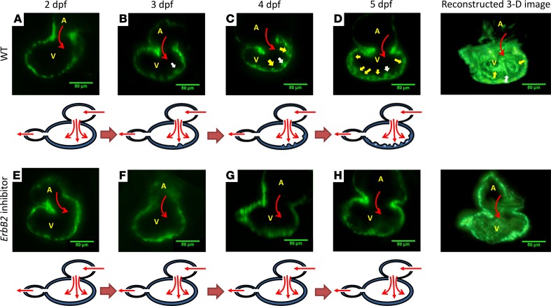
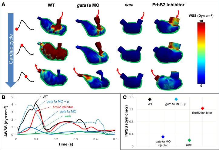
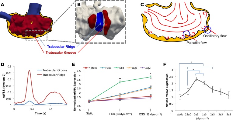
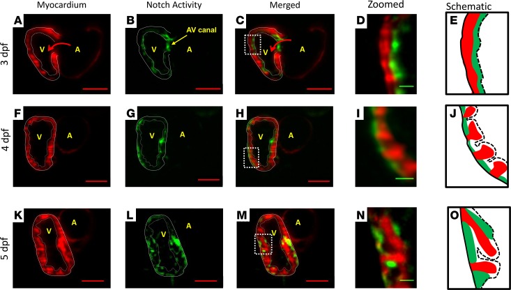
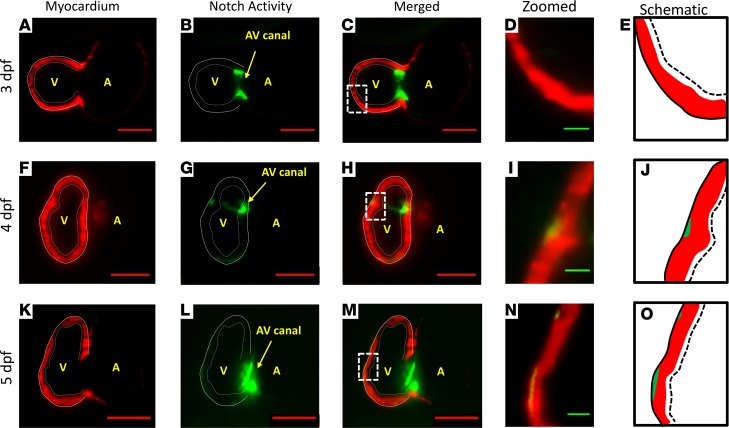
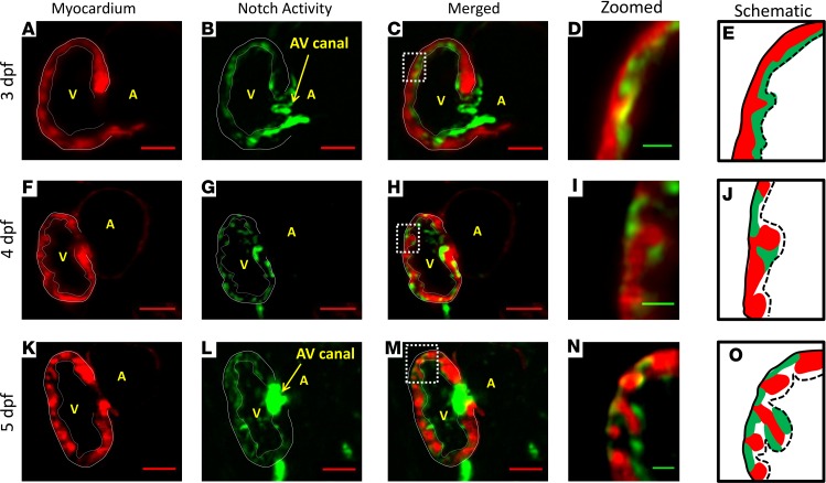
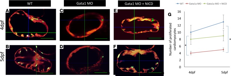
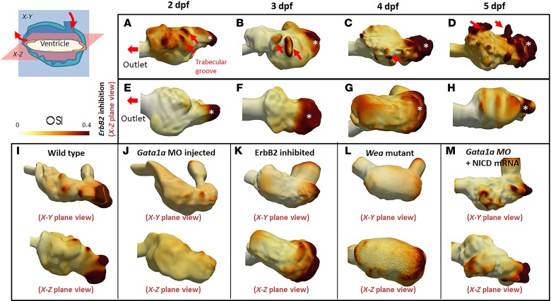
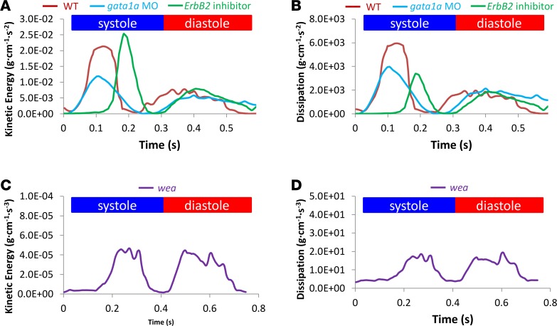
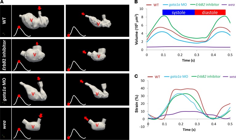
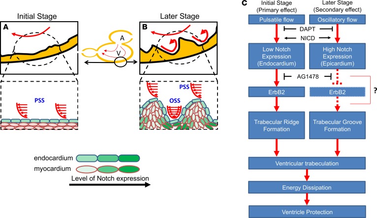
Hemodynamic shear force has been implicated as modulating Notch signaling-mediated cardiac trabeculation. Whether the spatiotemporal variations in wall shear stress (WSS) coordinate the initiation of trabeculation to influence ventricular contractile function remains unknown. Using light-sheet fluorescent microscopy, we reconstructed the 4D moving domain and applied computational fluid dynamics to quantify 4D WSS along the trabecular ridges and in the groves. In WT zebrafish, pulsatile shear stress developed along the trabecular ridges, with prominent endocardial Notch activity at 3 days after fertilization (dpf), and oscillatory shear stress developed in the trabecular grooves, with epicardial Notch activity at 4 dpf. Genetic manipulations were performed to reduce hematopoiesis and inhibit atrial contraction to lower WSS in synchrony with attenuation of oscillatory shear index (OSI) during ventricular development. γ-Secretase inhibitor of Notch intracellular domain (NICD) abrogated endocardial and epicardial Notch activity. Rescue with NICD mRNA restored Notch activity sequentially from the endocardium to trabecular grooves, which was corroborated by observed Notch-mediated cardiomyocyte proliferations on WT zebrafish trabeculae. We also demonstrated in vitro that a high OSI value correlated with upregulated endothelial Notch-related mRNA expression. In silico computation of energy dissipation further supports the role of trabeculation to preserve ventricular structure and contractile function. Thus, spatiotemporal variations in WSS coordinate trabecular organization for ventricular contractile function.

Keywords: Cardiology; Development; Embryonic development; Heart failure.


Conflict of interest statement

Conflict of interest: The authors have declared that no conflict of interest exists.

Figures

Figure 1
Figure 1. ErbB2-dependent trabeculation during cardiac morphogenesis.
(A) In WT zebrafish embryos, trabecular ridges were absent at 2 dpf. (B) A prominent trabecular ridge developed across the atrioventricular (AV) canal at 3 dpf. Atrial blood flow (red arrows) through the AV canal directly affected the endocardium. White arrows indicate the initial trabecular ridge. Yellow arrows indicate other trabecular ridges. (C) Additional trabecular ridges (yellow arrows) developed on both sides of the initial trabecular ridge at 4 dpf. (D) Trabeculation organized to form an interwoven network at 5 dpf. (E–H) In response to ErbB2 inhibitor (AG1478), trabeculation remained absent in the ventricular wall throughout the cardiac developmental stages. The ventricular wall thickness in response to ErbB2 inhibitor was reduced as compared with that in WT zebrafish embryos. A, atrium; V, ventricle. Scale bar: 50 μm.
Figure 2
Figure 2. Time-dependent 3D computational fluid dynamics simulation of the endocardial wall shear stress.
(A) Computational fluid dynamics (CFD) simulation (3D + time) was constructed from light-sheet imaging of zebrafish embryos in response to 4 genetic manipulations: gata1a morpholino (MO), wea mutants, ErbB2 inhibitor, and coinjection of gata1a MO with NICD mRNA. The CFD simulation revealed spatial and temporal variations in ventricular wall shear stress (WSS) in synchrony with the changes in ventricular morphology during a cardiac cycle. (B) Over the entire WT embryo ventricle, averaged WSS (AWSS) was higher than that in nontrabeculated ventricles from the 3 other groups. Despite ErbB2 inhibitor treatment, AWSS was still higher in WT embryos than in those injected with gata1a MO and wea mutants. When the ventricular cavity of the gata1a MO–injected model was demarcated to simulate WSS with WT blood viscosity, the AWSS value was restored to that of WT. (C) Time-averaged WSS (TWSS) in the WT embryos was higher than that in those injected with gata1a MO (lower blood viscosity) and wea mutants (lower cardiac contractility), whereas the ErbB2-inhibited embryos developed a similar TWSS, as compared with the WT zebrafish embryos. In silico simulation to restore to the WT blood viscosity in the gata1a MO–injected embryos normalized the TWSS to that of the WT zebrafish embryos.
Figure 3
Figure 3. Spatiotemporal variations in wall shear stress in trabecular ridges and grooves modulate Notch-related gene expression.
(A) Time-dependent 3D computational fluid dynamics (CFD) simulation revealed distinct spatial variations in ventricular wall shear stress (WSS), as highlighted in red. (B) Distinct shear stress profiles developed in the trabecular ridges versus grooves. (C) Trabecular ridges were exposed to pulsatile shear stress (PSS), while trabecular grooves were exposed to oscillatory shear stress (OSS). An elevated oscillatory shear index (OSI) developed as a result of flow trapped between the two trabecular ridges. (D) Trabecular ridges were exposed to substantially higher AWSS than trabecular grooves. This observation prompted the investigation into the initiation of trabecular ridges in response to high WSS. (E) Using a previously reported dynamic flow system (56), we demonstrated that OSS induced Notch-related mRNA expression to a greater extent than did PSS. (F) Notch1 receptor expression was highest under oscillatory shear stress (0 ± 3 dyn∙cm–2 with 0 net flow). Notch1 expression was attenuated in response to a gradual increase in net forward flow (1 ± 3, 2 ± 3, 3 ± 3, and 5 ± 3 dyn/cm2) (t test, *P < 0.05, n = 3).
Figure 4
Figure 4. Sequential Notch activity from endocardium to trabecular grooves in the WT zebrafish embryo.
Our 4D LSFM imaging captured sequential Notch1b activation (green) from endocardium to epicardium in the transgenic Tg(Tp-1:GFP;cmlc:mcherry) line. (A–E) At 3 dpf, Notch1b activity localized to the endocardial layer and AV canal. (F–J) At 4 dpf, Notch activity located primarily in the epicardium versus in the endocardium. Epicardial Notch1b activity and trabecular ridges organized into an alternating pattern. (K–O) At 5 dpf, trabeculae developed into a network structure. Notch activity was prominent in both the endocardium and trabecular grooves. Red scale bar: 50 μm. Green scale bar: 10 μm. Dotted lines in E, J, and O indicate endocardium. Solid lines in E, J, and O indicate epicardium. n = 3.
Figure 5
Figure 5. Inhibition of Notch activity and trabeculation in response to γ-secretase inhibition.
(A–E) At 3 dpf, DAPT treatment reduced Notch activity in the endocardium, except for in the AV canal region, in the transgenic Tg(Tp-1:GFP;cmlc:mcherry) line. (F–J) At 4 dpf, Notch activity remained absent in the endocardium. A small area of Notch activity appeared in the epicardium (green arrow). (K–O) At 5 dpf, additional small areas of Notch activation appeared in the epicardium, while trabeculation remained absent. Red scale bar: 50 μm. Green scale bar: 10 μm. Dotted lines in E, J, and O indicate endocardium. Solid lines in E, J, and O indicate epicardium. n = 3.
Figure 6
Figure 6. NICD mRNA injection rescued Notch activation and trabeculation in the DAPT-treated embryos.
(A–E) At 3 dpf, NICD injection rescued Notch activation in the endocardium of DAPT-treated transgenic Tg(Tp-1:GFP;cmlc:mcherry) embryos. (F–J) At 4 dpf, Notch activity appeared to be more prominent in the trabecular grooves than in the endocardium, reminiscent of that in WT embryos. (K–O) At 5 dpf, Notch activity was present in both endocardium and trabecular grooves but not in myocardium. Red scale bar: 50 μm. Green scale bar: 10 μm. Dotted lines in E, J, and O indicate endocardium. Solid lines in E, J, and O indicate epicardium. n = 5.
Figure 7
Figure 7. FUCCI system localized wall shear stress–mediated proliferating cardiomyocytes in the trabecular ridges.
(A and B) FUCCI system was used to visualize myocardial proliferation via a double-transgenic zebrafish line that was generated by crossing Tg(cmlc2:mCherry) fish and Tg(cmlc2:Venus-hGeminin)pd58 fish. Proliferating cardiomyocytes (green nuclei) were present at the trabecular ridges. (C and D) In response to gata1a MO, the number of proliferating cardiomyocytes was attenuated. (E and F) Coinjection of gata1a MO with NICD mRNA partially restored cardiomyocyte proliferation. (G) The graph statistically quantified the numbers of proliferating cardiomyocytes in response to gata1a MO and to NICD mRNA rescue (n = 4, t test, *P < 0.05).
Figure 8
Figure 8. Elevated OSI values developed in trabecular ridges and are dependent on ventricular surface roughness; OSI revealed distinct differences in the WT versus ErbB2 inhibitor treatment and roughness of ventricular surface.
(A and B) At 2 and 3 dpf, elevated OSI values were interspersed between trabecular ridges and grooves in the WT embryos. (C and D) At 4 and 5 dpf, high OSI values associated with formation of the trabecular network. (E–G) However, ErbB2 inhibition to attenuate trabeculation abrogated the interspersed OSI at 2, 3, and 4 dpf. (H) Despite a distinct OSI in the WT embryos at 5 dpf, slight interspersion of OSI values appeared in the ErbB2-inhibited group, with consistent absence of the trabecular network. (I) High OSI values were generated in trabecular ridges at 4 dpf. (J) OSI values were substantially reduced after lowering of blood viscosity. (K) ErbB2-inhibited zebrafish showed an attenuated trabecular wall, which stymied oscillatory flow at ventricular walls. (L) Due to a lack of atrial contraction, blood flow acting on the ventricle was low. This generated minimal OSI values. (M) After coinjection of NICD mRNA and gata1a MO, OSI values were reminiscent of those of WT zebrafish due to restored trabeculation.
Figure 9
Figure 9. Effects of trabeculation on kinetic energy and energy dissipation in the ventricle at 4 dpf.
(A) Gata1a MO injection reduced viscosity and WSS, resulting in a lower kinetic energy (KE) profile than in WT embryos during a cardiac cycle. However, the ErbB2 inhibition model, which reduces trabeculation, resulted in similarly high KE. (B) Both gata1a MO injection and ErbB2 inhibition reduced energy dissipation in the ventricle, compared with that in WT embryos. (C) Wea mutants, which lack the atrial contraction needed for initiation of trabeculation, had a profound reduction in KE. (D) Wea mutants also had a profound reduction in energy dissipation, associated with ventricular remodeling (Figure 2A).
Figure 10
Figure 10. Genetic manipulations of trabeculation influenced ventricular remodeling and strain rates.
(A) Changes in 3D fluid domain at end diastole and systole were recaptured by light-sheet imaging and were subsequently reconstructed. The 3D ventricular contours reflect the trabeculated endocardium in the WT embryos and the nontrabeculated endocardium in the genetic models. The red dots indicate the instantaneous moment at which ventricular volume was reconstructed during the cardiac cycle. (B) Time-dependent changes in ventricular volume were compared in response to genetic manipulations. ErbB2 inhibitor–mediated attenuation in trabeculation resulted in an increase in ventricular volume, as compared with that in the WT zebrafish embryos. Gata1a MO–mediated reduction in shear stress reduced ventricular volume. Wea mutation resulted in a ventricular volume of nearly 0. (C) Genetic manipulations to inhibit trabeculation also resulted in a reduction in ventricular strain rates (P < 0.01, n = 3).
Figure 11
Figure 11. Schematic of spatial and temporal determinants of shear stress and endocardial trabeculation.
(A) WSS activates endocardial Notch signaling. (B) The development of trabecular ridges promotes flow recirculation, yielding OSS, which, in turn, induces Notch activity in the trabecular grooves. (C) Coordination of PSS-induced endocardial Notch and OSS-mediated Notch in the grooves may organize ventricular trabeculation. While endocardial Notch activates myocardial Erbb2 expression to initiate trabecular ridge formation, Notch activity in the grooves may cause lateral inhibition of Erbb2 expression and resultant trabeculation (6). DAPT treatment inhibits NICD release, whereas NICD expression rescues Notch signaling and trabeculation. Trabeculae may serve to dissipate kinetic energy, thus preventing ventricular remodeling and dysfunction.

References

    1. Hove JR, Köster RW, Forouhar AS, Acevedo-Bolton G, Fraser SE, Gharib M. Intracardiac fluid forces are an essential epigenetic factor for embryonic cardiogenesis. Nature. 2003;421(6919):172–177. doi: 10.1038/nature01282. - DOI - PubMed
    1. Wang X, Ha T. Defining single molecular forces required to activate integrin and notch signaling. Science. 2013;340(6135):991–994. doi: 10.1126/science.1231041. - DOI - PMC - PubMed
    1. Gordon WR, et al. Mechanical allostery: evidence for a force requirement in the proteolytic activation of Notch. Dev Cell. 2015;33(6):729–736. doi: 10.1016/j.devcel.2015.05.004. - DOI - PMC - PubMed
    1. Lee J, et al. Moving domain computational fluid dynamics to interface with an embryonic model of cardiac morphogenesis. PLoS ONE. 2013;8(8):e72924. doi: 10.1371/journal.pone.0072924. - DOI - PMC - PubMed
    1. Peshkovsky C, Totong R, Yelon D. Dependence of cardiac trabeculation on neuregulin signaling and blood flow in zebrafish. Dev Dyn. 2011;240(2):446–456. doi: 10.1002/dvdy.22526. - DOI - PubMed

Publication types

MeSH terms