. 2018 Apr 16;8(2):53-61.
doi: 10.1556/1886.2018.00005. eCollection 2018 Jun 25.

Peroral Low-Dose Toxoplasma gondii Infection of Human Microbiota-Associated Mice - A Subacute Ileitis Model to Unravel Pathogen-Host Interactions

Affiliations

Peroral Low-Dose Toxoplasma gondii Infection of Human Microbiota-Associated Mice - A Subacute Ileitis Model to Unravel Pathogen-Host Interactions

Markus M Heimesaat et al. Eur J Microbiol Immunol (Bp). .

Abstract

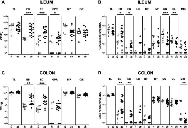
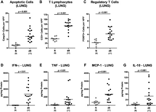
Within 1 week following high-dose Toxoplasma gondii infection, mice develop lethal necrotizing ileitis. However, data from a subacute T. gondii-induced ileitis model are scarce. Therefore, mice harboring a human gut microbiota were perorally infected with one cyst of T. gondii. Within 9 days post-infection, the intestinal microbiota composition shifted towards higher loads of commensal enterobacteria and enterococci. Following T. gondii infection, mice were clinically only mildly affected, whereas ≈60% of mice displayed fecal blood and mild-to-moderate ileal histopathological changes. Intestinal inflammation was further characterized by increased apoptotic intestinal epithelial cells, which were accompanied by elevated proliferating gut epithelial cell numbers. As compared to naive controls, infected mice displayed elevated numbers of intestinal T lymphocytes and regulatory T-cells and increased pro-inflammatory mediator secretion. Remarkably, T. gondii-induced apoptotic and pro-inflammatory immune responses were not restricted to the gut, but could also be observed in extra-intestinal compartments including kidney, liver, and lung. Strikingly, low-dose T. gondii infection resulted in increased serum levels of pro- and anti-inflammatory cytokines. In conclusion, the here presented subacute ileitis model following peroral low-dose T. gondii infection of humanized mice allows for detailed investigations of the molecular mechanism underlying the "ménage à trois" of pathogens, human gut microbiota, and immunity.

Keywords: Toxoplasma gondii; extra-intestinal; fecal microbiota transplantation; host immunity; host-pathogen interactions; human microbiota; intestinal; secondary abiotic mice; subacute ileitis mouse model; systemic immune responses.


Figures

Figure 1.
Figure 1.
Microbiota composition of human donor feces. Before fecal microbiota transplantation of secondary abiotic (i.e., gnotobiotic) mice for three consecutive days, main intestinal bacterial groups were quantitatively assessed in human donor fecal suspensions. (A) Numbers of viable enterobacteria (EB), enterococci (EC), Gram-positive rods (GPR), Bacteroides/Prevotella species (B/P), Clostridium/Eubacterium species (C/E), and the total bacterial load (TL) were determined by culture and expressed as colony-forming units (CFU) per milliliter suspension. (B) 16S rRNA of the main intestinal bacterial commensals including enterobacteria (EB), enterococci (EC), lactobacilli (LB), bifidobacteria (Bif), Bacteroides/Prevotella species (B/P), Clostridium coccoides group (Clocc), Clostridium leptum group (Clept), Mouse Intestinal Bacteroides (MIB), and the total eubacterial load (TL) were analyzed by quantitative RT-PCR and expressed as gene numbers per ng DNA. Data shown are representative for three independent experiments.
Figure 2.
Figure 2.
Intestinal microbiota composition in low-dose T. gondii-infected human microbiota-associated mice. Human microbiota-associated (hma) mice were perorally infected with one cyst of T. gondii strain ME49 to induce non-lethal subacute ileitis as described in Materials and Methods. Main intestinal bacterial groups abundant in the ileum (A, B) and the colon lumen (C, D) of hma mice were quantitatively assessed applying both culture (A, C) and culture-independent (i.e., molecular 16S rRNA based; B, D) methods 9 days following ileitis induction (ILE, filled circles; n = 17). Noninfected hma mice served as controls (N, open circles; n = 9). Total bacterial loads (TLs) as well as numbers of enterobacteria (EB), enterococci (EC), Gram-positive rods (GPR), Bacteroides/Prevotella species (B/P), and Clostridium/Eubacterium species (C/E) are expressed as colony-forming units per gram feces (CFU/g). 16S rRNAs of the total eubacterial loads (TL), as well as of the main intestinal bacterial groups including enterobacteria (EB), enterococci (EC), lactobacilli (LB), bifidobacteria (Bif), Bacteroides/Prevotella species (B/P), Clostridium coccoides group (Clocc), Clostridium leptum group (Clept), and Mouse Intestinal Bacteroides (MIB), are expressed as gene numbers per ng DNA. Medians (black bars) and significance levels (p-values) determined by Mann–Whitney U test are indicated. Data shown are pooled from three independent experiments.
Figure 3.
Figure 3.
Clinical and histopathological sequelae following low-dose T. gondii infection of human microbiota-associated mice. Human microbiota-associated (hma) mice were perorally infected with one cyst of T. gondii strain ME49 to induce non-lethal subacute ileitis as described in Materials and Methods (ILE; filled symbols). Noninfected hma mice served as controls (N, open symbols). Clinical and microscopic intestinal changes were assessed at day 9 following ileitis induction. (A) Abundance of blood was determined in fecal samples by the Guajac (Haemoccult) method. Means, standard deviations, and numbers of analyzed mice are indicated. (B) Histopathological changes were determined in H&E-stained ileal paraffin sections applying a standardized scoring system (see Materials and Methods). Scores 4 (dotted line) indicate severe inflammation with necrosis. Numbers of animals (in parentheses), means, and significance levels (p-values) determined by Mann–Whitney U test are indicated. Data shown were pooled from three independent experiments.
Figure 4.
Figure 4.
Intestinal apoptotic and proliferating cell responses following low-dose T. gondii infection of human microbiota-associated mice. Human microbiota-associated (hma) mice were perorally infected with one cyst of T. gondii strain ME49 to induce non-lethal subacute ileitis as described in Materials and Methods (ILE; filled symbols). Noninfected hma mice served as controls (N, open symbols). The average numbers of (A, B) ileal and (C, D) colonic apoptotic cells (positive for caspase3 [Casp3]; A, C) and proliferating cells (positive for Ki67; B, D) from six high power fields (HPF, 400× magnification) per animal were determined microscopically in immunohistochemically stained intestinal paraffin sections at day 9 post-ileitis induction. Numbers of animals (in parentheses), means, and significance levels (p-values) determined by Mann–Whitney U test are indicated. Data shown were pooled from three independent experiments.
Figure 5.
Figure 5.
Intestinal T cell responses following low-dose T. gondii infection of human microbiota-associated mice. Human microbiota-associated (hma) mice were perorally infected with one cyst of T. gondii strain ME49 to induce non-lethal subacute ileitis as described in Materials and Methods (ILE; filled symbols). Noninfected hma mice served as controls (N, open symbols). The average numbers of (A, B) ileal and (C, D) colonic T lymphocytes (positive for CD3; A, C) and regulatory T cells (positive for FOXP3; B, D) from six high power fields (HPF, 400× magnification) per animal were determined microscopically in immunohistochemically stained intestinal paraffin sections at day 9 post-ileitis induction. Numbers of animals (in parentheses), means, and significance levels (p-values) determined by Mann–Whitney U test are indicated. Data shown were pooled from three independent experiments.
Figure 6.
Figure 6.
Intestinal pro-inflammatory mediator responses following low-dose T. gondii infection of human microbiota-associated mice. Human microbiota-associated (hma) mice were perorally infected with one cyst of T. gondii strain ME49 to induce non-lethal subacute ileitis as described in Materials and Methods (ILE; filled symbols). Noninfected hma mice served as controls (N, open symbols). At day 9 post-ileitis induction, secretion of distinct pro-inflammatory mediators (as indicated) were determined in ex vivo biopsies derived from distinct intestinal compartments such as (A) ileum, (B) colon, and (C, D, and E) mesenteric lymph nodes (MLN). Numbers of animals (in parentheses), medians, and significance levels (p-values) determined by Mann–Whitney U test are indicated. Data shown were pooled from three independent experiments.
Figure 7.
Figure 7.
Apoptosis, T cells, and cytokines in kidneys following low-dose T. gondii infection of human microbiota-associated mice. Human microbiota-associated (hma) mice were perorally infected with one cyst of T. gondii strain ME49 to induce non-lethal subacute ileitis as described in Materials and Methods (ILE; filled symbols). Noninfected hma mice served as controls (N, open symbols). At day 9 post-ileitis induction, the average numbers of (A) apoptotic cells (positive for caspase3 [Casp3]), (B) T lymphocytes (positive for CD3), and (C) regulatory T cells (positive for FOXP3) from six high power fields (HPF, 400× magnification) per animal were determined microscopically in immunohistochemically stained paraffin sections of ex vivo kidney biopsies. Furthermore, secretion of pro-inflammatory cytokines such as (D) IFN-γ, (E) TNF, (F) MCP-1, and (G) IL-6 were determined in ex vivo kidney biopsies. Numbers of animals (in parentheses), means, and significance levels (p-values) determined by Mann–Whitney U test are indicated. Data shown were pooled from three independent experiments.
Figure 8.
Figure 8.
Apoptosis, T cells, and cytokines in livers following low-dose T. gondii infection of human microbiota-associated mice. Human microbiota-associated (hma) mice were perorally infected with one cyst of T. gondii strain ME49 to induce non-lethal subacute ileitis as described in Materials and Methods (ILE; filled symbols). Noninfected hma mice served as controls (N, open symbols). At day 9 post-ileitis induction, the average numbers of (A) apoptotic cells (positive for caspase3 [Casp3]), (B) T lymphocytes (positive for CD3), and (C) regulatory T cells (positive for FOXP3) from six high power fields (HPF, 400× magnification) per animal were determined microscopically in immunohistochemically stained paraffin sections of ex vivo liver biopsies. Furthermore, secretion of pro-inflammatory cytokines such as (D) IFN-γ and (E) TNF as well as of the anti-inflammatory cytokine (F) IL-10 were determined in ex vivo liver biopsies. Numbers of animals (in parentheses), means, and significance levels (p-values) determined by Mann–Whitney U test are indicated. Data shown were pooled from three independent experiments.
Figure 9.
Figure 9.
Apoptosis, T cells, and cytokines in lungs following low-dose T. gondii infection of human microbiota-associated mice. Human microbiota-associated (hma) mice were perorally infected with one cyst of T. gondii strain ME49 to induce non-lethal subacute ileitis as described in Materials and Methods (ILE; filled symbols). Noninfected hma mice served as controls (N, open symbols). At day 9 post-ileitis induction, the average numbers of (A) apoptotic cells (positive for caspase3 [Casp3]), (B) T lymphocytes (positive for CD3), and (C) regulatory T cells (positive for FOXP3) from six high power fields (HPF, 400× magnification) per animal were determined microscopically in immunohistochemically stained paraffin sections of ex vivo lung biopsies. Furthermore, secretion of pro-inflammatory cytokines such as (D) IFN-γ, (E) TNF, and (F) MCP-1, as well as of the anti-inflammatory cytokine (G) IL-10 were determined in ex vivo lung biopsies. Numbers of animals (in parentheses), means and significance levels (p-values) determined by Mann–Whitney U test are indicated. Data shown were pooled from three independent experiments.
Figure 10.
Figure 10.
Splenic pro-inflammatory mediator responses following low-dose T. gondii infection of human microbiota-associated mice. Human microbiota-associated (hma) mice were perorally infected with one cyst of T. gondii strain ME49 to induce non-lethal subacute ileitis as described in methods (ILE; filled symbols). Noninfected hma mice served as controls (N, open symbols). At day 9 post-ileitis induction, secretion of (A) IFN-γ, (B) TNF, (C) MCP-1, and (D) nitric oxide were determined in splenic ex vivo biopsies. Numbers of animals (in parentheses), means and significance levels (p-values) determined by Mann–Whitney U test are indicated. Data shown were pooled from three independent experiments.
Figure 11.
Figure 11.
Systemic cytokine responses following low-dose T. gondii infection of human microbiota-associated mice. Human microbiota-associated (hma) mice were perorally infected with one cyst of T. gondii strain ME49 to induce non-lethal subacute ileitis as described in methods (ILE; filled symbols). Noninfected hma mice served as controls (N, open symbols). At day 9 post-ileitis induction, systemic secretion of (A) IFN-γ, (B) TNF, (C) MCP-1, (D) IL-6, and (E) IL-10 were determined in serum samples. Numbers of animals (in parentheses), means, and significance levels (p-values) determined by Mann–Whitney U test are indicated. Data shown were pooled from three independent experiments.

References

    1. McLeod R, Skamene E, Brown CR, Eisenhauer PB, Mack DG. Genetic regulation of early survival and cyst number after peroral Toxoplasma gondii infection of A x B/B x A recombinant inbred and B10 congenic mice. J Immunol. 1989;143:3031–4. - PubMed
    1. McLeod R, Eisenhauer P, Mack D, Brown C, Filice G, Spitalny G. Immune responses associated with early survival after peroral infection with Toxoplasma gondii. J Immunol. 1989;142:3247–55. - PubMed
    1. Munoz M, Liesenfeld O, Heimesaat MM. Immunology of Toxoplasma gondii. Immunol Rev. 2011;240:269–85. - PubMed
    1. Liesenfeld O, Kosek J, Remington JS, Suzuki Y. Association of CD4+ T cell-dependent, interferon-gamma-mediated necrosis of the small intestine with genetic susceptibility of mice to peroral infection with Toxoplasma gondii. J Exp Med. 1996;184:597–607. - PMC - PubMed
    1. Liesenfeld O, Kang H, Park D, Nguyen TA, Parkhe CV, Watanabe H, et al. TNF-alpha, nitric oxide and IFN-gamma are all critical for development of necrosis in the small intestine and early mortality in genetically susceptible mice infected perorally with Toxoplasma gondii. Parasite Immunol. 1999;21:365–76. - PubMed

LinkOut - more resources