

Ochronosis of the aortic valve
- PMID: 29997987
- PMCID: PMC6006137
- DOI: 10.21037/jtd.2018.05.16
Ochronosis of the aortic valve
Abstract
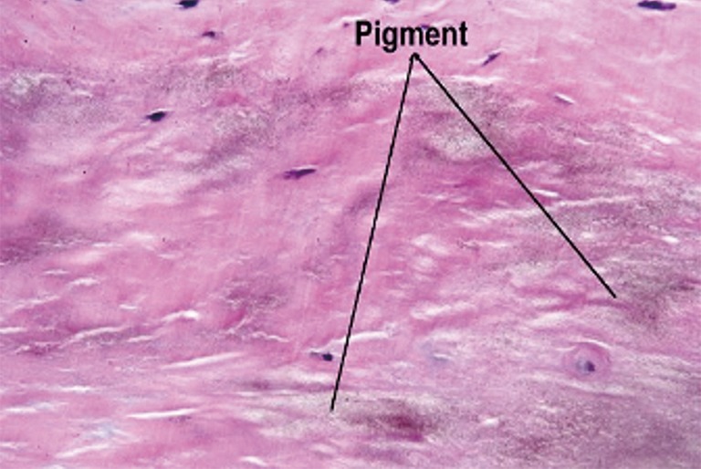
Ochronosis is the bluish-black discolouration of connective tissue, including heart valves, joints, kidney and the skin. It is due to the deposition of homogentisic acid (HGA) commonly found in alkaptonuria. Ochronosis in the aortic valve is a rare occurrence and there is limited data available on the most appropriate choice of valve prosthesis in these patients. This case involves a 72-year-old male with symptomatic aortic stenosis and on echocardiogram a severe calcific trileaflet aortic stenosis with normal ejection fraction. Intraoperative aortic cannulation was routine and uncomplicated, and bluish-black discolouration of aortic valve was noted. Thorough decalcification was undertaken and a bioprosthetic valve was chosen in accordance with patient's age and preference. There were no complications post-operatively and the patient reported being well. Ochronosis affecting the aortic valve is a rare condition and there is limited data on the recurrence rate as well as the natural history of the disease. This case reports aims to provide data to facilitate further research to better understand the natural history of aortic valve ochronosis and rates of recurrence following bioprosthetic aortic valve replacement (AVR).
Keywords: Ochronosis; alkaptonuria; aortic valve replacement (AVR); bioprosthetic valve.
Conflict of interest statement
Conflicts of Interest: The authors have no conflicts of interest to declare.
Figures

References
-
- Gaines JJ, Jr, Pai GM. Cardiovascular ochronosis. Arch Pathol Lab Med 1987;111:991-4. - PubMed
Publication types
LinkOut - more resources
Full Text Sources
Other Literature Sources