

An Unusual Case of Facial Steatocystoma Multiplex: A Clinicopathologic and Dermoscopic Report
- PMID: 29998099
- PMCID: PMC6031952
- DOI: 10.1159/000488584
An Unusual Case of Facial Steatocystoma Multiplex: A Clinicopathologic and Dermoscopic Report
Abstract
Background: Steatocystoma multiplex is a benign skin disorder originating from the sebaceous and nevoid ducts. Commonly classified under hamartomas, they are distributed over the trunk, neck, axillae, and groin.
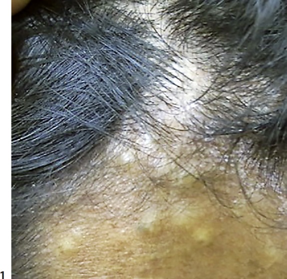
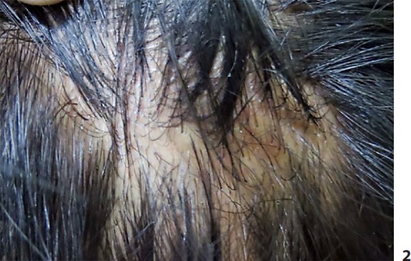
Methods: A 28-year-old male patient complained of multiple, asymptomatic skin-colored nodules over the face of 10-year duration. Clinical examination confirmed the historic findings of nontender, polysized, flesh-colored papules and nodules over the said distribution.
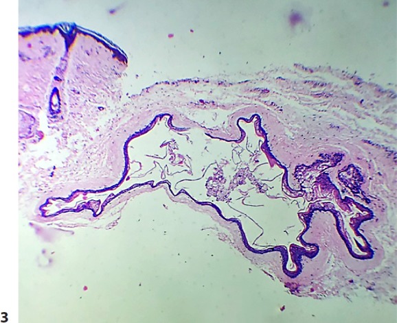
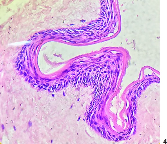
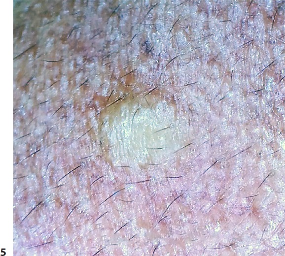
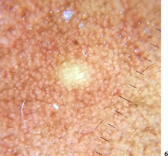
Results: On histopathology, a cyst was noted in the mid-dermis, lined by stratified squamous, agranular epithelium, which contained degenerated keratin. Nonpolarized dermoscopy showed a structureless, cream-colored area, and polarized dermoscopy revealed a distinctive, well-circumscribed, yellowish hue which was superimposed over the facial pseudoreticular pigmentary pattern. The findings were compatible with steatocystoma multiplex, and the patient was taken up for radiofrequency ablation.
Conclusion: Herein, we report a rare variant of steatocystoma multiplex limited to the face and scalp subjected to dermatoscopy and characteristic histological correlation. To the best of our knowledge and following a literature search, dermoscopic features of this condition have not been reported thus far.
Keywords: Dermoscopy; Facial steatocystoma multiplex; Hamartoma; Sebaceous gland.
Figures
References
-
- Alsabbagh MM. Steatocystoma multiplex: A review. J Dermatol Dermatol Surg. 2016;20:91–99.
-
- Cho S, Chang SE, Choi JH, et al. Clinical and histologic features of 64 cases of steatocystoma multiplex. J Dermatol. 2002;29:152–156. - PubMed
-
- Thomas VD, Swanson NA, Lee KK, Benign epithelial tumors, hamartomas, and hyperplasias . Fitzpatrick's Dermatology in General Medicine. In: Wolff K, Goldsmith LA, Katz SI, Gilchrest BA, editors; Paller AS, Leffell DJ, editors. ed 7. New York: McGraw-Hill; 2008. pp. pp 1065–1066.
-
- Plewig G, Wolff HH, Braun-Falco O. Steatocystoma multiplex: anatomic reevaluation, electron microscopy, and autoradiography. Arch Dermatol Res. 1982;272(363) - PubMed
Publication types
LinkOut - more resources
Full Text Sources
Other Literature Sources
