

Function and Regulation of Agrobacterium tumefaciens Cell Surface Structures that Promote Attachment
- PMID: 29998422
- PMCID: PMC6330146
- DOI: 10.1007/82_2018_96
Function and Regulation of Agrobacterium tumefaciens Cell Surface Structures that Promote Attachment
Abstract
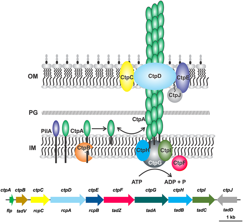
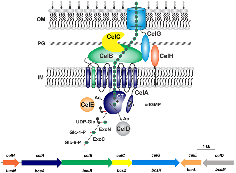
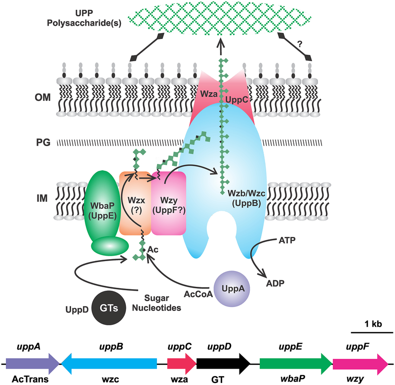
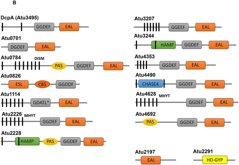
Agrobacterium tumefaciens attaches stably to plant host tissues and abiotic surfaces. During pathogenesis, physical attachment to the site of infection is a prerequisite to infection and horizontal gene transfer to the plant. Virulent and avirulent strains may also attach to plant tissue in more benign plant associations, and as with other soil microbes, to soil surfaces in the terrestrial environment. Although most A. tumefaciens virulence functions are encoded on the tumor-inducing plasmid, genes that direct general surface attachment are chromosomally encoded, and thus this process is not obligatorily tied to virulence, but is a more fundamental capacity. Several different cellular structures are known or suspected to contribute to the attachment process. The flagella influence surface attachment primarily via their propulsive activity, but control of their rotation during the transition to the attached state may be quite complex. A. tumefaciens produces several pili, including the Tad-type Ctp pili, and several plasmid-borne conjugal pili encoded by the Ti and At plasmids, as well as the so-called T-pilus, involved in interkingdom horizontal gene transfer. The Ctp pili promote reversible interactions with surfaces, whereas the conjugal and T-pili drive horizontal gene transfer (HGT) interactions with other cells and tissues. The T-pilus is likely to contribute to physical association with plant tissues during DNA transfer to plants. A. tumefaciens can synthesize a variety of polysaccharides including cellulose, curdlan (β-1,3 glucan), β-1,2 glucan (cyclic and linear), succinoglycan, and a localized polysaccharide(s) that is confined to a single cellular pole and is called the unipolar polysaccharide (UPP). Lipopolysaccharides are also in the outer leaflet of the outer membrane. Cellulose and curdlan production can influence attachment under certain conditions. The UPP is required for stable attachment under a range of conditions and on abiotic and biotic surfaces. Other factors that have been reported to play a role in attachment include the elusive protein called rhicadhesin. The process of surface attachment is under extensive regulatory control and can be modulated by environmental conditions, as well as by direct responses to surface contact. Complex transcriptional and post-transcriptional control circuitry underlies much of the production and deployment of these attachment functions.
Keywords: Attachment; Biofilms; Cell surface structures; Regulation.
Figures


Similar articles
-
The Ctp type IVb pilus locus of Agrobacterium tumefaciens directs formation of the common pili and contributes to reversible surface attachment.J Bacteriol. 2014 Aug 15;196(16):2979-88. doi: 10.1128/JB.01670-14. Epub 2014 Jun 9. J Bacteriol. 2014. PMID: 24914181 Free PMC article.
-
Genetic and environmental factors affecting T-pilin export and T-pilus biogenesis in relation to flagellation of Agrobacterium tumefaciens.J Bacteriol. 2000 Jul;182(13):3705-16. doi: 10.1128/JB.182.13.3705-3716.2000. J Bacteriol. 2000. PMID: 10850985 Free PMC article.
-
Elevated temperature differentially affects virulence, VirB protein accumulation, and T-pilus formation in different Agrobacterium tumefaciens and Agrobacterium vitis strains.J Bacteriol. 2001 Dec;183(23):6852-61. doi: 10.1128/JB.183.23.6852-6861.2001. J Bacteriol. 2001. PMID: 11698374 Free PMC article.
-
Mechanisms and regulation of polar surface attachment in Agrobacterium tumefaciens.Curr Opin Microbiol. 2009 Dec;12(6):708-14. doi: 10.1016/j.mib.2009.09.014. Epub 2009 Oct 29. Curr Opin Microbiol. 2009. PMID: 19879182 Free PMC article. Review.
-
Exopolysaccharides of Agrobacterium tumefaciens.Curr Top Microbiol Immunol. 2018;418:111-141. doi: 10.1007/82_2018_100. Curr Top Microbiol Immunol. 2018. PMID: 29992358 Review.
Cited by
-
Inhibition of growth, biofilm formation, virulence, and surface attachment of Agrobacterium tumefaciens by cinnamaldehyde derivatives.Front Microbiol. 2022 Oct 11;13:1001865. doi: 10.3389/fmicb.2022.1001865. eCollection 2022. Front Microbiol. 2022. PMID: 36304952 Free PMC article.
-
Sticky decisions: The multilayered regulation of adhesin production by bacteria.PLoS Genet. 2023 Mar 2;19(3):e1010648. doi: 10.1371/journal.pgen.1010648. eCollection 2023 Mar. PLoS Genet. 2023. PMID: 36862629 Free PMC article. No abstract available.
-
Role of Caulobacter Cell Surface Structures in Colonization of the Air-Liquid Interface.J Bacteriol. 2019 Aug 22;201(18):e00064-19. doi: 10.1128/JB.00064-19. Print 2019 Sep 15. J Bacteriol. 2019. PMID: 31010900 Free PMC article.
-
Control of Biofilm Formation by an Agrobacterium tumefaciens Pterin-Binding Periplasmic Protein Conserved Among Pathogenic Bacteria.bioRxiv [Preprint]. 2023 Nov 21:2023.11.18.567607. doi: 10.1101/2023.11.18.567607. bioRxiv. 2023. Update in: Proc Natl Acad Sci U S A. 2024 Jun 18;121(25):e2319903121. doi: 10.1073/pnas.2319903121. PMID: 38014264 Free PMC article. Updated. Preprint.
-
Preparation of an Active Dressing by In Situ Biosynthesis of a Bacterial Cellulose-Graphene Oxide Composite.Polymers (Basel). 2022 Jul 14;14(14):2864. doi: 10.3390/polym14142864. Polymers (Basel). 2022. PMID: 35890640 Free PMC article.
References
-
- Aldridge P, & Hughes KT (2002) Regulation of flagellar assembly. Curr Opin Microbiol 5:160–165 - PubMed
-
- Aly KA, Baron C (2007) The VirB5 protein localizes to the T-pilus tips in Agrobacterium tumefaciens. Microbiology 153:3766–3775 - PubMed
-
- Arioli T, Peng L, Betzner AS et al. (1998) Molecular analysis of cellulose biosynthesis in Arabidopsis. Science 279:717–720 - PubMed
Publication types
MeSH terms
Substances
Grants and funding
LinkOut - more resources
Full Text Sources
Other Literature Sources
