Review
. 2018;58(2):61-67.
doi: 10.3960/jslrt.18014.

The histopathology of myeloma in the bone marrow

Review

The histopathology of myeloma in the bone marrow

Masahiko Fujino. J Clin Exp Hematop. 2018.

Abstract

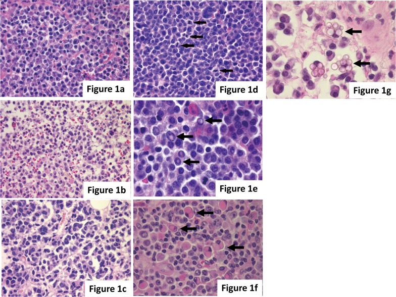
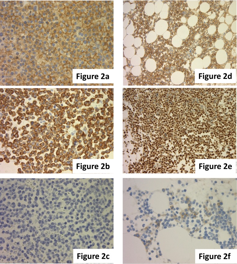
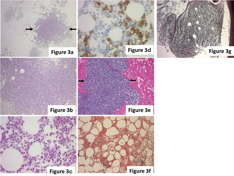
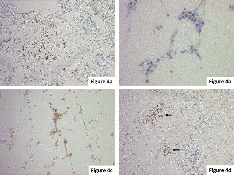
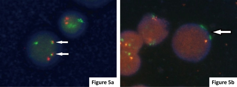
Myeloma is characterized by the neoplastic proliferation of monoclonal plasma cells. A diagnosis of myeloma is based on the criteria proposed by the International Myeloma Working Group and the pathological findings.Myeloma cells are classified into four types: mature, immature, pleomorphic, and plasmablastic. There are three patterns in which myeloma infiltrates bone marrow - nodular, interstitial, and diffuse. Dutcher bodies are highly specific to neoplastic myeloma cells. On immunohistochemical staining, the specificity of CD138 is high for plasma cells. As a clear image is often not obtained from the immunohistochemical staining of the immunoglobulin light chain, in situ hybridization is recommended. Abnormal expression of CD56 is seen in 70-80% of cases by flow cytometry analysis. CD56 expression definitively indicates myeloma, suggesting its high diagnostic value. Evaluation of the infiltration pattern, monoclonality, and abnormal antigen expression of plasma cells is more important than the plasmocytic ratio to determine whether a case is reactive or neoplastic.Multiple gene abnormalities function in the onset and progression of myeloma. In our department, we analyze CCND1, FGFR3, MAF, and del (17p13) by FISH for all myeloma cases. None of the cases with genetic abnormalities were recognized by G-banding. Therefore, FISH is more effective than G-banding for the evaluation of genetic abnormalities in myeloma.

Keywords: Bone marrow; FISH method; Genetic abnormality; Myeloma; Pathological finding.


Conflict of interest statement

CONFLICT OF INTEREST: The author declares no conflict of interest in this manuscript.

Figures

Fig. 1
Fig. 1
(a-d) Cytological features of myeloma cells exhibiting characteristics of the (a) mature type, (b) immature type, (c) pleomorphic type to (d) plasmablastic type. Mitotic figures (arrows) are shown in myeloma cells. (e-g) Arrows indicate (e) Dutcher bodies, (f) Russel bodies, and (g) Mott cells in myeloma cells. (a-g) HE staining.
Fig. 2
Fig. 2
(a-c) Slide showing (a) CD138(+), (b) kappa-ISH (+), and (c) lambda-ISH (-) myeloma cells. (d-f) Slide showing the abnormal expression of (d) CD56, (e) cyclin D1, and (f) CD117 in myeloma cells. (a, d-f) Immunohistochemical staining. (b, c) ISH.
Fig. 3
Fig. 3
(a-d) The infiltration pattern of myeloma. (a) Nodular pattern. Myeloma cells form a nodular lesion (arrows), and the border with the surrounding hematopoietic cells is clear. (b) Diffuse pattern. Myeloma cells exhibit diffused infiltration into the bone marrow, and hematopoietic cells are markedly reduced. (c, d) Interstitial pattern. (c) Myeloma cells are scattered between normal hematopoietic cells with occasional small clusters, but identification of neoplastic cells based on morphology is difficult. (d) CD138(+) myeloma cells are easily identified. (e-g) Secondary changes with myeloma. (e) Interstitial acidophilic change. The stroma exhibits acidophilic changes reflecting hyperproteinemia. Arrows indicate a nodular lesion of myeloma. (f) Amyloid deposition. Amyloid deposition, indicated by the orange stain, is broadly observed in the stroma. (g) Myelofibrosis (grade 2). Reticular fibers are diffusely increased in the nodular lesion of myeloma cells. In this case, the genetic abnormality FGFR3-IGH was identified by FISH. (a-c, e) HE staining. (d) Immunohistochemical staining. (f) Congo red staining. (g) Reticular staining.
Fig. 4
Fig. 4
(a-c) This case was (a) CD138(+), and a few (c) kappa-ISH (+) plasma cells infiltrated into inter-fat marrow spaces; the diagnosis was myeloma. (d) In this case, small clusters of CD138(+) myeloma cells were identified (arrows). The clusters replaced hematopoietic cells and the distribution is unequal in the bone marrow. (a, d) Immunohistochemical staining. (b) HE staining. (c) ISH.
Fig. 5
Fig. 5
FISH analysis of myeloma cells. (a) FGFR3-IGH: The presence of t(4;14) was noted in this case showing 1 red FGFR3 signal, 1 green IGH signal, and 2 yellow fusion signals (arrows). (b) TP53 deletion: The presence of del (17p) was identified in this case showing 1 red TP53 signal and 2 green CEP 17 signals (arrows).

Similar articles

Cited by

References

    1. Rajkumar SV, Dimopoulos MA, Palumbo A, et al. International Myeloma Working Group updated criteria for the diagnosis of multiple myeloma. Lancet Oncol. 2014; 15: e538-e548. 10.1016/S1470-2045(14)70442-5 - DOI - PubMed
    1. Greipp PR, Leong T, Bennett JM, et al. Plasmablastic morphology--an independent prognostic factor with clinical and laboratory correlates: Eastern Cooperative Oncology Group (ECOG) myeloma trial E9486 report by the ECOG Myeloma Laboratory Group. Blood. 1998; 91: 2501-2507. - PubMed
    1. Bartl R, Frisch B, Fateh-Moghadam A, et al. Histologic classification and staging of multiple myeloma. A retrospective and prospective study of 674 cases. Am J Clin Pathol. 1987; 87: 342-355. 10.1093/ajcp/87.3.342 - DOI - PubMed
    1. Seili-Bekafigo I, Valković T, Babarović E, et al. Myeloma cell morphology and morphometry in correlation with clinical stages and survival. Diagn Cytopathol. 2013; 41: 947-954. 10.1002/dc.22986 - DOI - PubMed
    1. Lin P, Owens R, Tricot G, et al. Flow cytometric immunophenotypic analysis of 306 cases of multiple myeloma. Am J Clin Pathol. 2004; 121: 482-488. 10.1309/74R4TB90BUWH27JX - DOI - PubMed

MeSH terms