

Transsynaptic tracing to dissect supraspinal serotonergic input regulating the bladder reflex in rats
- PMID: 29999191
- PMCID: PMC6211582
- DOI: 10.1002/nau.23762
Transsynaptic tracing to dissect supraspinal serotonergic input regulating the bladder reflex in rats
Abstract
Aims: This study was designed to determine specific cell groups of the raphe nuclei (RN) that give rise to supraspinal serotonergic projections regulating the bladder reflex.
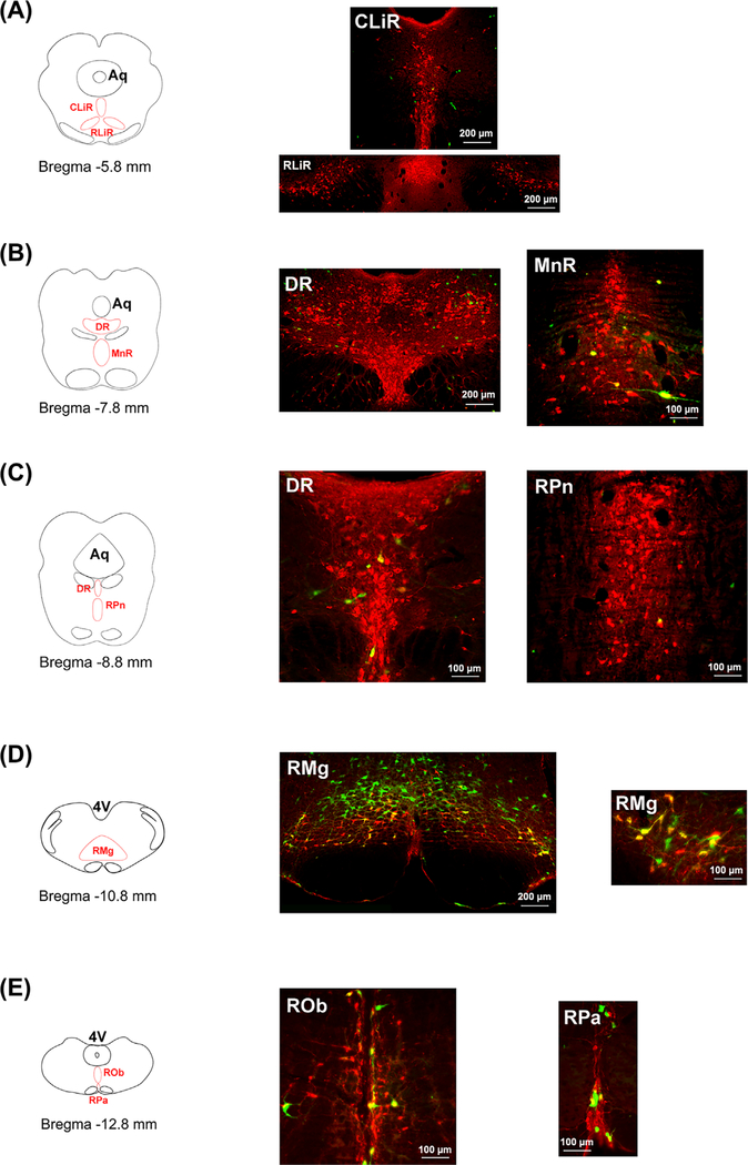
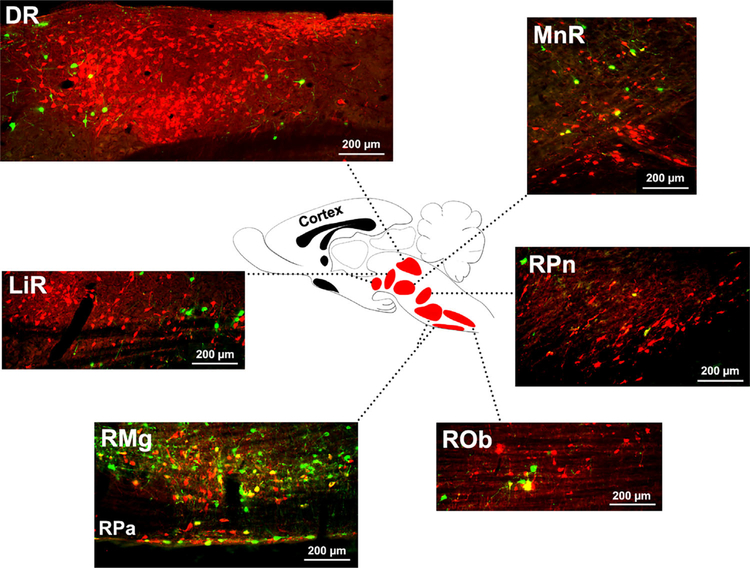
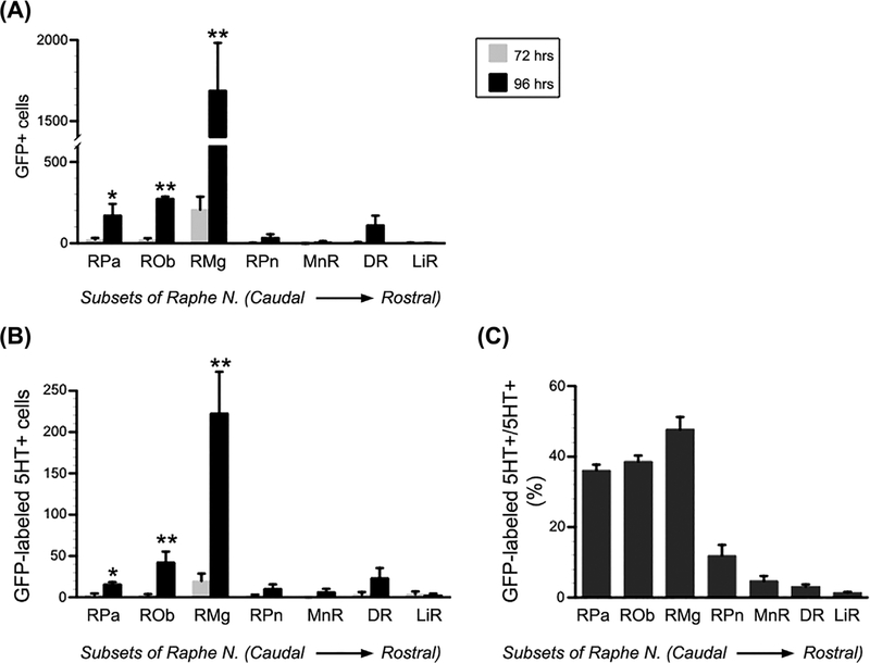
Methods: Anesthetized rats underwent surgery to open the abdomen and expose the bladder. A total of 6 µL transsynaptic neuronal tracer pseudorabies virus (PRV-152), encoding for green fluorescent protein (GFP), was injected into the bladder detrusor. After 72 or 96 h, animals were perfused and the brain was dissected for processing transverse and sagittal sections. Subsequently, fluorescent immunohistochemistry for GFP and Serotonin (5-hydroxytryptamine [5-HT]) was performed in the brain sections. Under the microscope, each RN subset was characterized individually from caudal to rostral according to the atlas. GFP+ or GFP/5-HT double labeled neurons in each subset were quantified for statistical analysis.
Results: At 72-h post-infection, very few GFP+ or GFP/5-HT double-labeled neurons appeared in the brainstem and beyond. In contrast, many labeled neurons were found at these levels after 96 h. Quantitative analysis showed that the majority of infected 5-HT+ neurons were located in the pallidus, obscurus, and magnus nuclei. Conversely, very few infected neurons were found in other raphe subsets, that is the pontis, median, dorsal, or linear nuclei. Overall, the raphe magnus had the highest number of GFP-labeled and GFP/5-HT double-labeled cells.
Conclusions: The caudal subsets of RN, especially the raphe magnus, are the main sources of serotonergic input to the lower spinal cord controlling bladder activity.
Keywords: detrusor; pseudorabies virus; retrograde tracing; serotonin; urinary.
© 2018 Wiley Periodicals, Inc.
Figures
References
-
- Griffiths D, Derbyshire S, Stenger A, Resnick N. Brain control ofnormal and overactive bladder. J Urol. 2005;174:1862–1867. - PubMed
-
- Dahlstrom A, Fuxe K. Localization of monoamines in the lowerbrain stem. Experientia. 1964;20:398–399. - PubMed
-
- Ryall RW, DeGroat WC. The microelectrophoretic administrationof noradrenaline, 5-hydroxytryptamine, acetylcholine and glycine to sacral parasympathetic preganglionic neurones. Brain Res. 1972;37:345–347. - PubMed
-
- Steers WD, de Groat WC. Effects of m-chlorophenylpiperazine onpenile and bladder function in rats. Am J Physiol. 1989;257: R1441–R1449. - PubMed
Publication types
MeSH terms
Substances
Grants and funding
LinkOut - more resources
Full Text Sources
Other Literature Sources
