

Uterine PEComas: A Morphologic, Immunohistochemical, and Molecular Analysis of 32 Tumors
- PMID: 30001237
- PMCID: PMC6133752
- DOI: 10.1097/PAS.0000000000001119
Uterine PEComas: A Morphologic, Immunohistochemical, and Molecular Analysis of 32 Tumors
Abstract
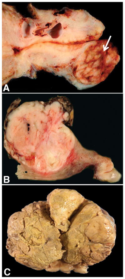
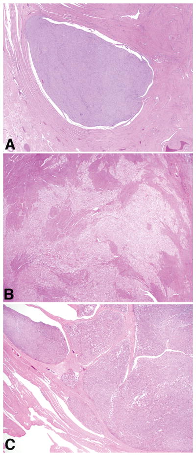
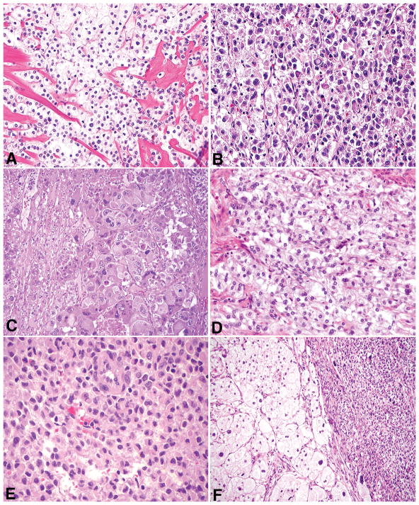
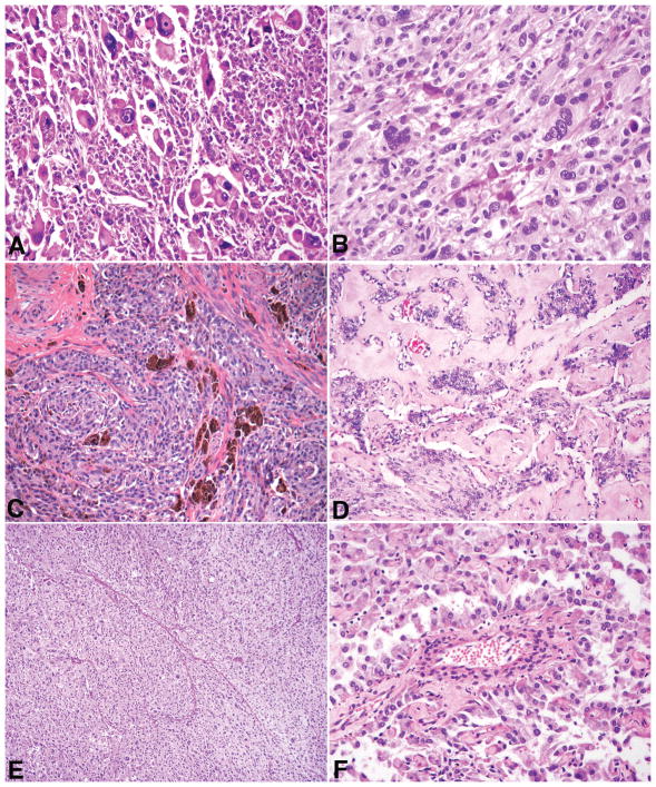
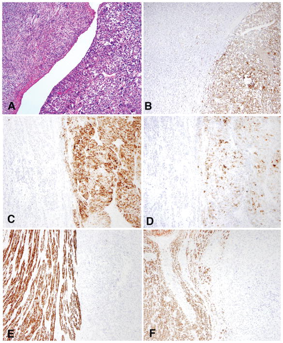
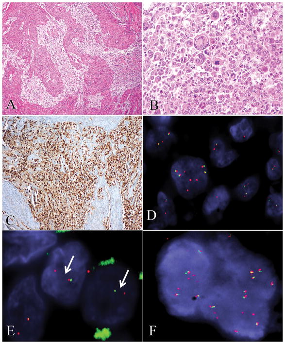
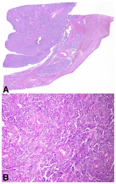
Uterine perivascular epithelioid cell tumors (PEComas) are rare neoplasms that may show overlapping morphology and immunohistochemistry with uterine smooth muscle tumors. In this study, we evaluated the morphologic, immunohistochemical, and molecular features of 32 PEComas, including 11 with aggressive behavior. Two distinct morphologies were observed: classic (n=30) and those with a lymphangioleiomyomatosis appearance (n=2). In the former, patients ranged from 32 to 77 (mean: 51) years and 13% had tuberous sclerosis. Tumors ranged from 0.2 to 17 (mean: 5.5) cm with 77% arising in the corpus. Epithelioid cells were present in 100% and a spindled component was seen in 37%. Nuclear atypia was low (53%), intermediate (17%), or high (30%). Mitoses ranged from 0 to 36 (mean: 6) and 0 to 133 (mean: 19) per 10 and 50 high-power fields, with atypical mitoses present in 30%. Thin and delicate vessels were noted in 100%, clear/eosinophilic and granular cytoplasm in 93%, stromal hyalinization in 73%, necrosis in 30%, and lymphovascular invasion in 10%. All tumors were positive for HMB-45, cathepsin K, and at least one muscle marker, with most expressing melan-A (77%) and/or MiTF (79%). A PSF-TFE3 fusion was identified in one while another showed a RAD51B-OPHN1 fusion. Follow-up ranged from 2 to 175 (mean: 41) months, with 63% of patients alive and well, 20% dead of disease, 13% alive with disease, and 3% dead from other causes. In the latter group (n=2), patients were 39 and 49 years old, one had tuberous sclerosis, while the other had pulmonary lymphangioleiomyomatosis. Both tumors expressed HMB-45, cathepsin K, and muscle markers, but lacked TFE3 and RAD51B rearrangements. The 2 patients are currently alive and well. Application of gynecologic-specific criteria (≥4 features required for malignancy: size ≥5 cm, high-grade atypia, mitoses >1/50 high-power fields, necrosis, and lymphovascular invasion) for predicting outcome misclassified 36% (4/11) of aggressive tumors; thus, a modified algorithm with a threshold of 3 of these features is recommended to classify a PEComa as malignant.
Keywords: perivascular epithelioid cell tumor; PEComa; uterus; tuberous sclerosis; diagnostic criteria; TFE3; RAD51B.
Conflict of interest statement
The authors have disclosed that they have no significant relationships with, or financial interest in, any commercial companies pertaining to this article.
Figures
References
-
- Vang R, Kempson RL. Perivascular epithelioid cell tumor (‘PEComa’) of the uterus: a subset of HMB-45-positive epithelioid mesenchymal neoplasms with an uncertain relationship to pure smooth muscle tumors. Am J Surg Pathol. 2002;26:1–13. - PubMed
-
- Folpe AL, Mentzel T, Lehr HA, et al. Perivascular epithelioid cell neoplasms of soft tissue and gynecologic origin: a clinicopathologic study of 26 cases and review of the literature. Am J Surg Pathol. 2005;29:1558–1575. - PubMed
-
- Schoolmeester JK, Howitt BE, Hirsch MS, et al. Perivascular epithelioid cell neoplasm (PEComa) of the gynecologic tract: clinicopathologic and immunohistochemical characterization of 16 cases. Am J Surg Pathol. 2014;38:176–188. - PubMed
Publication types
MeSH terms
Substances
Grants and funding
LinkOut - more resources
Full Text Sources
Other Literature Sources
Medical
