. 2018 Jul 12;13(7):e0200474.
doi: 10.1371/journal.pone.0200474. eCollection 2018.

Azithromycin therapy reduces cardiac inflammation and mitigates adverse cardiac remodeling after myocardial infarction: Potential therapeutic targets in ischemic heart disease

Affiliations

Azithromycin therapy reduces cardiac inflammation and mitigates adverse cardiac remodeling after myocardial infarction: Potential therapeutic targets in ischemic heart disease

Ahmed Al-Darraji et al. PLoS One. .

Abstract

Introduction: Acute myocardial infarction (MI) is a primary cause of worldwide morbidity and mortality. Macrophages are fundamental components of post-MI inflammation. Pro-inflammatory macrophages can lead to adverse cardiac remodeling and heart failure while anti-inflammatory/reparative macrophages enhance tissue healing. Shifting the balance between pro-inflammatory and reparative macrophages post-MI is a novel therapeutic strategy. Azithromycin (AZM), a commonly used macrolide antibiotic, polarizes macrophages towards the anti-inflammatory phenotype, as shown in animal and human studies. We hypothesized that AZM modulates post-MI inflammation and improves cardiac recovery.

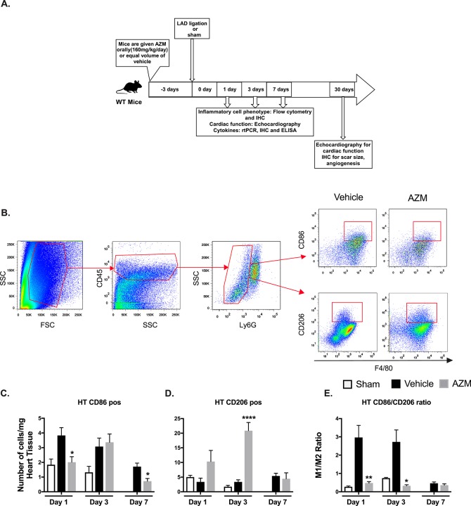
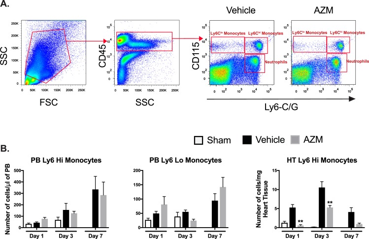
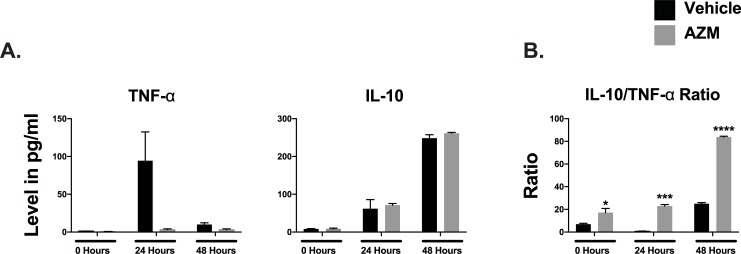
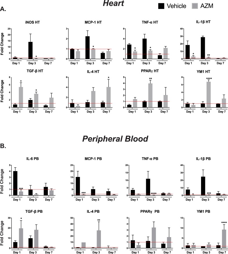
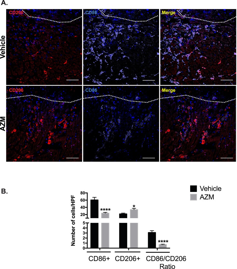
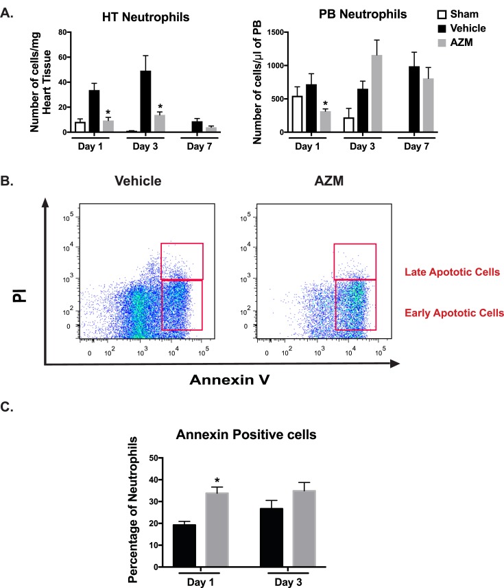
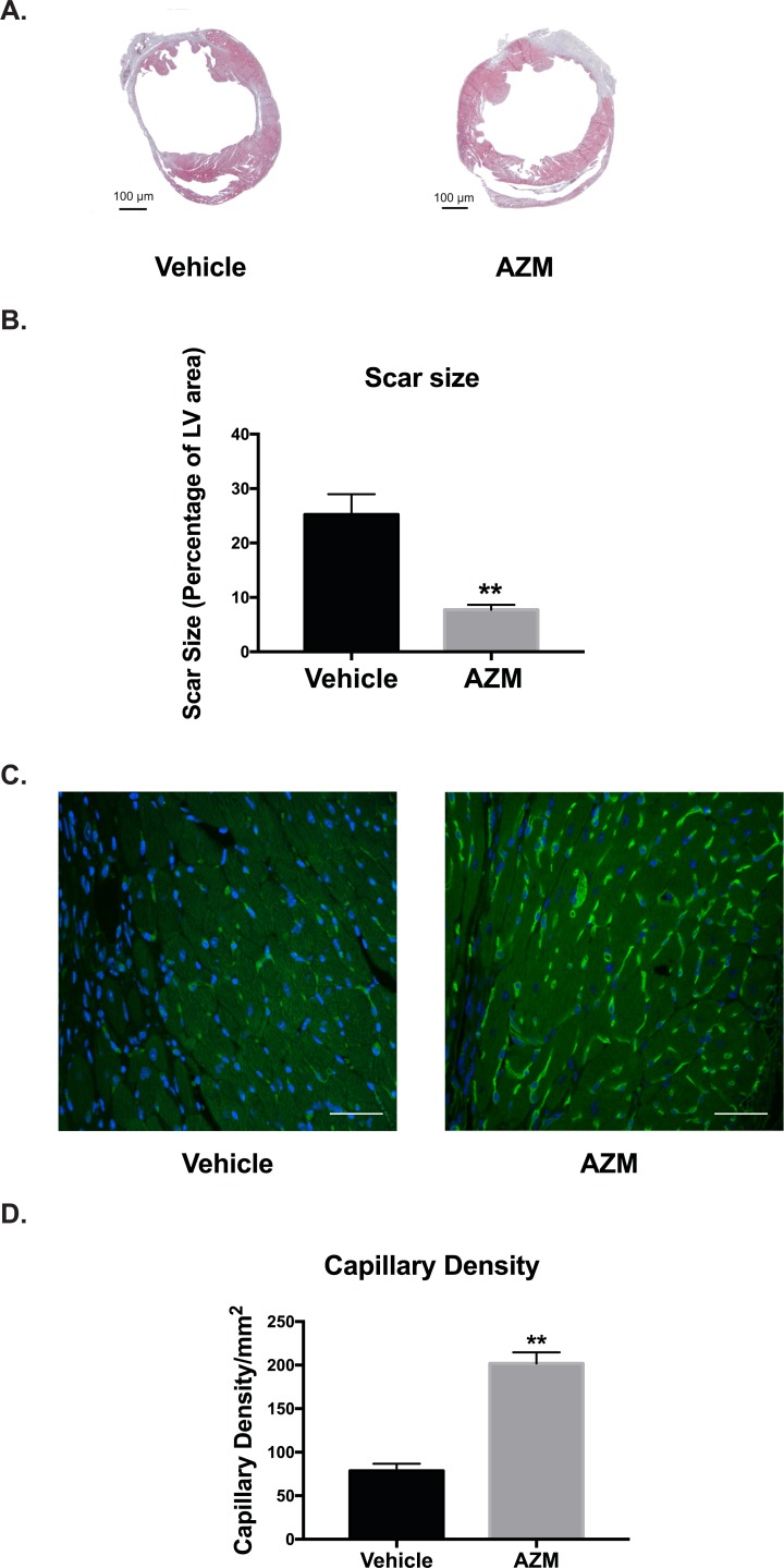
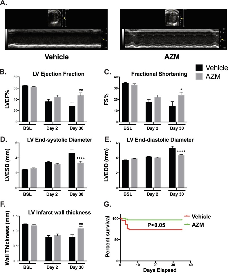
Methods and results: Male WT mice (C57BL/6, 6-8 weeks old) were treated with either oral AZM (160 mg/kg/day) or vehicle (control) starting 3 days prior to MI and continued to day 7 post-MI. We observed a significant reduction in mortality with AZM therapy. AZM-treated mice showed a significant decrease in pro-inflammatory (CD45+/Ly6G-/F4-80+/CD86+) and increase in anti-inflammatory (CD45+/Ly6G-/F4-80+/CD206+) macrophages, decreasing the pro-inflammatory/anti-inflammatory macrophage ratio in the heart and peripheral blood as assessed by flow cytometry and immunohistochemistry. Macrophage changes were associated with a significant decline in pro- and increase in anti-inflammatory cytokines. Mechanistic studies confirmed the ability of AZM to shift macrophage response towards an anti-inflammatory state under hypoxia/reperfusion stress. Additionally, AZM treatment was associated with a distinct decrease in neutrophil count due to apoptosis, a known signal for shifting macrophages towards the anti-inflammatory phenotype. Finally, AZM treatment improved cardiac recovery, scar size, and angiogenesis.

Conclusion: Azithromycin plays a cardioprotective role in the early phase post-MI through attenuating inflammation and enhancing cardiac recovery. Post-MI treatment and human translational studies are warranted to examine the therapeutic applications of AZM.


Conflict of interest statement

The authors have declared that no competing interests exist.

Figures

Fig 1
Fig 1. AZM therapy shifts macrophages towards the anti-inflammatory phenotype in the heart after myocardial infarction.
Experimental design for the in vivo study (Panel A). Representative FACS plots demonstrating the gating strategy for pro-inflammatory (CD45+/Ly6G+/F4-80+/CD86+) and anti-inflammatory (CD45+/Ly6G+/F4-80+/CD206+) macrophages (Panel B). Quantitative analyses of pro-inflammatory subpopulations (CD86+) and anti-inflammatory subpopulations (CD206+) are presented in Panels C and D, respectively, at different time points following MI in AZM and vehicle treated groups. There is a significant reduction in the inflammatory macrophages in the first day after MI in AZM-treated mice, which is associated with a significant increase in anti-inflammatory ones. These changes translate into a significant reduction in the pro-/anti-inflammatory (CD86+/ CD206+) ratio at days 1 and 3 after MI (Panel E) (n = 4 MI and 3 sham mice/group/time point, *P<0.05, **P<0.01 and ****P<0.001 compared to vehicle controls). Data presented as mean ± SEM. AZM, azithromycin; IHC, immunohistochemistry; LAD, left anterior descending coronary artery; rtPCR, real-time Polymerase Chain Reaction.
Fig 2
Fig 2. AZM therapy reduces cardiac inflammatory monocytes after MI.
Representative FACS plots demonstrating the gating strategy for Ly6Chi (CD45+/CD115hi/Ly6-Chi) and Ly6Clo (CD45+/CD115hi/Ly6-Clo) monocytes (Panel A). Quantitative analyses of monocyte subpopulations in PB and heart tissue demonstrating no significant changes in PB monocyte subpopulations but significant reduction in cardiac Ly6Chi population throughout the different time points after MI in AZM treated mice (Panel B) (n = 4 MI and 3 sham mice/group/time point, *P<0.05 and **P<0.01 compared to vehicle controls). Data presented as mean ± SEM. AZM, azithromycin; PB, peripheral blood.
Fig 3
Fig 3. AZM therapy reduces the production of inflammatory cytokines in macrophages subjected to ischemia/reperfusion injury.
Quantitative analyses of pro-inflammatory cytokine, TNF-α, and the anti-inflammatory cytokine, IL-10, production from J774 macrophages subjected to 24 hypoxia followed by 24 and 48 hours of reperfusion. The analysis demonstrates reduction of TNF- α compared to vehicle both at 24 and 48 hours. No significant changes were noted in IL-10 production. Overall, the IL-10/TNF-α ratio was significantly higher in macrophages treated with AZM (Panel B) (two independent experiments and 4 replicates/time point, *P<0.05, ***P<0.001 and **** P<0.0001 compared to vehicle controls). Data presented as mean ± SEM. AZM, azithromycin; IL-10, interleukin 10; TNF-α, tumor necrosis factor-alpha.
Fig 4
Fig 4. AZM treatment exerts immunomodulatory effects on cytokines expression following MI.
mRNA expression of pro-inflammatory cytokines in HT (Panel A) and PB (Panel B), demonstrate significant reduction in gene expression of these cytokines in the early inflammatory phase following injury with AZM therapy compared to vehicle controls (red line demarcates the level of gene expression in sham operated mice). The mRNA expression of anti-inflammatory cytokines is augmented with AZM therapy compared to vehicle control (n = 4 mice/group/time point, *P<0.05, **P<0.01, ***P<0.001 and **** P<0.0001 compared to vehicle controls). Data presented as mean ± SEM. AZM, azithromycin; HT, heart; IL-1β, interleukin 1 beta; IL-6, interleukin 6; IL-4, interleukin 4; iNOS, inducible nitric oxide synthase; MCP-1, monocyte chemoattractant protein-1; PB, peripheral blood; PPARγ, peroxisome proliferator-activated receptor gamma; TGF-1β, tissue growth factor 1 beta; TNF-α, tumor necrosis factor-alpha; YM1 (Chil3), chitinase-like 3.
Fig 5
Fig 5. AZM treatment enhances alternative macrophage activation in the peri-infarct border of the injured heart.
Immunohistochemical assessment of the content of pro-inflammatory (CD86+) and reparative macrophages (CD206+) markers 3 days post-MI. Panel A shows representative images from vehicle- and AZM-treated mice demonstrating higher density of CD86+ compared to CD206+ cells in the peri-infarct border in vehicle-treated mice. White line demarcates the infarct border. Panel B shows quantitative assessment of CD86+, CD206+ and the markers ratio 3 days post-MI. The difference in pro-inflammatory and anti-inflammatory macrophages lead to a significant shift towards an anti-inflammatory state and the reduction in their ratio in AZM-treated mice (n = 4 animals/group, *P<0.05 and ****P<0.0001 compared to vehicle controls). Scale bars represent 50 μm. Data presented as mean ± SEM. AZM, azithromycin; HPF, high power field.
Fig 6
Fig 6. Cardiac neutrophils are reduced with AZM treatment due to apoptosis.
Panel A summarizes the quantitative assessment of neutrophil (CD45 +/CD115lo/Ly6G-Clo) numbers and demonstrates significant reduction in the AZM-treated group relative to controls in the heart and peripheral blood during the early inflammatory stage post-MI. Panel B illustrates the flow cytometry analyses of neutrophil populations stained against annexin V and PI showing higher percentage of early apoptotic neutrophils in AZM-treated mice. Panel C summarizes the quantitative assessment of early apoptotic neutrophils and demonstrates significantly higher percentage in the heart in the AZM-treated group during the early stage following MI (n = 4 mice/group/time point, *P<0.05 compared to vehicle controls). Data presented as mean ± SEM. AZM, azithromycin; HT, heart; PB, peripheral blood; PI, propodium Iodide.
Fig 7
Fig 7. AZM reduces apoptosis post-infarction.
Panel A shows representative light microscope images of cleaved caspase-3 staining for infarcted and border regions in AZM- and vehicle-treated mice 3 days after MI. Quantitative analyses (Panel B) of apoptosis reveal a remarkable reduction in caspase-3 activation in the infarct and border regions of AZM-treated group compared to the control group (n = 4 animals/group, **P<0.01 compared to vehicle controls). Scale bars represent 100 μm. Data presented as mean ± SEM. AZM, azithromycin; HPF, high power field.
Fig 8
Fig 8. AZM treatment reduces scar size and enhances angiogenesis after myocardial injury.
Representative Masson's trichrome staining at 30 days after myocardial injury in vehicle- and AZM-treated groups (Panel A). Quantitative analysis of scars as percentage of LV area shows significant reduction in AZM group relative to the control group (Panel B) (vehicle-treated, n = 15 vs. AZM-treated, n = 12, **P<0.01 compared to vehicle controls). Representative isolectin staining (Green) for capillary density in the peri-infarct region in AZM- and vehicle-treated animals demonstrates higher capillary density in AZM group compared to control (Panel C). Quantitative analysis of capillary density confirms the higher angiogenesis rate and capillary density in AZM-treated group (Panel D) (n = 4 animals/group, **P<0.01 compared to vehicle controls). Scale bars represent 50 μm. Data presented as mean ± SEM. AZM, azithromycin; MI, acute myocardial infarction; LV, left ventricular.
Fig 9
Fig 9. AZM treatment improves chronic cardiac remodeling and survival post-MI.
30 day following MI, transthoracic echocardiography using M-Mode (Panel A) and 2D echocardiography was performed on AZM- and vehicle-treated animals to evaluate left ventricular function and remodeling parameters. Quantitative analyses demonstrate significant recovery in LV function as assessed by ejection fraction (LVEF) (Panel B) and fractional shortening (FS) (Panel C). Data also shows significant improvements in LV adverse remodeling parameters such as end-systolic diameter (LVESD) (Panel D) and end-diastolic diameter (LVEDD) (Panel E). Additionally, we observed significantly thicker infarct walls suggesting of enhanced recovery and regeneration (Panel F). Survival curves of AZM and vehicle-treated mice 30 days post-MI demonstrate a significant improvement in survival with AZM treatment (Panel G). (Echo data: vehicle-treated, n = 15 vs. AZM-treated, n = 12; survival data: vehicle-treated, n = 48 and AZM-treated, n = 53; *P<0.05, **P<0.01 and ****P<0.001 compared to vehicle control). Data presented as mean ± SEM. AZM, azithromycin; MI, acute myocardial infarction.

References

    1. van der Laan AM, Nahrendorf M, Piek JJ. Healing and adverse remodelling after acute myocardial infarction: role of the cellular immune response. Heart. 2012;98(18):1384–90. 10.1136/heartjnl-2012-301623 . - DOI - PubMed
    1. Epelman S, Liu PP, Mann DL. Role of innate and adaptive immune mechanisms in cardiac injury and repair. Nat Rev Immunol. 2015;15(2):117–29. 10.1038/nri3800 ; PubMed Central PMCID: PMCPMC4669103. - DOI - PMC - PubMed
    1. Dutta P, Nahrendorf M. Monocytes in myocardial infarction. Arterioscler Thromb Vasc Biol. 2015;35(5):1066–70. 10.1161/ATVBAHA.114.304652 ; PubMed Central PMCID: PMCPMC4409536. - DOI - PMC - PubMed
    1. de Couto G, Liu W, Tseliou E, Sun B, Makkar N, Kanazawa H, et al. Macrophages mediate cardioprotective cellular postconditioning in acute myocardial infarction. J Clin Invest. 2015;125(8):3147–62. 10.1172/JCI81321 ; PubMed Central PMCID: PMCPMC4563759. - DOI - PMC - PubMed
    1. Protti A, Mongue-Din H, Mylonas KJ, Sirker A, Sag CM, Swim MM, et al. Bone marrow transplantation modulates tissue macrophage phenotype and enhances cardiac recovery after subsequent acute myocardial infarction. J Mol Cell Cardiol. 2016;90:120–8. 10.1016/j.yjmcc.2015.12.007 ; PubMed Central PMCID: PMCPMC4727788. - DOI - PMC - PubMed

Publication types

MeSH terms