. 2018 Jul 12;25(1):56.
doi: 10.1186/s12929-018-0455-z.

IL-33 receptor (ST2) deficiency downregulates myeloid precursors, inflammatory NK and dendritic cells in early phase of sepsis

Affiliations

IL-33 receptor (ST2) deficiency downregulates myeloid precursors, inflammatory NK and dendritic cells in early phase of sepsis

Zivan M Babic et al. J Biomed Sci. .

Abstract

Background: Sepsis is a life-threatening disease mediated by profound disturbances in systemic inflammatory response to infection. IL-33 is multifunctional regulator of numerous aspects of innate and adaptive immune response. The aim of this article was to further evaluate the role of IL-33 receptor (ST2) in different pathways of innate immunity during early polymicrobial sepsis.

Methods: Polymicrobial sepsis was induced using cecal ligation and puncture (CLP) model in ST2 deficient (ST2-/-) and wild type BALB/c mice. Peritoneal and spleen cells were isolated for further phenotyping. Apoptosis was determined by immunohistochemistry and flow cytometry.

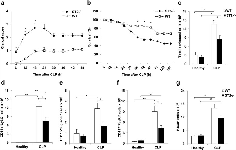
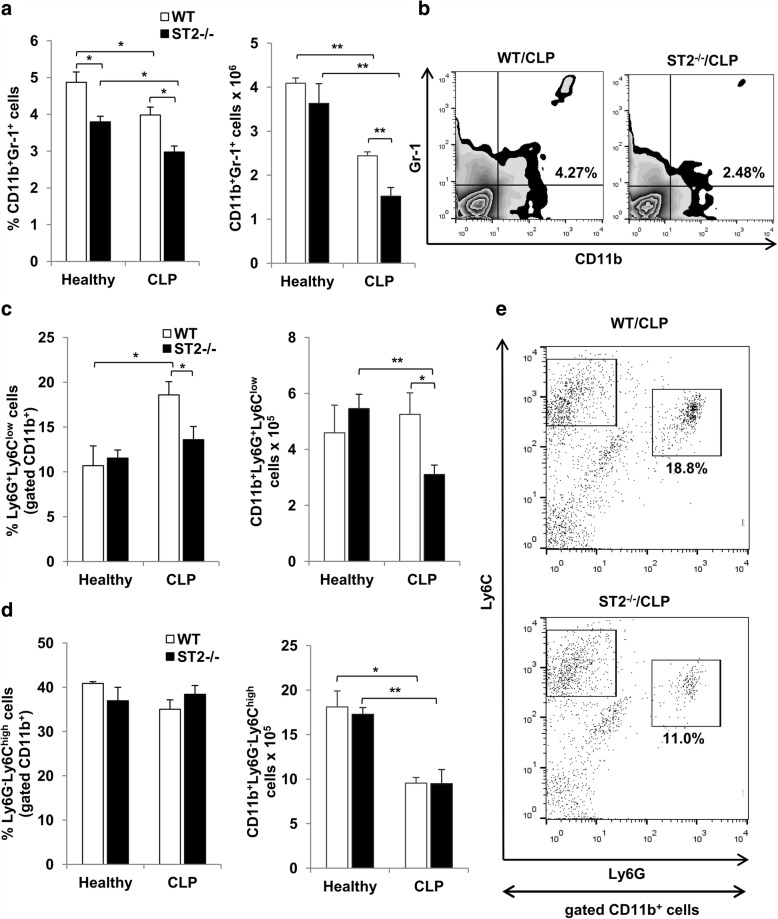
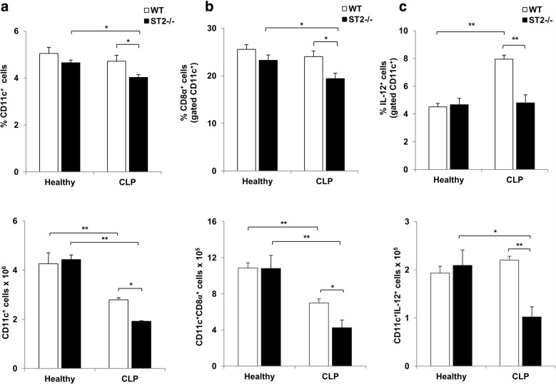
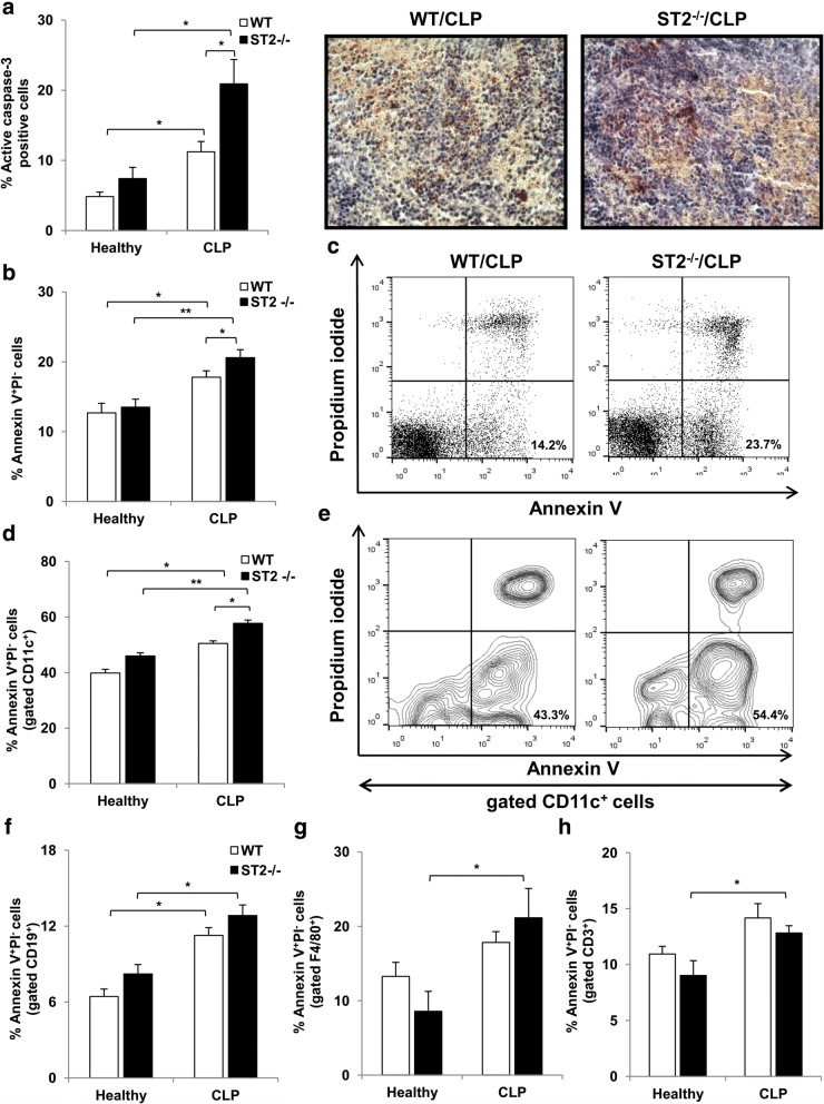
Results: Deletion of ST2 leads to increased susceptibility to early manifestations of sepsis as evaluated by clinical signs and survival. These are accompanied by decrease in the total number of neutrophils, eosinophils and mast cells in peritoneal cavity 12 h after CLP. In early sepsis there was also low number of precursors of myeloid cells in particular CD11b+Ly6G+Ly6Clow cells in spleen of ST2-/- mice. Although the number of NK cells in the spleen was similar, there were significant differences in the presence of inflammatory IFN-γ and IL-17 producing NK cells. Further, ST2 deletion affects the phenotype and maturation of dendritic cell in sepsis. The total number of dendritic cells in the spleen was lower as well as IL-12 expressing dendritic cells. Finally, there was higher frequency of active caspase-3 positive and early apoptotic cells, in particular CD11c positive cells, in spleen of septic ST2-/- mice.

Conclusion: Taken together, our data provide the evidence that ST2 deficiency in early phase of sepsis downregulates myeloid precursors, inflammatory NK and dendritic cells.

Keywords: Dendritic cells; IL-33; Myeloid precursors; NK cells; ST2; Sepsis.


Conflict of interest statement

Ethics approval and consent to participate

All animal procedures were approved by the Ethical Committee of the Faculty of Medical Sciences, University of Kragujevac, Serbia (No 01–10873/3).

Consent for publication

Not applicable.

Competing interests

The authors declare that they have no competing interests.

Publisher’s Note

Springer Nature remains neutral with regard to jurisdictional claims in published maps and institutional affiliations.

Figures

Fig. 1
Fig. 1
Deletion of ST2 accelerates CLP induced polymicrobial sepsis and affects granulocytes influx into peritoneal cavity. Clinical score and survival rate of septic ST2−/− and WT mice was monitored every 6 h. a ST2−/− mice exhibit significantly increased clinical score during first 18 h following CLP. b Survival rate of septic ST2−/− and WT mice was analyzed by Fisher’s exact test for each time point. ST2−/− mice had significantly decreased survival rate at 36, 42 and 48 h following CLP (n = 22 mice per group). c Significantly decreased number of peritoneal cells in septic ST2−/− in comparison with WT mice 12 h following CLP. The number of peritoneal neutrophils (CD11b+Ly6G+) (d), eosinophils (CD11b+Siglec-F+) (e), mast cells (CD117+FcεRI+) (f) and macrophages (g) 12 h following CLP. Data are presented as mean ± SE, n = 4–7 mice per group. *p < 0.05, **p < 0.01
Fig. 2
Fig. 2
ST2 deficiency is associated with decreased number of myeloid precursors during early sepsis. a The frequencies and total number of splenic CD11b+Gr-1+ cells were analyzed by flow cytometry 12 h following CLP. b Representative plots show CD11b+Gr-1+ cells in septic ST2−/− and WT mice. c The percentage and total number of splenic CD11b+Ly6G+Ly6Clow granulocytic myeloid precursors. d The percentage and total number of splenic CD11b+Ly6GLy6Chigh monocytic myeloid precursors. e Representative plots denote the frequencies of granulocytic and monocytic myeloid precursors in spleen of septic ST2−/− and WT mice. Data are presented as mean ± SE, n = 4–7 mice per group. *p < 0.05, **p < 0.01
Fig. 3
Fig. 3
Decreased number of inflammatory NK cells in septic ST2−/− mice. a The frequencies and total number of splenic NK (CD49b+CD3) cells were analyzed by flow cytometry 12 h following CLP. The percentage and total number of splenic IFN-γ (b) and IL-17 (c) positive NK (CD49b+CD3) cells. d The representative plots denote decreased expression of IFN-γ and IL-17 among the population of NK (CD49b+CD3) cells derived from spleen of septic ST2−/− in comparison with WT mice. Data are presented as mean ± SE, n = 4–5 mice per group. *p < 0.05
Fig. 4
Fig. 4
ST2 deficiency is associated with decreased number of inflammatory dendritic cells during early sepsis. a The percentage and total number of splenic CD11c+ dendritic cells was determined by flow cytometry 12 h following CLP. b The percentage and total number of CD8α+ splenic CD11c+ dendritic cells. c The percentage and total number of IL-12 expressing CD11c+ splenic dendritic cells. Data are presented as mean ± SE, n = 4–7 mice per group. *p < 0.05, **p < 0.01
Fig. 5
Fig. 5
ST2 deficiency enhances early apoptosis of CD11c+ cells in sepsis. a The percentage of active caspase-3 positive spleen cells is assessed by immunohistochemistry. The results are presented as a mean percentage of positive stained nuclei per field (5 random fields, 4–5 tissues per group). Representative images show active caspase-3 positive nuclei in spleen of septic ST2−/− and WT mice (magnification at × 400). b The percentage of early apoptotic Annexin V+PI spleen cells is assessed 12 h after CLP by anti-mouse Annexin V antibody and PI staining. c Representative plots denote the percentage of early apoptotic Annexin V+PI cells of total spleen cells in septic ST2−/− and WT mice. d The percentage of early apoptotic Annexin V+PI spleen cells among CD11c+ cells. e Representative plots denote the percentage of early apoptotic Annexin V+PI cells of CD11c+ spleen cells in septic ST2−/− and WT mice. The percentage of early apoptotic Annexin V+PI spleen cells among B cells (CD19+) (f), macrophages (F4/80+) (g) and T cells (CD3+) (h) was shown. Data are presented as mean ± SE, n = 4–7 mice per group. *p < 0.05, **p < 0.01

References

    1. Vincent JL, Opal SM, Marshall JC, Tracey KJ. Sepsis definitions: time for change. Lancet. 2013;381(9868):774–775. doi: 10.1016/S0140-6736(12)61815-7. - DOI - PMC - PubMed
    1. Munford RS, Pugin J. Normal responses to injury prevent systemic inflammation and can be Immunosuppressive. Am J Respir Crit Care Med. 2001;163(2):316–321. doi: 10.1164/ajrccm.163.2.2007102. - DOI - PubMed
    1. Xiao W, Mindrinos MN, Seok J, Cuschieri J, Cuenca AG, Gao H, Hayden DL, Hennessy L, Moore EE, Minei JP, et al. A genomic storm in critically injured humans. J Exp Med. 2011;208(13):2581–2590. doi: 10.1084/jem.20111354. - DOI - PMC - PubMed
    1. Stearns-Kurosawa DJ, Osuchowski MF, Valentine C, Kurosawa S, Remick DG. The pathogenesis of sepsis. Annu Rev Pathol. 2011;6:19–48. doi: 10.1146/annurev-pathol-011110-130327. - DOI - PMC - PubMed
    1. Hotchkiss RS, Monneret G, Payen D. Sepsis-induced immunosuppression: from cellular dysfunctions to immunotherapy. Nat Rev Immunol. 2013;13(12):862–874. doi: 10.1038/nri3552. - DOI - PMC - PubMed

MeSH terms