. 2018 Oct;17(10):1979-1990.
doi: 10.1074/mcp.RA118.000689. Epub 2018 Jul 12.

Proteomic Analysis of NCK1/2 Adaptors Uncovers Paralog-specific Interactions That Reveal a New Role for NCK2 in Cell Abscission During Cytokinesis

Affiliations

Proteomic Analysis of NCK1/2 Adaptors Uncovers Paralog-specific Interactions That Reveal a New Role for NCK2 in Cell Abscission During Cytokinesis

Kévin Jacquet et al. Mol Cell Proteomics. 2018 Oct.

Abstract

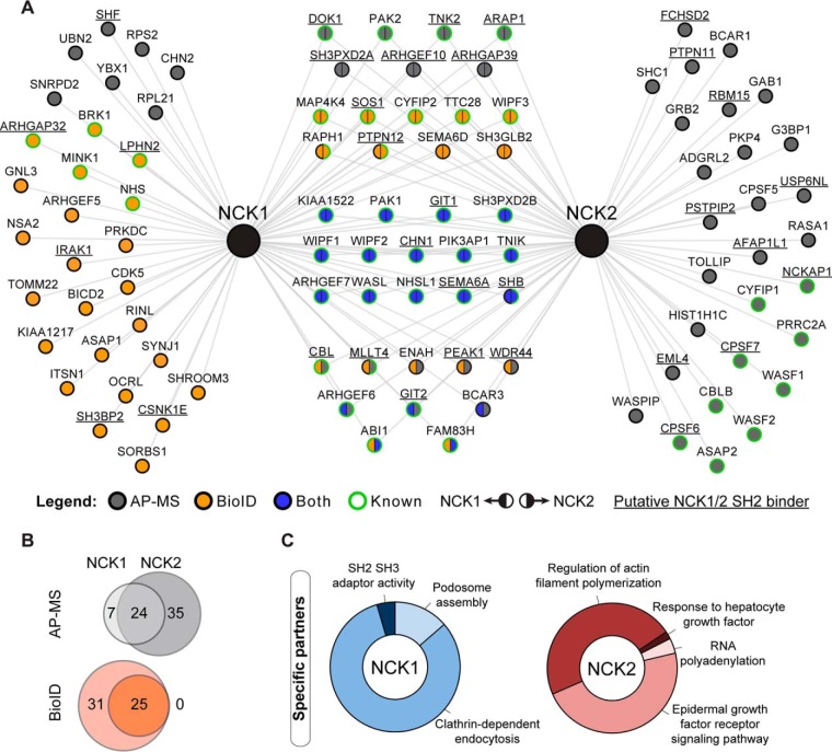
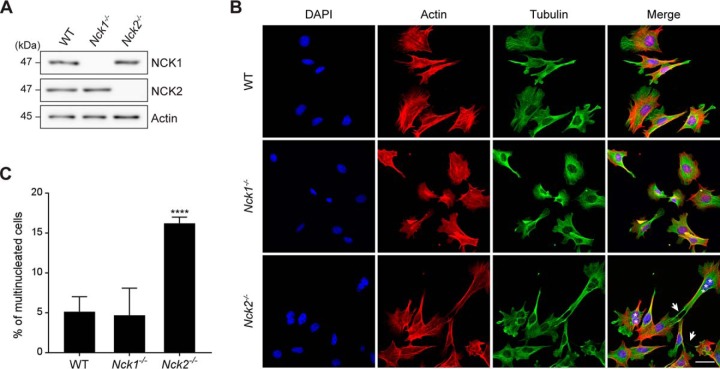
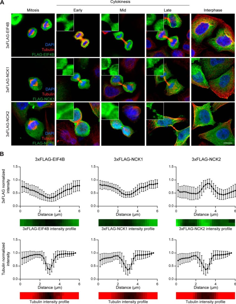
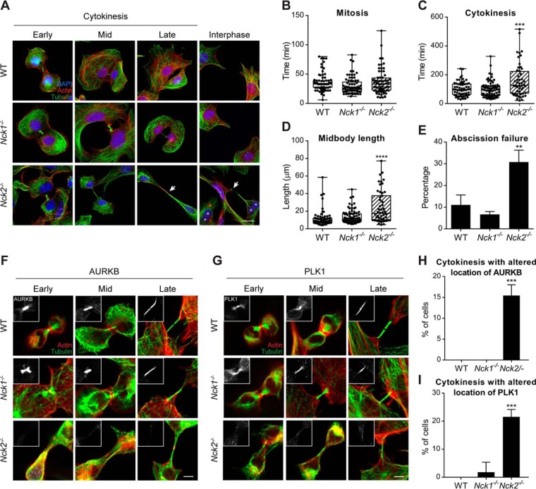
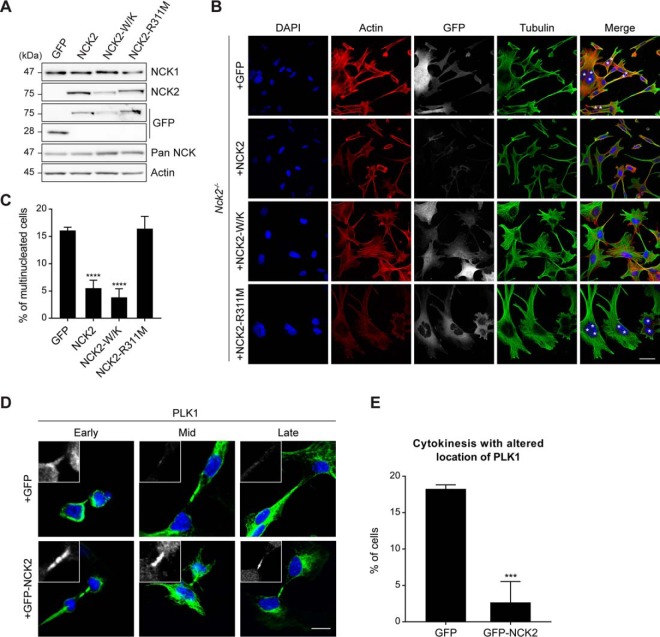
Signals from cell surface receptors are often relayed via adaptor proteins. NCK1 and NCK2 are Src-Homology (SH) 2 and 3 domain adaptors that regulate processes requiring a remodeling of the actin cytoskeleton. Evidence from gene inactivation in mouse suggests that NCK1 and NCK2 are functionally redundant, although recent reports support the idea of unique functions for NCK1 and NCK2. We sought to examine this question further by delineating NCK1- and NCK2-specific signaling networks. We used both affinity purification-mass spectrometry and BioID proximity labeling to identify NCK1/2 signaling networks comprised of 98 proteins. Strikingly, we found 30 proteins restricted to NCK1 and 28 proteins specifically associated with NCK2, suggesting differences in their function. We report that Nck2-/-, but not Nck1-/- mouse embryo fibroblasts (MEFs) are multinucleated and display extended protrusions reminiscent of intercellular bridges, which correlate with an extended time spent in cytokinesis as well as a failure of a significant proportion of cells to complete abscission. Our data also show that the midbody of NCK2-deficient cells is not only increased in length, but also altered in composition, as judged by the mislocalization of AURKB, PLK1 and ECT2. Finally, we show that NCK2 function during cytokinesis requires its SH2 domain. Taken together, our data delineate the first high-confidence interactome for NCK1/2 adaptors and highlight several proteins specifically associated with either protein. Thus, contrary to what is generally accepted, we demonstrate that NCK1 and NCK2 are not completely redundant, and shed light on a previously uncharacterized function for the NCK2 adaptor protein in cell division.

Keywords: Affinity proteomics; Cell biology; Cell division; Imaging; Mass Spectrometry; Networks; Phosphorylation; Protein-Protein Interactions; SH2/SH3 Domains; Signal Transduction.


Figures

Fig. 1.
Fig. 1.
NCK adaptor proteins AP-MS and BioID interactome reveals targets specifically associated with NCK1 or NCK2. A, NCK1/2-associated proteins were identified using AP-MS and BioID and combined to map the NCK1/2 interaction landscape. Previously reported interactions (known) from the BioGRID database were manually annotated. Putative NCK1/2 SH2 binders (underlined) were identified using SH2PepInt. B, Venn diagram representation of the overlap between NCK1- and NCK2-associated proteins found via AP-MS or BioID. C, Gene ontology (GO) and functional pathway analysis via ClueGO of NCK1- and NCK2-specific interactors.
Fig. 2.
Fig. 2.
Nck2−/− cells are multi-nucleated and display long protrusions. A, Mouse embryonic fibroblasts (MEFs) bearing Nck1 or Nck2 gene inactivation were analyzed by Western blotting to confirm NCK1/2 protein expression. B, Wild-type, Nck1−/− and Nck2−/− MEFs were analyzed by immunofluorescence for actin (red), tubulin (green) and DAPI (blue) to assess cellular morphology. Multi-nucleation (asterisks) and intercellular bridges (arrows) are indicated. Representative images are presented (scale bar = 40 μm). C, The penetrance of the multi-nucleation phenotype was calculated for each genotype. Mean values and standard deviation from three independent experiments with >100 cells each are presented (WT, n = 374; Nck1−/−, n = 325; Nck2−/−, n = 372) (****p ≤ 0.0001, Fisher's exact test).
Fig. 3.
Fig. 3.
NCK2 but not NCK1 localizes to the midbody during cytokinesis. A, HeLa cells were transiently transfected with 3xFLAG-EIF4B (control), 3xFLAG-NCK1 or 3xFLAG-NCK2 and synchronized. Cells were fixed at the cytokinesis stage and immunostained for tubulin (red), 3xFLAG (green) and DAPI (blue) to analyze 3xFLAG-tagged chimeras localization during cell division, as indicated. Representative images are shown (scale bar: 10 μm). B, Quantification of pixel intensity as a function of position within the midbody of images processed as in (A). Distance was measured from a 6 μm line selection over the midbody. Intensity of the 3xFLAG (top panels, green) or tubulin (bottom panels, red) staining is reported following normalization over the most intense pixel. Mean values and standard deviation from three independent experiments with >10 cells each are shown (3xFLAG-EIF4B cells: FLAG staining n = 34, tubulin n = 38; 3xFLAG-NCK1 cells: FLAG n = 33, tubulin n = 40; 3xFLAG-NCK2 cells: FLAG n = 30, tubulin n = 42).
Fig. 4.
Fig. 4.
NCK2 is required to complete cytokinesis and abscission. A, Wild-type, Nck1−/− and Nck2−/− MEFs were fixed at different stages of cytokinesis and analyzed by immunofluorescence for actin (red), tubulin (green) and DAPI (blue) to evaluate midbody morphology. Multi-nucleation is indicated with asterisks and extended midbodies with arrows. Representative images are presented (scale bar: 20 μm). B–E, Wild-type, Nck1−/− and Nck2−/− MEFs were stained with SIR-ACTIN and analyzed by live imaging (supplemental Fig. S2). Average time spent in mitosis (B), in cytokinesis (C), average midbody length (D) and abscission failure (E) were calculated for each genotype, from three independent experiments with >20 cells each (WT, n = 70; Nck1−/−, n = 76; Nck2−/−, n = 63) (**p ≤ 0.01; *** p ≤ 0.001; **** p ≤ 0.0001; unpaired t test). F–G, Wild-type, Nck1−/− and Nck2−/− MEFs were fixed at different stages of cytokinesis and analyzed by immunofluorescence for actin (red), tubulin (green) and AURKB (insert, panel F) or PLK1 (insert, panel G). Representative images from four experiments are shown (scale bar: 5 μm). H–I, The penetrance of the AURKB/PLK1 mislocalization was calculated for each genotype. Cells in which the AURKB or PLK1 signal at the midbody (identified via the tubulin signal) was barely detectable or not detected at all, relative to WT cells under identical acquisition settings, were considered to display altered staining. Mean values and standard deviation from four independent experiments with >10 cells each are presented (WT, n = 53; Nck1−/−, n = 59; Nck2−/−, n = 81) (*** p ≤ 0.001; Fisher's exact test).
Fig. 5.
Fig. 5.
NCK2 regulation of cytokinesis is dependent on its SH2 domain. A, Mouse embryonic fibroblasts (MEFs) bearing Nck2 gene inactivation were infected with GFP, GFP-NCK2 wild-type, SH3-inactive (W38K, W148K, 234K; W/K) or SH2-inactive (R311M) mutants. Lysates were analyzed by Western blotting to confirm GFP or GFP-NCK2 protein expression. B, Nck2−/− MEFs expressing GFP or GFP-NCK2 wild-type and mutants (as indicated) were analyzed by immunofluorescence for actin, tubulin, DAPI and GFP to assess cellular morphology. Multi-nucleation is indicated with asterisks. Representative images are presented (scale bar: 40 μm). C, The penetrance of the multi-nucleation phenotype was calculated for each tentative rescue condition. Mean values and standard deviation from three independent experiments with >100 cells each are presented (+GFP, n = 409; +GFP-NCK2, n = 335; +GFP-NCK2-W/K, n = 328; +GFP-NCK2-R311M, n = 392) (****p ≤ 0.0001; Fisher's exact test). D, Nck2−/− MEFs expressing GFP or GFP-NCK2 wild-type were fixed at different stages of cytokinesis and analyzed by immunofluorescence for tubulin (green), DAPI (blue) and PLK1 (insert panel, white) to assess cellular morphology. Representative images from four experiments are shown (scale bar: 10 μm). E, The penetrance of the PLK1 mislocalization was calculated for each condition. Mean values and standard deviation from four independent experiments with >20 cells each are presented (+GFP, n = 120; +GFP-NCK2, n = 106) (*** p ≤ 0.001; Fisher's exact test).

Similar articles

Cited by

References

    1. Buday L., Wunderlich L., and Tamas P. (2002) The Nck family of adapter proteins: regulators of actin cytoskeleton. Cell Signal 14, 723–731 - PubMed
    1. Frese S., Schubert W. D., Findeis A. C., Marquardt T., Roske Y. S., Stradal T. E., and Heinz D. W. (2006) The phosphotyrosine peptide binding specificity of Nck1 and Nck2 Src homology 2 domains. J. Biol. Chem. 281, 18236–18245 - PubMed
    1. Chen M., She H., Davis E. M., Spicer C. M., Kim L., Ren R., Le Beau M. M., and Li W. (1998) Identification of Nck family genes, chromosomal localization, expression, and signaling specificity. J. Biol. Chem. 273, 25171–25178 - PubMed
    1. Li W., Fan J., and Woodley D. T. (2001) Nck/Dock: an adapter between cell surface receptors and the actin cytoskeleton. Oncogene 20, 6403–6417 - PubMed
    1. Rohatgi R., Nollau P., Ho H. Y., Kirschner M. W., and Mayer B. J. (2001) Nck and phosphatidylinositol 4,5-bisphosphate synergistically activate actin polymerization through the N-WASP-Arp2/3 pathway. J. Biol. Chem. 276, 26448–26452 - PubMed

Publication types