. 2018 Sep;178(1):247-257.
doi: 10.1104/pp.18.00475. Epub 2018 Jul 12.

MAP KINASE PHOSPHATASE1 Controls Cell Fate Transition during Stomatal Development

Affiliations

MAP KINASE PHOSPHATASE1 Controls Cell Fate Transition during Stomatal Development

Farzaneh Tamnanloo et al. Plant Physiol. 2018 Sep.

Abstract

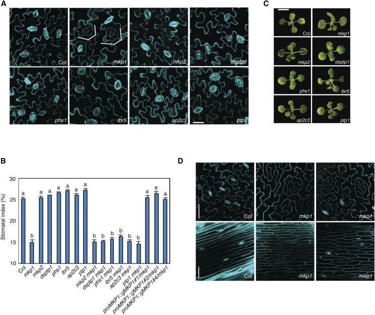
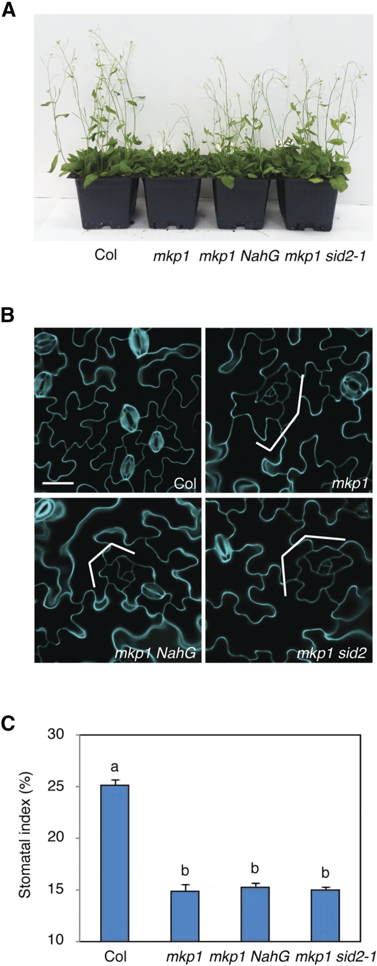
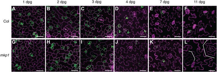
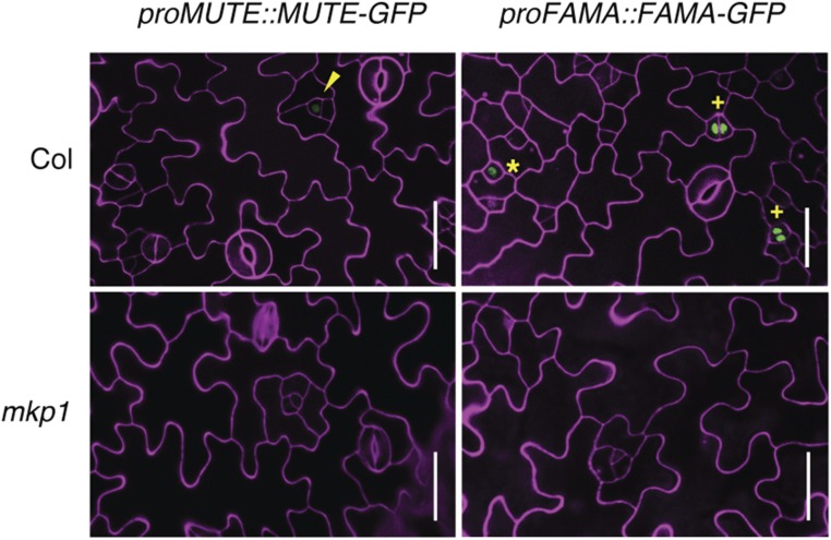
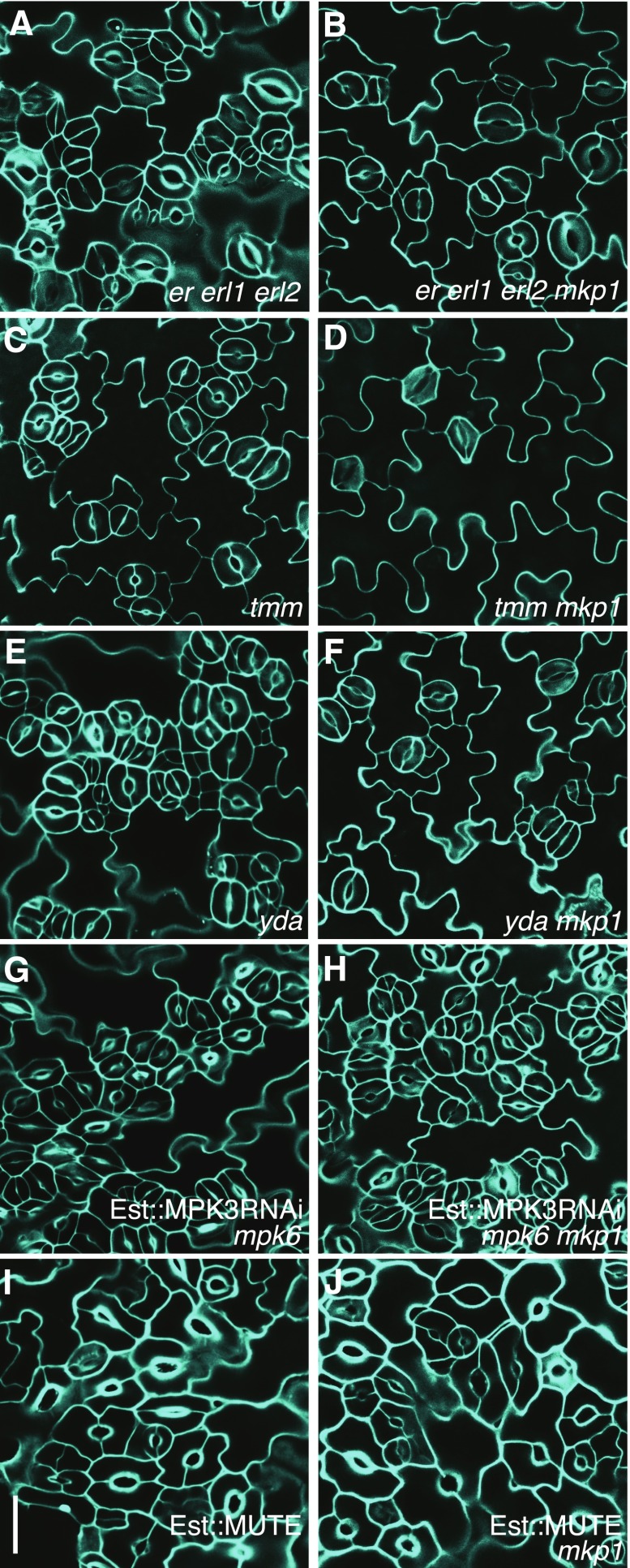
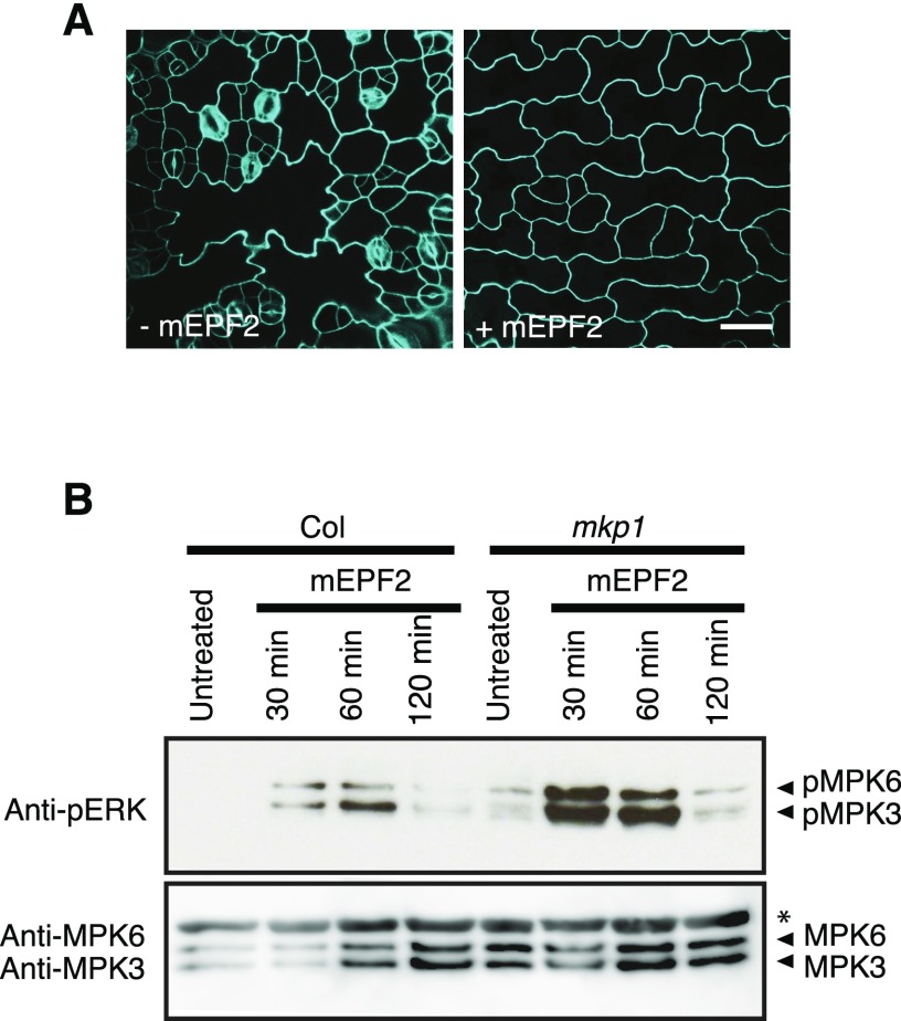
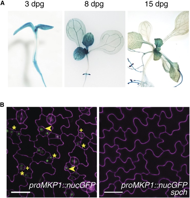
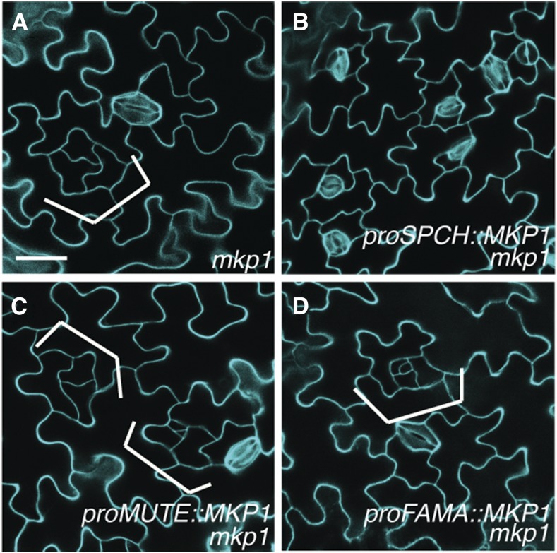
Stomata on the plant epidermis control gas and water exchange and are formed by MAPK-dependent processes. Although the contribution of MAP KINASE3 (MPK3) and MPK6 (MPK3/MPK6) to the control of stomatal patterning and differentiation in Arabidopsis (Arabidopsis thaliana) has been examined extensively, how they are inactivated and regulate distinct stages of stomatal development is unknown. Here, we identify a dual-specificity phosphatase, MAP KINASE PHOSPHATASE1 (MKP1), which promotes stomatal cell fate transition by controlling MAPK activation at the early stage of stomatal development. Loss of function of MKP1 creates clusters of small cells that fail to differentiate into stomata, resulting in the formation of patches of pavement cells. We show that MKP1 acts downstream of YODA (a MAPK kinase kinase) but upstream of MPK3/MPK6 in the stomatal signaling pathway and that MKP1 deficiency causes stomatal signal-induced MAPK hyperactivation in vivo. By expressing MKP1 in the three discrete cell types of stomatal lineage, we further identified that MKP1-mediated deactivation of MAPKs in early stomatal precursor cells directs cell fate transition leading to stomatal differentiation. Together, our data reveal the important role of MKP1 in controlling MAPK signaling specificity and cell fate decision during stomatal development.


Figures

Figure 1.
Figure 1.
MKP1 positively regulates stomatal development. A, Representative confocal images of 10-d-old abaxial cotyledons of the following genotypes: wild type (Col), mkp1, mkp2, dsptp1, phs1, ibr5, ap2c3, and ptp1. The mkp1 single mutant, but not other phosphatase mutants, showed stomatal development defects, including fewer stomata and islands of small arrested cells, indicated by brackets. Cells were outlined by propidium iodide staining (cyan), and images were taken under the same magnification. Scale bar, 30 µm. B, Quantitative analysis of 10-d-old abaxial cotyledon epidermis. Stomatal index (SI) of phosphatase single mutants, double mutants, and three independent complemented lines expressed as the percentage of the number of stomata to the total number of epidermal cells. Genotypes with nonsignificantly different phenotypes were grouped together with the same letter (P < 0.05, Tukey’s HSD test after one-way ANOVA). n = 14 to 15 for each genotype. The experiments were repeated three times with similar results. Bars, means. Error bars, se. C, The growth phenotypes of 2-week-old seedlings of indicated genotypes grown on 0.5× MS plates. Scale bar in Col, 0.5 cm, and others are at the same scale. The representative images were selected from at least five replicates. D, Representative confocal images of 5-week-old abaxial rosette leaf (top) and 7-week-old stem epidermis (bottom) from Col and mkp1. Cells were outlined by propidium iodide staining (cyan), and images were taken under the same magnification. Scale bars, 50 µm.
Figure 2.
Figure 2.
Elevated salicylic acid levels are not responsible for the stomatal phenotype of mkp1. A, Five-week-old plants of wild type (Col), mkp1, mkp1 nahG, and mkp1 sid2. B, Representative confocal images of 10-d-old abaxial cotyledons of wild type, mkp1, mkp1 NahG, and mkp1 sid2 mutants. Both mkp1 nahG and mkp1 sid2 mutants exhibit stomatal development defects (significant reduction of stomata and islands of small arrested cells, indicated by brackets) similar to those of the mkp1 single mutant. Cells were outlined by propidium iodide staining (cyan), and images were taken under the same magnification. Scale bar, 30 µm. C, Stomatal index (SI) of the cotyledon abaxial epidermis from 10-d-old seedlings of Col, mkp1, mkp1 NahG, and mkp1 sid2 mutants, expressed as the percentage of the number of stomata to the total number of epidermal cells. Genotypes with nonsignificantly different phenotypes were grouped together with a letter (P < 0.05, Tukey’s HSD test after one-way ANOVA). n = 14 to 15 for each genotype. The experiments were repeated three times with similar results. Bars, means. Error bars, se.
Figure 3.
Figure 3.
Time-course analysis of epidermal development in mkp1. Confocal images of the abaxial epidermis of wild-type and mkp1 cotyledons. mkp1 seedlings conferred an epidermal phenotype with arrested stomatal precursors, resulting in suppression of stomatal cell fate and formations of expanded SLGC-like clusters becoming lobed pavement cells (brackets). proTMM::GUS-GFP (green) was used to monitor stomatal lineage cells. The representative images were selected from at least five replicates. Scale bars, 30 µm. A to F, Wild-type epidermis at 1, 2, 3, 4, 7, and 11 dpg, respectively. G to L, mkp1 epidermis at 1, 2, 3, 4, 7, and 11 dpg, respectively.
Figure 4.
Figure 4.
Expression of stomatal cell lineage markers in mkp1. Confocal images of abaxial cotyledon epidermis of wild-type (3–5 dpg, top) and mkp1 (10–11 dpg, bottom) seedlings carrying the translational fusion of proMUTE::MUTE-GFP (left) and proFAMA::FAMA-GFP (right). Mature meristemoids are indicated by arrowheads, GMCs by asterisks, and immature guard cells by plus signs. At least three transgenic lines for each construct were used, and similar results were obtained. The representative images were selected from at least five replicates. Scale bar, 30 µm.
Figure 5.
Figure 5.
MKP1 acts downstream of ERECTA family members, TMM, and YDA but upstream of MPK3 and MPK6 in the stomatal development signaling pathway. A to J, Confocal images of abaxial cotyledon epidermis from 7-d-old seedlings of the following genotypes: er erl1 erl2 (A), er erl1 erl2 mkp1 (B), tmm (C), tmm mkp1 (D), yda (E), yda mkp1 (F), induced Est::MPK3RNAi in mpk6 (G), induced Est::MPK3RNAi in mpk6 mkp1 (H), induced Est::MUTE (I), and induced Est::MUTE in mkp1 background (J). At least three transgenic lines for each construct in G to J were used, and similar results were obtained. The representative images were selected from at least five replicates. Scale bar, 30 µm.
Figure 6.
Figure 6.
EPF2 triggers MAPK hyperactivation in mkp1 seedlings. A, Confocal images of the abaxial epidermis of cotyledons of epf2 seedlings grown with or without 2 µm mature EPF2 peptide (mEPF2) treatment. Peptide application experiments were performed at least three times. Scale bar, 30 µm. B, Seedlings of wild type (Col) and mkp1 treated with mEPF2 (1 µm) for the indicated time periods. Activated MAPKs were detected in the Col and mkp1 seedlings by the immunoblots using antipERK antibody (top). The blots were reprobed with anti-MPK3 and MPK6 antibodies to detect MPK3 and MPK6 protein levels on the same samples (bottom). The asterisk indicates a nonspecific band. The molecular weights of MPK3 and MPK6 proteins detected using anti-MPK3 and MPK6 antibodies were consistent with the activated MAPKs (top), suggesting that they are likely MPK3 and MPK6 activated by mEPF2. Experiments were conducted more than three times with similar results, and representative data from one such experiment are shown.
Figure 7.
Figure 7.
Expression patterns of MKP1. A, proMKP1::GUS expression (blue) at 3, 8, and 15 dpg. B, Confocal image of proMKP1::nucGFP (green) in developing wild-type and spch abaxial cotyledon epidermis. Meristemoids are indicated by asterisks, GMCs by arrowheads, and immature guard cells by plus signs. At least three transgenic lines for each construct were used, and similar results were obtained. The representative images were selected from at least five replicates. Scale bars, 30 µm.
Figure 8.
Figure 8.
Early stomatal-cell-specific expression of MKP1 is sufficient for restoring normal stomatal development. A to D, Confocal images of abaxial cotyledon epidermis from 10-d-old seedlings of mkp1 (A), mkp1 expressing proSPCH::MKP1 (B), proMUTE::MKP1 (C), and proFAMA::MKP1 (D). Brackets indicate small cell clusters with arrested or dedifferentiated meristemoids. At least three transgenic lines for each construct in B to D were used, and similar results were obtained. The representative images were selected from at least five replicates and images were taken under the same magnification. Scale bar, 30 µm.

Similar articles

Cited by

References

    1. Andreasson E, Ellis B (2010) Convergence and specificity in the Arabidopsis MAPK nexus. Trends Plant Sci 15: 106–113 - PubMed
    1. Bartels S, Anderson JC, González Besteiro MA, Carreri A, Hirt H, Buchala A, Métraux JP, Peck SC, Ulm R (2009) MAP kinase phosphatase1 and protein tyrosine phosphatase1 are repressors of salicylic acid synthesis and SNC1-mediated responses in Arabidopsis. Plant Cell 21: 2884–2897 - PMC - PubMed
    1. Bartels S, González Besteiro MA, Lang D, Ulm R (2010) Emerging functions for plant MAP kinase phosphatases. Trends Plant Sci 15: 322–329 - PubMed
    1. Bergmann DC, Lukowitz W, Somerville CR (2004) Stomatal development and pattern controlled by a MAPKK kinase. Science 304: 1494–1497 - PubMed
    1. Brock AK, Willmann R, Kolb D, Grefen L, Lajunen HM, Bethke G, Lee J, Nürnberger T, Gust AA (2010) The Arabidopsis mitogen-activated protein kinase phosphatase PP2C5 affects seed germination, stomatal aperture, and abscisic acid-inducible gene expression. Plant Physiol 153: 1098–1111 - PMC - PubMed

MeSH terms

Substances

LinkOut - more resources