Review
. 2018 Sep;18(9):533-548.
doi: 10.1038/s41568-018-0038-z.

Every step of the way: integrins in cancer progression and metastasis

Affiliations
Review

Every step of the way: integrins in cancer progression and metastasis

Hellyeh Hamidi et al. Nat Rev Cancer. 2018 Sep.

Erratum in

Abstract

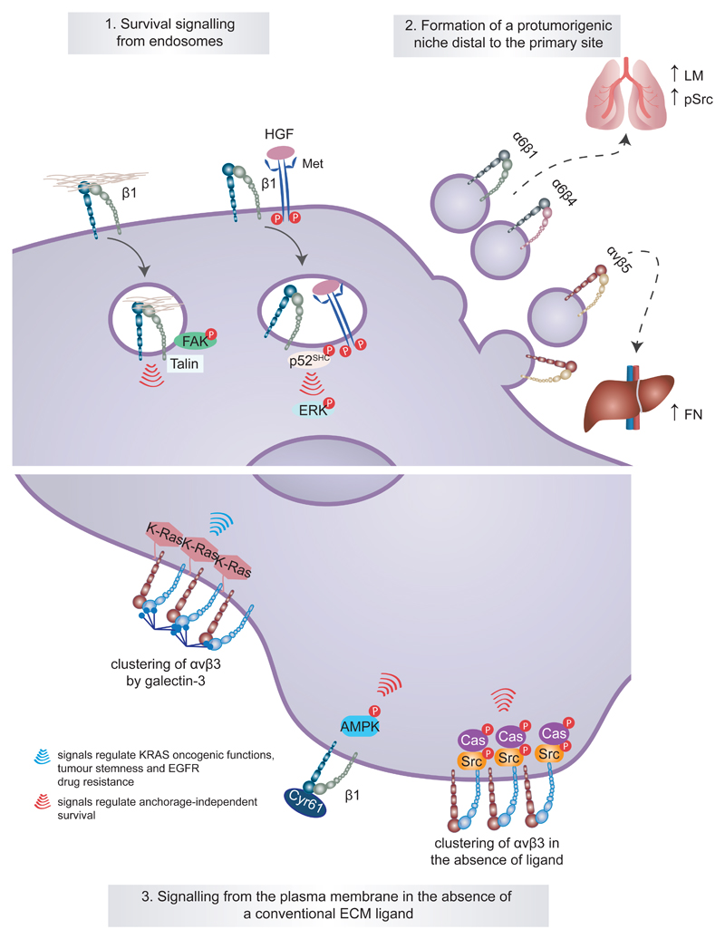
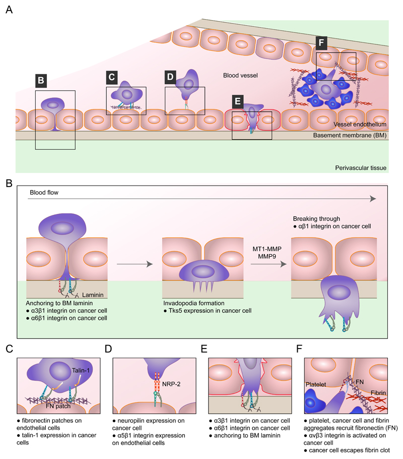
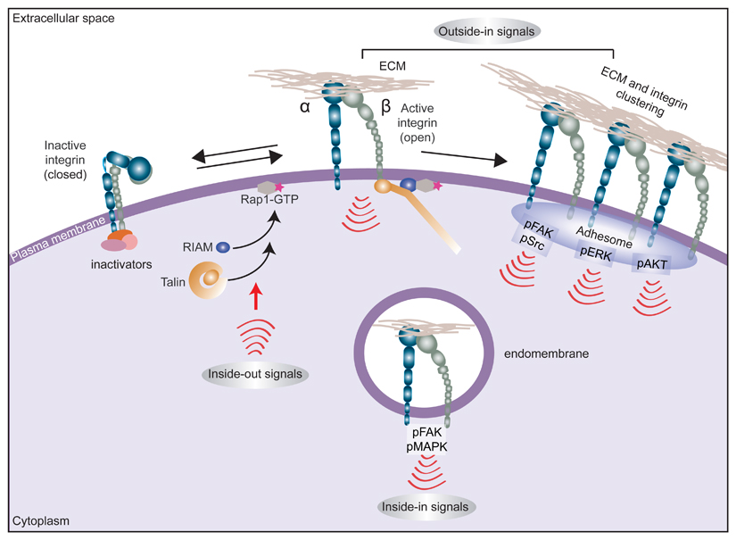
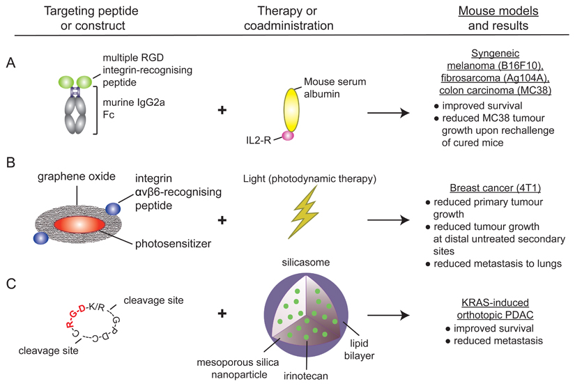
Cell adhesion to the extracellular matrix is fundamental to tissue integrity and human health. Integrins are the main cellular adhesion receptors that through multifaceted roles as signalling molecules, mechanotransducers and key components of the cell migration machinery are implicated in nearly every step of cancer progression from primary tumour development to metastasis. Altered integrin expression is frequently detected in tumours, where integrins have roles in supporting oncogenic growth factor receptor (GFR) signalling and GFR-dependent cancer cell migration and invasion. In addition, integrins determine colonization of metastatic sites and facilitate anchorage-independent survival of circulating tumour cells. Investigations describing integrin engagement with a growing number of versatile cell surface molecules, including channels, receptors and secreted proteins, continue to lead to the identification of novel tumour-promoting pathways. Integrin-mediated sensing, stiffening and remodelling of the tumour stroma are key steps in cancer progression supporting invasion, acquisition of cancer stem cell characteristics and drug resistance. Given the complexity of integrins and their adaptable and sometimes antagonistic roles in cancer cells and the tumour microenvironment, therapeutic targeting of these receptors has been a challenge. However, novel approaches to target integrins and antagonism of specific integrin subunits in stringently stratified patient cohorts are emerging as potential ways forward.


Conflict of interest statement

Competing interests

The authors declare no competing interests.

Figures

Fig. 1
Fig. 1. Integrin involvement in many of the steps of cancer progression.
Integrin expression and/or function have been implicated in nearly every stage of cancer development from primary tumour formation to cancer cell extravasation and formation of a metastatic niche (parts 14). In addition, integrin signalling has been linked to the acquisition of drug resistance (part 5). This fact, together with the vital roles of integrins in cancer, has rendered integrins and integrin-dependent functions attractive therapeutic targets in the fight against cancer (part 6). CAF, cancer-associated fibroblast; ECM, extracellular matrix; EMT, epithelial-to-mesenchymal transition; FAK, focal adhesion kinase; RTK, receptor tyrosine kinase; TGFβ, transforming growth factor-β.
Fig. 2
Fig. 2. Unconventional integrin signalling contributes to cancer cell survival, stemness and drug resistance.
Classical integrin signalling has been described to be restricted to the plasma membrane and to require intact integrin–extracellular matrix (ECM) ligand engagement. However, it is now clear that unconventional modes of integrin signalling exist and contribute to cancer cell survival and disease progression. For example, active integrin signalling has been shown to be maintained away from the plasma membrane following integrin endocytosis in the presence of an ECM ligand or hepatocyte growth factor (HGF)-stimulated receptor tyrosine kinase MET and to contribute to anchorage-independent growth and survival of cancer cells through different pathways (part 1). A more extreme example of long-range integrin signalling has been illustrated in tumour exosomes and is associated with disease advancement. Tumour exosomal α6β1 integrin and α6β4 integrin appear to target metastatic cells to the lung, whereas exosomal αvβ5 integrin is linked to liver metastasis. At these defined sites, the exosomes prepare a premetastatic niche by triggering the expression of specific ECM components (higher laminin and elevated SRC phosphorylation (pSRC) in lung fibroblasts and elevated fibronectin in the liver) and pro-inflammatory S100 proteins in the target tissue (part 2). Integrin signalling from the plasma membrane can also be unconventional and occur in the absence of an ECM ligand or be promoted by the interaction of non-structural ECM proteins such as galectin 3 and cysteine-rich angiogenic inducer 61 (CYR61) with the integrin extracellular domains (part 3). AMPK, AMP-activated protein kinase; CAS, CRK-associated substrate; EGFR, epidermal growth factor receptor; FAK, focal adhesion kinase; P, phosphorylation; p52-SHC, p52 isoform of SHC-transforming protein 1.
Fig. 3
Fig. 3. Integrins in extravasation.
The top panel shows a schematic of integrin-mediated extravasation of circulating tumour cells (CTCs) (part a). The mechanisms involved in this process are α3β1 integrin-mediated and α6β1 integrin-mediated cancer cell adhesion to subendothelial laminin, which is required for successful transendothelial migration. β1 integrin is also a prerequisite for tumour cells to fully clear the endothelial layer and invade into the basement membrane (part b). The presence of fibronectin patches on endothelial cells promotes cancer cell adhesion to the vessel wall in a manner dependent on the integrin activator talin 1 (part c). Endothelial cells also express integrins. Endothelial α5 integrin directly binds to neuropilin 2 (NRP2), a receptor for vascular endothelial growth factor (VEGF) and the semaphorin family of proteins, on cancer cells, and this interaction promotes cancer cell attachment to the endothelium and subsequent extravasation (part d). Pre-existing patches of exposed basement membrane can promote CTC arrest on the vascular wall in a mechanism whereby exposed laminin is engaged by α3β1 integrin on the tumour cell (part e). Features of the blood clotting cascade can promote integrin-mediated cancer cell invasive protrusion and extravasation, such as local recruitment of plasma fibronectin to trigger αvβ3 integrin activation (part f). MMP, matrix metalloproteinase; MT1, membrane type 1; TKS5, tyrosine kinase substrate with five SH3 domains (also known as SH3PXD2A).
None
None

References

    1. Humphries JD, Byron A, Humphries MJ. Integrin ligands at a glance. J Cell Sci. 2006;119:3901–3903. - PMC - PubMed
    1. Arruda Macêdo JK, Fox JW, de Souza Castro M. Disintegrins from snake venoms and their applications in cancer research and therapy. Curr Protein Pept Sci. 2015;16:532–548. - PMC - PubMed
    1. Hussein HAM, et al. Beyond RGD: virus interactions with integrins. Arch Virol. 2015;160:2669–2681. - PMC - PubMed
    1. De Franceschi N, Hamidi H, Alanko J, Sahgal P, Ivaska J. Integrin traffic - the update. J Cell Sci. 2015;128:839–852. - PMC - PubMed
    1. Horton ER, et al. Definition of a consensus integrin adhesome and its dynamics during adhesion complex assembly and disassembly. Nat Cell Biol. 2015;17:1577–1587. - PMC - PubMed

Publication types