

Frequency and Significance of Perforating Venous Insufficiency in Patients with Chronic Venous Insufficiency of Lower Extremity
- PMID: 30002576
- PMCID: PMC6039150
- DOI: 10.5152/eurasianjmed.2018.18338
Frequency and Significance of Perforating Venous Insufficiency in Patients with Chronic Venous Insufficiency of Lower Extremity
Abstract
Objective: The aim of this study was to reveal the frequency and impact of perforating venous insufficiency (PVI) in chronic venous insufficiency (CVI) of lower extremity (LE).
Materials and methods: Between 2012 and 2017, a total of 1154 patients [781 females (67.68%) and 373 males (32.32%), 228 (19.76%) unilateral and 926 (80.24%) bilateral LE] were examined using Doppler ultrasound (US). A total of 2080 venous systems of LEs [31.4% male (n=653) and 68.6% female (n=1427); 1056 left LEs (50.77%) and 1024 right LEs (49.23%)] were examined. All patients had symptoms of venous insufficiency (VI).
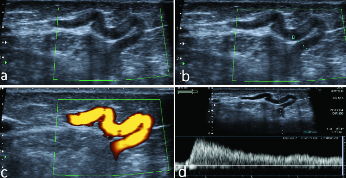
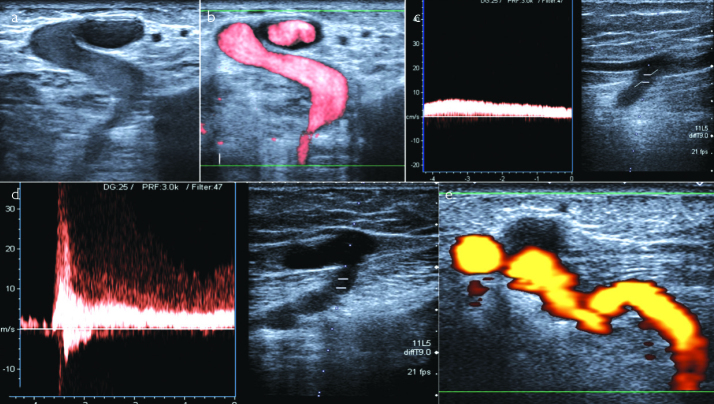
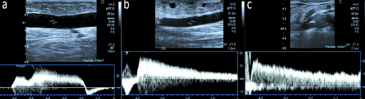
Results: PVI was revealed in 27.5% (n=571) of LEs. Varicose veins (VVs) related with perforating vein (PV) were revealed in 44.7% of LEs (n=929). PVI was observed in 50.91% of patients with chronic deep venous thrombosis (DVT), 64.41% with deep venous insufficiency (DVI), 59.81% with great saphenous vein (GSV) insufficiency, 68.49% with small saphenous vein (SSV) insufficiency, 58.65% with accessory GSV insufficiency, and 58.77% with PV associated with VVs. There was a statistically significant relationship between PVI and chronic DVT, DVI, GSV, SSV, and accessory GSV insufficiency (p<0.001). A significant relationship was observed between the increase in PV diameter and the presence of PVI (p<0.001).
Conclusion: PVI is quite common in combined VI, and PV evaluation should be a part of LE venous system examination.
Keywords: Chronic venous insufficiency; doppler ultrasound; perforating venous insufficiency; varicose veins.
Conflict of interest statement
Conflict of Interest: Authors have no conflict of interest to declare.
Figures
References
-
- Thorisson HM, Pollak JS, Scoutt L. The role of ultrasound in the diagnosis and treatment of chronic venous insufficiency. Ultrasound Q. 2007;23:137–50. doi: 10.1097/01.ruq.0000277035.82208.bf. - DOI - PubMed
-
- Liu CH, Wu CJ, Yu CY, Liu CH, Chang CW, Huang GS. Evaluation of lower limb varicose vein by ultrasonic venous duplex examination. J Med Ultrasound. 2013;21:76–80. doi: 10.1016/j.jmu.2013.04.009. - DOI
-
- Coleridge-Smith P, Labropoulos N, Partsch H, Myers K, Nicolaides A, Cavezzi A. Duplex ultrasound investigation of the veins in chronic venous disease of the lower limbs-UIP consensus document. Part I. Basic principles. Eur J Vasc Endovasc Surg. 2006;31:83–92. doi: 10.1016/j.ejvs.2005.07.019. - DOI - PubMed
-
- Mozes G, Gloviczki P. Venous embryology and anatomy. In: Bergan JJ, Paquette NB, editors. The vein book. 2th ed. San Diego, California: Elsevier Academic Press; 2007. pp. 20–24. - DOI
LinkOut - more resources
Full Text Sources
Other Literature Sources