Review
. 2018 Jun 28:9:1486.
doi: 10.3389/fimmu.2018.01486. eCollection 2018.

Apoptotic Cell-Derived Extracellular Vesicles: More Than Just Debris

Affiliations
Review

Apoptotic Cell-Derived Extracellular Vesicles: More Than Just Debris

Sarah Caruso et al. Front Immunol. .

Abstract

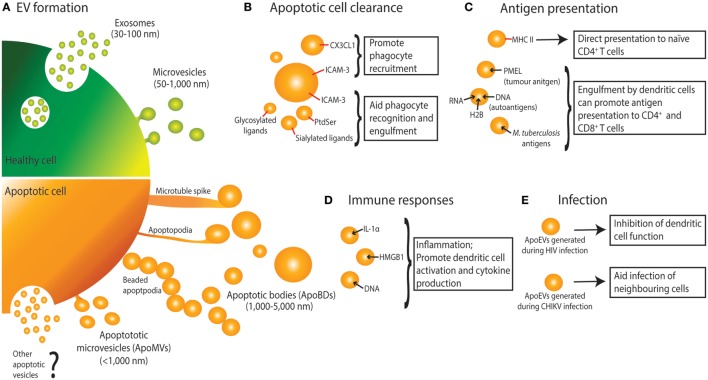
The many functions of extracellular vesicles (EVs) like exosomes and microvesicles released from healthy cells have been well characterized, particularly in relation to their roles in immune modulation. Apoptotic bodies, a major class of EV released as a product of apoptotic cell disassembly, and other types of EVs released from dying cells are also becoming recognized as key players in this emerging field. There is now increasing evidence to suggest that EVs produced during apoptosis have important immune regulatory roles, a concept relevant across different disease settings including autoimmunity, cancer, and infection. Therefore, this review focuses on how the formation of EVs during apoptosis could be a key mechanism of immune modulation by dying cells.

Keywords: apoptosis; apoptotic bodies; apoptotic microvesicles; extracellular vesicles; immunomodulation.


Figures

Figure 1
Figure 1
Extracellular vesicle (EV) formation and immune functions of apoptotic cell-derived EVs (ApoEVs). (A) Healthy cells form two main types of EV, namely exosomes that are release via exocytosis of multivesicular bodies, and microvesicles that are shed from the plasma membrane. During apoptosis, dying cells can also release ApoEVs. Apoptotic cells can undergo morphological changes including membrane blebbing, thin membrane protrusion formation (microtubule spikes, apoptopodia, and beaded-apoptopodia), and generation of distinct apoptotic bodies (ApoBDs). Apoptotic cells can also release EVs that are similar in size as microvesicles (ApoMVs), however, it is unclear if ApoMVs are generated via the same mechanism as microvesicles from healthy cells. Whether apoptotic cells can generate vesicles that are similar to exosomes is undetermined. (B) ApoEVs can harbor “find-me” signals (e.g., CX3CL1 and ICAM-3) to attract phagocytic cells, as well as “eat-me” signals [e.g., ICAM-3, phosphatidylserine (PtdSer), and sialylated and glycosylated ligands] to promote uptake by phagocytes. (C) ApoEVs have MHC II molecules on their surface, which is essential for direct antigen presentation to naïve CD4+ T cells and activation of immunological memory. ApoEVs can also carry antigen to professional antigen-presenting cells (e.g., dendritic cells). Antigens carried by ApoEVs include autoantigens, tumor antigens, and microbial antigens. (D) ApoEVs can promote inflammation by transporting proinflammatory cytokines such as IL-1α and damage-associated molecular patterns including DNA and HMGB1. (E) ApoEVs can aid HIV infection by inhibiting dendritic cell activation. Chikungunya virus (CHIKV) can hijack ApoEVs to propagate infection to neighboring cells.

References

    1. Akers JC, Gonda D, Kim R, Carter BS, Chen CC. Biogenesis of extracellular vesicles (EV): exosomes, microvesicles, retrovirus-like vesicles, and apoptotic bodies. J Neurooncol (2013) 113(1):1–11.10.1007/s11060-013-1084-8 - DOI - PMC - PubMed
    1. Robbins PD, Morelli AE. Regulation of immune responses by extracellular vesicles. Nat Rev Immunol (2014) 14(3):195–208.10.1038/nri3622 - DOI - PMC - PubMed
    1. Buzas EI, Gyorgy B, Nagy G, Falus A, Gay S. Emerging role of extracellular vesicles in inflammatory diseases. Nat Rev Rheumatol (2014) 10(6):356–64.10.1038/nrrheum.2014.19 - DOI - PubMed
    1. Thery C, Ostrowski M, Segura E. Membrane vesicles as conveyors of immune responses. Nat Rev Immunol (2009) 9(8):581–93.10.1038/nri2567 - DOI - PubMed
    1. Meckes DG, Jr, Raab-Traub N. Microvesicles and viral infection. J Virol (2011) 85(24):12844–54.10.1128/JVI.05853-11 - DOI - PMC - PubMed

LinkOut - more resources