. 2018 Jun;14(4):735-744.
doi: 10.5114/aoms.2017.71076. Epub 2018 Mar 28.

MicroRNA-135b-5p prevents oxygen-glucose deprivation and reoxygenation-induced neuronal injury through regulation of the GSK-3β/Nrf2/ARE signaling pathway

Affiliations

MicroRNA-135b-5p prevents oxygen-glucose deprivation and reoxygenation-induced neuronal injury through regulation of the GSK-3β/Nrf2/ARE signaling pathway

Qiang Duan et al. Arch Med Sci. 2018 Jun.

Abstract

Introduction: MicroRNAs (miRNAs) are emerging as critical regulators in the pathological process of cerebral ischemia/reperfusion injury. miRNAs play an important role in regulating neuronal survival. miR-135b-5p has been reported as an important miRNA in regulating cell apoptosis. However, the role of miR-135b-5p in regulating neuronal survival remains poorly understood. Here, we aimed to investigate the role of miR-135b-5p in cerebral ischemia/ reperfusion using an in vitro model of oxygen-glucose deprivation and reoxygenation-(OGD/R) induced neuron injury.

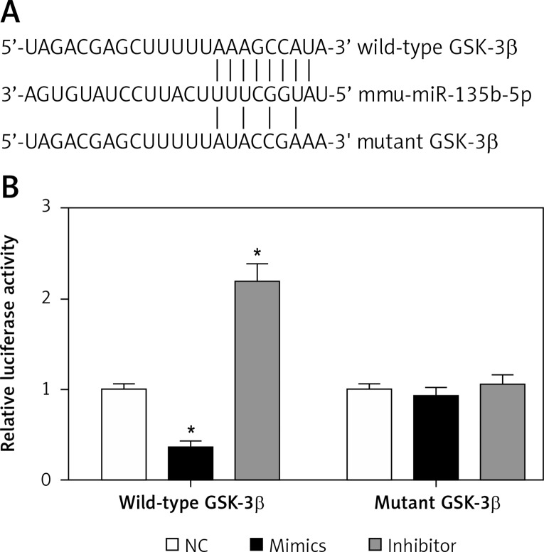
Material and methods: miRNA, mRNA and protein expression was detected by real-time quantitative polymerase chain reaction and Western blot. Cell viability was detected by cell counting kit-8 and lactate dehydrogenase assays. Cell apoptosis was detected by caspase-3 activity assay. Oxidative stress was determined using commercial kits. The target of miR-135b-5p was confirmed by dual-luciferase reporter assay.

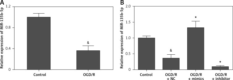
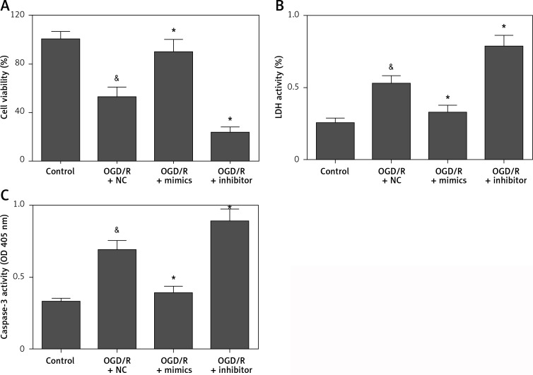
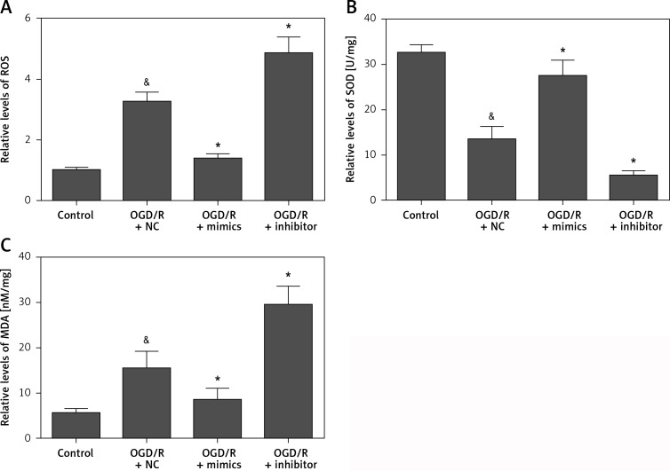
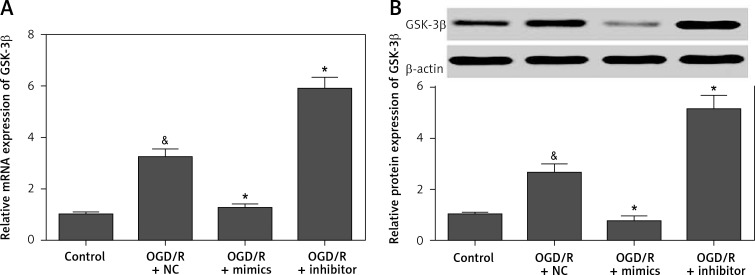
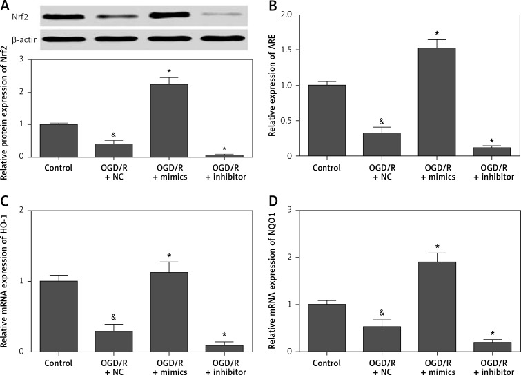
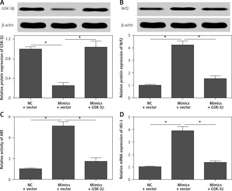
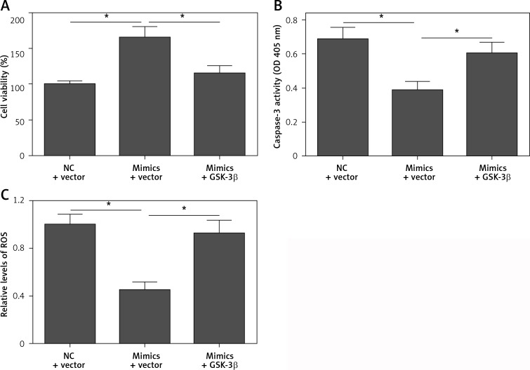
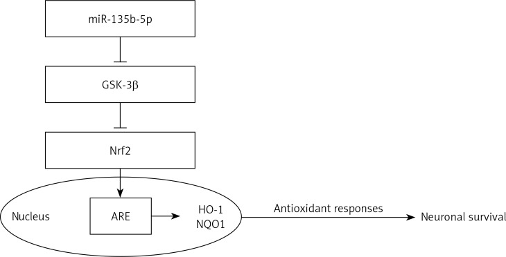
Results: We found that miR-135b-5p expression was significantly decreased in hippocampal neurons receiving OGD/R treatment. Overexpression of miR-135b-5p markedly alleviated OGD/R-induced cell injury and oxidative stress, whereas suppression of miR-135b-5p showed the opposite effects. We observed that miR-135b-5p directly targeted the 3'-untranslated region of glycogen synthase kinase-3β (GSK-3β). We found that miR-135b-5p negatively regulates the expression of GSK-3β in hippocampal neurons. Moreover, miR-135b-5p overexpression promotes activation of nuclear factor erythroid 2-related factor 2 (Nrf2)/antioxidant response element (ARE) signaling. However, the restoration of GSK-3β expression significantly reversed the protective effects of miR-135b-5p overexpression.

Conclusions: Overall, our results suggest that miR-135b-5p protects neurons against OGD/R-induced injury through downregulation of GSK-3β and promotion of the Nrf2/ARE signaling pathway-mediated antioxidant responses.

Keywords: GSK-3β; Nrf2; ischemia/reperfusion injury; miR-135b-5p.


Conflict of interest statement

The authors declare no conflict of interest.

Figures

Figure 1
Figure 1
Expression of miR-135b-5p in hippocampal neurons receiving OGD/R treatment. A – RT-qPCR analysis of miR-135b-5p expression in HT22 cells with OGD/R treatment. Cells cultured in normal condition were used as a control. B – RT-qPCR analysis of miR-135b-5p expression in HT22 cells transfected with miR-135b-5p mimics or inhibitor for 24 h followed by OGD/R treatment &p < 0.05 vs. control and *p < 0.05 vs. OGD/R + NC.
Figure 2
Figure 2
Overexpression of miR-135b-5p attenuates OGD/R-induced injury. HT22 cells were transfected with miR-135b-5p mimics or inhibitor for 24 h and then subjected to OGD/R treatment. A – Cell viability was measured by CCK-8 assay. B – Cell death was evaluated by LDH assay. C – Cell apoptosis was detected by caspase-3 activity assay &p < 0.05 vs. control and *p < 0.05 vs. OGD/R + NC.
Figure 3
Figure 3
Overexpression of miR-135b-5p inhibits OGD/R-induced oxidative stress. HT22 cells were transfected with miR-135b-5p mimics or inhibitor for 24 h and then subjected to OGD/R treatment. A – ROS levels were measured by DCFH-DA assay. Levels of SOD (B) and MDA (C) were measured using commercial kits &p < 0.05 vs. control and *p < 0.05 vs. OGD/R + NC.
Figure 4
Figure 4
miR-135b-5p targets the 3′-UTR of GSK-3β. A – Diagram of the predicted miR-135b-5p binding site in the 3′-UTR of GSK-3β. B – A dual-luciferase reporter assay was performed to examine whether miR-135b-5p directly binds to the GSK-3β 3′-UTR in HT22 cells *p < 0.05 vs. NC.
Figure 5
Figure 5
miR-135b-5p negatively regulates GSK-3β expression. HT22 cells were transfected with miR-135b-5p mimics or inhibitor for 24 h and then subjected to OGD/R treatment. A – The mRNA expression of GSK-3β was detected by RT-qPCR. B – Protein expression of GSK-3β was detected by Western blot &p < 0.05 vs. control and *p < 0.05 vs. OGD/R + NC.
Figure 6
Figure 6
Overexpression of miR-135b-5p promotes activation of the Nrf2/ARE signaling pathway. HT22 cells were transfected with ARE reporter vector and miR-135b-5p mimics or inhibitor for 24 h and then subjected to OGD/R treatment. A – Protein expression of Nrf2 was detected by Western blot analysis. B – ARE activity was detected by dual-luciferase reporter assays. The mRNA expression of HO-1 (C) and NQO1 (D) was detected by RT-qPCR &p < 0.05 vs. control and *p < 0.05 vs. OGD/R + NC.
Figure 7
Figure 7
Restoration of GSK-3β expression reverses the effect of miR-135b-5p overexpression on Nrf2/ARE signaling. HT22 cells were cotransfected with pcDNA3.1/GSK-3β vector and miR-135b-5p mimics for 24 h and then subjected to OGD/R treatment. Expression of GSK-3β (A) and Nrf2 (B) was detected by Western blot. C – ARE activity was detected by dual-luciferase reporter assays. D – The mRNA expression of HO-1 was detected by RT-qPCR *p < 0.05.
Figure 8
Figure 8
Restoration of GSK-3β expression reverses the protective effect of miR-135b-5p overexpression. HT22 cells were cotransfected with pcDNA3.1/GSK-3β vector and miR-135b-5p mimics for 24 h and then subjected to OGD/R treatment. A – Cell viability was measured by CCK-8 assay. B – Cell apoptosis was detected by caspase-3 activity assay. C – ROS levels were measured by DCFHDA assay *p < 0.05.
Figure 9
Figure 9
miR-135b-5p protects hippocampal neurons against OGD/R-induced injury by downregulation of GSK-3β and upregulation of Nrf2/ARE signaling pathway-mediated antioxidant responses

References

    1. Feigin VL, Forouzanfar MH, Krishnamurthi R, et al. Global and regional burden of stroke during 1990-2010: findings from the Global Burden of Disease Study 2010. Lancet. 2014;383:245–54. - PMC - PubMed
    1. Wechsler LR. Intravenous thrombolytic therapy for acute ischemic stroke. N Engl J Med. 2011;364:2138–46. - PubMed
    1. Sims NR, Muyderman H. Mitochondria, oxidative metabolism and cell death in stroke. Biochim Biophys Acta. 2010;1802:80–91. - PubMed
    1. Bartel DP. MicroRNAs: genomics, biogenesis, mechanism, and function. Cell. 2004;116:281–97. - PubMed
    1. Huang Y, Shen XJ, Zou Q, et al. Biological functions of microRNAs: a review. J Physiol Biochem. 2011;67:129–39. - PubMed