. 2018 Jun;14(4):910-919.
doi: 10.5114/aoms.2016.63743. Epub 2016 Nov 18.

2D and 3D cell cultures - a comparison of different types of cancer cell cultures

Affiliations

2D and 3D cell cultures - a comparison of different types of cancer cell cultures

Marta Kapałczyńska et al. Arch Med Sci. 2018 Jun.

Abstract

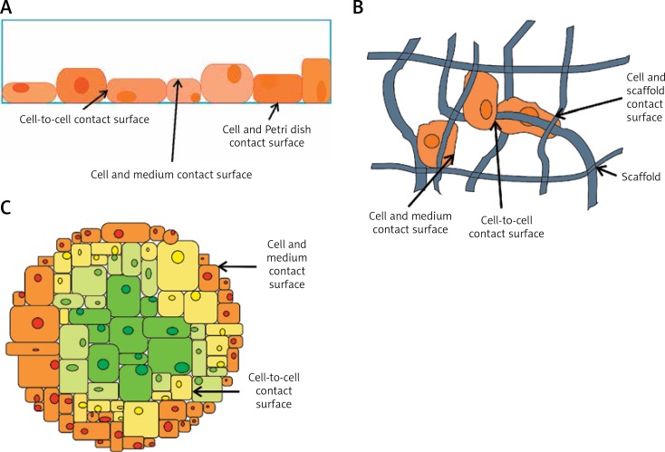
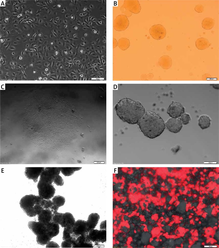
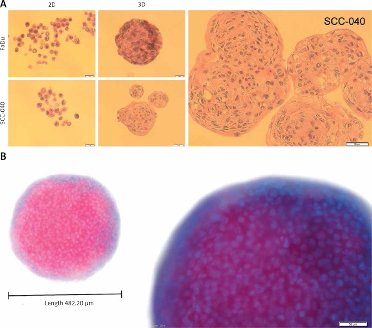
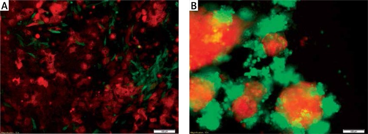
Cell culture is a widely used in vitro tool for improving our understanding of cell biology, tissue morphology, and mechanisms of diseases, drug action, protein production and the development of tissue engineering. Most research regarding cancer biology is based on experiments using two-dimensional (2D) cell cultures in vitro. However, 2D cultures have many limitations, such as the disturbance of interactions between the cellular and extracellular environments, changes in cell morphology, polarity, and method of division. These disadvantages led to the creation of models which are more closely able to mimic conditions in vivo. One such method is three-dimensional culture (3D). Optimisation of the culture conditions may allow for a better understanding of cancer biology and facilitate the study of biomarkers and targeting therapies. In this review, we compare 2D and 3D cultures in vitro as well as different versions of 3D cultures.

Keywords: 2D culture; 3D culture; cancer research; cell culture methods; co-culture.


Conflict of interest statement

The authors declare no conflict of interest.

Figures

Figure 1
Figure 1
Types of cell culture methods commonly used in research studies. A – Cells flattened in a monolayer on the bottom of the culture vessel. They are in contact with the culture vessel, neighbouring cells, and the culture medium. B – Cells attached to a scaffold are in contact with the scaffolding, neighbouring cells, and the culture medium. C – A group of cells suspended in the culture medium or cultivated in gel-like substance; the cells are in contact with neighbouring cells and with the culture medium
Figure 2
Figure 2
FaDu cell line cultured under various conditions. The FaDu cells were maintained in adherent conditions with standard medium (10% FBS) and next detached and placed as single cells in different (A–F) culture conditions in standard medium. A – flattened cells growing as a monolayer under 2D conditions (scale bar represents 100 μm); B – 3D structures in soft agar, single cells suspended in a gel are visible (scale bar represents 200 μm); C – adherent colonies formed between layers of soft agar (scale bar represents 200 μm); D – 3D structure formed on non-adherent plate (scale bar represents 100 μm); E – tissue-like structures formed by attached single spheres cultivated on ultra-low attachment plates (scale bar represents 200 μm); F – cells (red) cultured using 3D scaffold system with visible membrane pores (scale bar represents 100 μm)
Figure 3
Figure 3
Structural architecture of 3D spheroids. The SCC-040 and FaDu cells were maintained in adherent condition with standard medium (10% FBS) and next detached and placed as single cells on non-adherent plates in standard medium. The created spheroids were taken to make the formalin-fixed paraffin-embedded tissue sections (FFPET) and H&E staining as well as DAPI staining. A – cross section through the cells growing in 2D and 3D cultures of SCC-040 and FaDu cell lines, H&E staining (scale bars represent 20 μm and 50 μm, respectively); B – 3D structure stained with DAPI; blue – nuclei, pink – cells (scale bar represents 50 μm)
Figure 4
Figure 4
Co-culture of epithelial SCC-25 (red) and fibroblast MSU-1.1 (green) cell lines (scale bar represents 100 μm). A – cells cultured under 2D conditions are flattened and attached to the plate surface. The epithelial SCC-25 cells (red) have typical rhombus-like shape and MSU-1.1 cells (green) are spindle-like and surround SCC-25 cells; B – SCC-25 (red) and MSU-1.1 (green) cells cultured under 3D conditions changed their own morphology due to the lack of attachment. Cells lose their typical shape and aggregate, creating more (SCC-25) and less (MSU-1.1) compact structures

References

    1. Yamada K, Cukierman E. Modeling tissue morphogenesis and cancer in 3D. Cell. 2007;130:601–10. - PubMed
    1. Harrison R. Observations of the living developing nerve fiber. Anat Rec. 1907;1:116–28.
    1. Harrison RG. The outgrowth of the nerve fiber as a mode of protoplasmic movement. J Exp Zool. 1910;9:787–846. - PubMed
    1. Scudiero D, Shoemaker RH, Paull KD, et al. Evaluation of a soluble tetrazolium formazan assay for cell growth and drug sensitivity in culture using human and other tumor cell lines. Cancer Res. 1988;48:4827–33. - PubMed
    1. Jacoby W, Pasten I. Methods in Enzymology: Cell Culture. Vol. 58. New York: Academic Press; 1979.