Review
. 2018 Jul 7:9:22.
doi: 10.1186/s13100-018-0128-1. eCollection 2018.

L1 retrotransposition in the soma: a field jumping ahead

Affiliations
Review

L1 retrotransposition in the soma: a field jumping ahead

Geoffrey J Faulkner et al. Mob DNA. .

Abstract

Retrotransposons are transposable elements (TEs) capable of "jumping" in germ, embryonic and tumor cells and, as is now clearly established, in the neuronal lineage. Mosaic TE insertions form part of a broader landscape of somatic genome variation and hold significant potential to generate phenotypic diversity, in the brain and elsewhere. At present, the LINE-1 (L1) retrotransposon family appears to be the most active autonomous TE in most mammals, based on experimental data obtained from disease-causing L1 mutations, engineered L1 reporter systems tested in cultured cells and transgenic rodents, and single-cell genomic analyses. However, the biological consequences of almost all somatic L1 insertions identified thus far remain unknown. In this review, we briefly summarize the current state-of-the-art in the field, including estimates of L1 retrotransposition rate in neurons. We bring forward the hypothesis that an extensive subset of retrotransposition-competent L1s may be de-repressed and mobile in the soma but largely inactive in the germline. We discuss recent reports of non-canonical L1-associated sequence variants in the brain and propose that the elevated L1 DNA content reported in several neurological disorders may predominantly comprise accumulated, unintegrated L1 nucleic acids, rather than somatic L1 insertions. Finally, we consider the main objectives and obstacles going forward in elucidating the biological impact of somatic retrotransposition.

Keywords: Genomics; L1; LINE-1; Mosaicism; Neurobiology; Retrotransposon.


Conflict of interest statement

Not applicable.Not applicable.The authors declare that they have no competing interests.Springer Nature remains neutral with regard to jurisdictional claims in published maps and institutional affiliations.

Figures
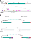
Fig. 1
Fig. 1
L1 retrotransposon structure and mobilization scenarios. a. A human L1-Ta element (top) is 6 kb in length and encodes two protein-coding open reading frames (ORF1 and ORF2) flanked by 5′ and 3′ UTRs. New L1 insertions are typically flanked by a 3′ polyadenine (An) tract as mRNA polyadenylation is critical to efficient L1 retrotransposition [61, 62]. An antisense open reading frame (ORF0, brown rectangle) is located in the 5′UTR and may facilitate retrotransposition [209]. ORF2p possesses endonuclease (EN) and reverse transcriptase (RT) activities [44, 45]. The L1 is transcribed from 5′ sense (canonical) [47] and antisense [208] promoters, as indicated by black arrows. Target-primed reverse transcription (TPRT) typically generates short target site duplications (TSDs, indicated by red triangles) flanking new L1 insertions [44, 46, 64, 66]. A closer view of the L1 5′UTR (bottom) indicates YY1 (purple rectangle), RUNX (brown rectangle) and SRY family (e.g. SOX2, pink rectangle) transcription factor binding sites [22, 69, 207]. Numerous CpG dinucleotides (orange bars) occur throughout this region and, at a point of sufficient density, form a CpG island (green line) that is regulated by a complex including MeCP2, HDAC1 and HDAC2 [27, 47, 75, 105]. b. Example L1 mobilization scenarios. Top: A donor L1 is transcribed from its canonical promoter, generates a polyadenylated mRNA, and is retrotransposed via TPRT, generating a new L1 insertion that is 5′ truncated. Middle: Transcription initiated by a promoter upstream of the donor L1 reads through into the L1 and generates a spliced (dotted line) mRNA. As a result, the new L1 insertion carries a 5′ transduction. Bottom: Transcription initiates as directed by the canonical promoter but reads through the L1 polyA signal to an alternative downstream signal. Reverse transcription and integration of this mRNA generates a 5′ truncated L1 insertion flanked by a 3′ transduction. Note: the monomeric promoters of the active mouse L1 subfamilies (TF, GF, A) are very different in their structure, and potentially their regulation, than the human L1-Ta promoter. Aspects of the figure are adapted from previous works [35, 290]
Fig. 2
Fig. 2
Interpreting results from the engineered L1-EGFP reporter assay. a. The L1-EGFP reporter gene [123] comprises a full-length human or mouse L1 (e.g. [41, 122, 291]) tagged with a cassette incorporating EGFP and its promoter in the opposite orientation to the L1, followed by an SV40 polyA signal. Transcription of the combined L1-EGFP reporter, followed by splicing (dotted line) of an intron in the EGFP gene, prepares the L1-EGFP mRNA for reverse transcription and integration into the genome via target-primed reverse transcription (TPRT). The L1-EGFP reporter has been introduced in vitro as a plasmid [–21, 126, 171] and also as a rodent transgene [8, 9, 21, 27, 116]. b. Successful TPRT-mediated retrotransposition of the engineered L1 mRNA yields an intact EGFP gene, leading to GFP+ cells (true positives). c. Mobilization of the engineered L1 mRNA may occur through TPRT but, due to severe 5′ truncation removing the L1 entirely, or 5′ inversion/deletion [95, 292] the EGFP gene may be incompetent at its 3′ end, and therefore retrotransposition results in GFP cells (false negatives). d. The engineered L1 mRNA may be retrotransposed, yielding a functional EGFP gene, but the EGFP promoter is epigenetically silenced [126], leading to GFP cells (false negatives). PCR-based assays targeting the EGFP splice junction can, however, identify instances where successful retrotransposition is not marked by EGFP expression [19, 46, 123, 126]. e. Finally, retrotransposition of the engineered L1 mRNA may simply have not occurred in GFP cells (true negatives)
Fig. 3
Fig. 3
Somatic retrotransposition can cause complex genomic mosaicism. a. Donor L1 expression and mobilization during development. A handful of L1 copies from each individual are highly active, or hot, when tested in vitro [38, 39]. Four scenarios for donor L1s mobilizing in vivo are illustrated here. Most L1s are repressed [105] during development and do not mobilize, except perhaps due to exceptional circumstances, such as the availability of an active upstream promoter (e.g. yellow donor L1) [36]. L1 promoter de-repression can however occur during development, either transiently (e.g. red and orange donor L1s) or durably (e.g. blue donor L1), leading to L1 mRNA and RNP accumulation. Retrotransposition enacted by the L1 machinery occurs as a function of donor L1 activity in a given spatiotemporal context (blue, red, orange and yellow arrowheads, matching each donor L1). b. The developmental timing of a given retrotransposition event impacts how many mature cells carry the new L1 insertion. Early embryonic L1 mobilization events (e.g. blue and red cells indicated by arrowheads and matching donor L1s by color) may be carried by numerous descendent cells, possibly in different tissues [18]. By contrast, L1 insertions arising later in development (indicated by orange, blue and yellow arrows) are more restricted in their spatiotemporal extent, and may be found in just one cell (e.g. a post-mitotic neurons). The resulting somatic genome mosaicism may disproportionately impact the brain [–, , , , –138], although further work is required to test whether other organs, such as the liver, also routinely carry somatic L1 insertions [72, 161]

References

    1. McClintock B. The origin and behavior of mutable loci in maize. Proc Natl Acad Sci U S A. 1950;36:344–355. doi: 10.1073/pnas.36.6.344. - DOI - PMC - PubMed
    1. Belzile F, Yoder JI. Pattern of somatic transposition in a high copy ac tomato line. Plant J. 1992;2:173–179. - PubMed
    1. Raizada MN, Nan GL, Walbot V. Somatic and germinal mobility of the RescueMu transposon in transgenic maize. Plant Cell. 2001;13:1587–1608. doi: 10.1105/tpc.13.7.1587. - DOI - PMC - PubMed
    1. Eickbush MT, Eickbush TH. Retrotransposition of R2 elements in somatic nuclei during the early development of Drosophila. Mob DNA. 2011;2:11. doi: 10.1186/1759-8753-2-11. - DOI - PMC - PubMed
    1. Li W, Prazak L, Chatterjee N, Gruninger S, Krug L, Theodorou D, Dubnau J. Activation of transposable elements during aging and neuronal decline in Drosophila. Nat Neurosci. 2013;16:529–531. doi: 10.1038/nn.3368. - DOI - PMC - PubMed