

Case Reports
doi: 10.1016/j.radcr.2018.05.015.
eCollection 2018 Aug.
Avulsion fracture of the tibial eminence in an adult with a unique mechanism of injury
Affiliations
- PMID: 30002785
- PMCID: PMC6040231
- DOI: 10.1016/j.radcr.2018.05.015
Item in Clipboard
Case Reports
Avulsion fracture of the tibial eminence in an adult with a unique mechanism of injury
Radiol Case Rep.
.
Abstract
Tibial eminence avulsion fractures are not infrequent in the pediatric population; however, they are rare in the adult population. These injuries typically occur in skeletally immature patients between the ages of 8 and 14 years. We report the unique clinical history, imaging findings, and operative results of a 48-year-old female who presented with severe knee pain. Imaging findings revealed an anterior tibial eminence fracture with an intact anterior cruciate ligament tendon attached to the avulsed fragment. The patient underwent knee arthroscopy, with direct repair of the tibial eminence fracture.
Keywords: ACL avulsion; Knee arthroscopy; MRI; Tibial eminence.
Figures
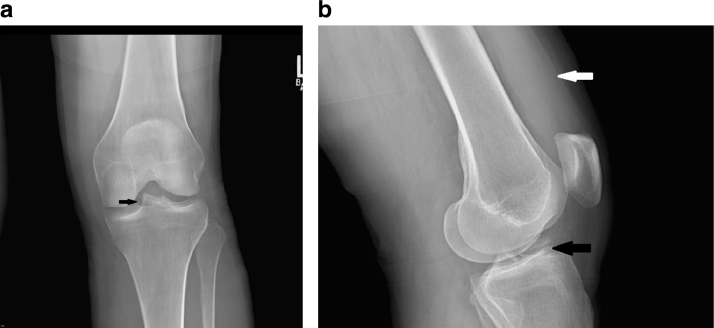
A 48-year-old female who presented with severe knee pain. Preliminary (a) AP view and (b) lateral view of the left knee were obtained demonstrating an avulsion fracture of the tibial eminence (black arrow). The lateral view (b) revealed a moderate size knee joint effusion (white arrow). AP, anteroposterior.
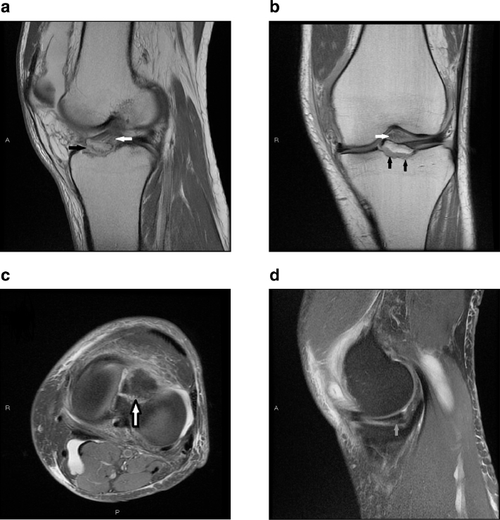
A 48-year-old female who presented with severe knee pain. (a) Sagittal proton density-weighted image and (b) coronal proton density-weighted image of the left knee revealed a tibial eminence avulsion fracture (black arrows) with preservation of the integrity of the ACL fibers (white arrows). (c) An axial T2-weighted image with fat suppression reveals the footprint of the avulsion fracture (hollow arrow) on the anterior tibial plateau. (d) Sagittal proton density-weighted image with fat-suppression reveals a medial meniscus tear (grey arrow). ACL, anterior cruciate ligament.
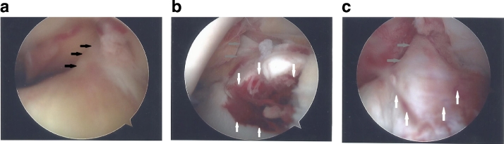
(a) An intraoperative images demonstrate an intact ACL (black arrows) on a lateral arthroscopic view. (b) An arthroscopic medial view demonstrates the avulsed tibial eminence (grey arrows). The fracture line is clearly evident (white arrows). (c) A frontal arthroscopic view postfracture reduction, demonstrates anatomic restoration of the tibial eminence (grey arrows). The fracture gap is reduced and the fracture line is less evident (white arrows). ACL, anterior cruciate ligament.
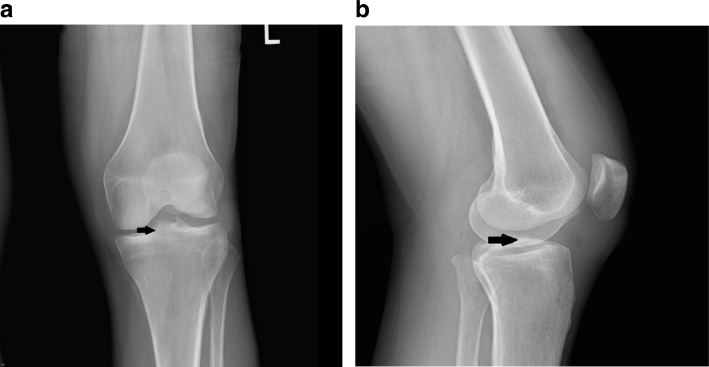
A 48-year-old female who presented with severe knee pain. (a) Postoperative AP view and (b) lateral view of the left knee were obtained which confirmed that anatomic restoration was achieved (black arrow). The fracture line is much less evident. AP, anteroposterior.
References
-
- Prince JS, Laor T, Bean JA. MRI of anterior cruciate ligament injuries and associated findings in the pediatric knee: changes with skeletal maturation. AJR. 2005;185:756–762. - PubMed
-
- Remer EM, Fitzgerald SW, Friedman H. Anterior cruciate ligament injury: MR imaging diagnosis and patterns of injury. RadioGraphics. 1992;12:901–915. - PubMed
-
- Shea KG, Apel PJ, Pfeiffer RP. Anterior cruciate ligament injury in paediatric and adolescent patients. Sports Med. 2003;33:455–471. - PubMed
-
- Millett PJ, Willis AA, Warren RF. Associated injuries in pediatric and adolescent anterior cruciate ligament tears: does a delay in treatment increase the risk of meniscal tear? Arthroscopy. 2002;18:955–999. - PubMed
-
- Anderson CN, Anderson AF. Tibial eminence fracture. Clin Sports Med. 2011;30:727–742. - PubMed
Publication types
LinkOut - more resources
Full Text Sources
Other Literature Sources
